 |
| About Bioline | All Journals | Testimonials | Membership | News |
|
||||||
|
||||||
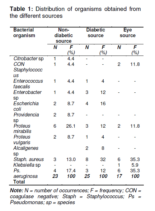
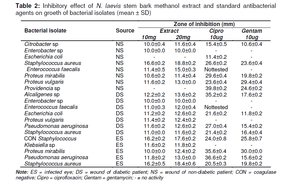
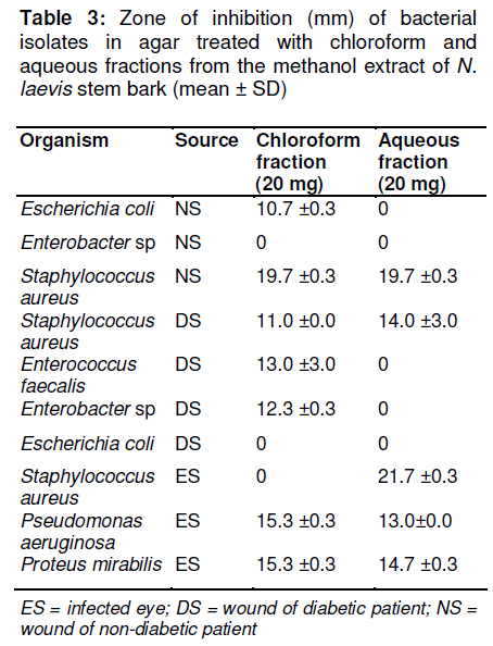
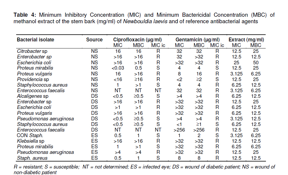
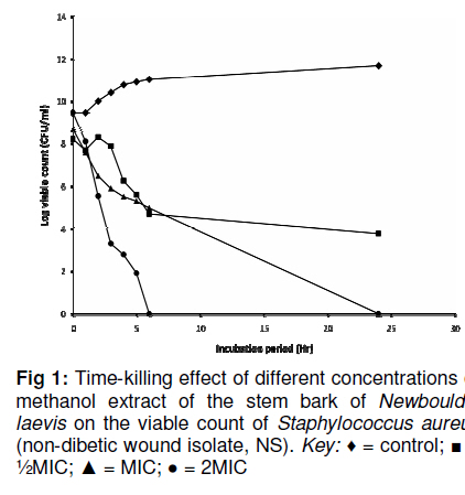
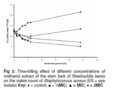
Tropical Journal of Pharmaceutical Research, Vol. 10, No. 2, April, 2011, pp. 211-218 Research Article Phytochemical and Antibacterial Evaluations of the Stem Bark of Newbouldia laevis against Isolates from Infected Wounds and Eyes JO Akerele1, BA Ayinde2* and J Ngiagah1 1Department of Pharmaceutical Microbiology, Received: 3 August 2010 Revised accepted: 2 February 2011 Code Number: pr11029 Abstract Purpose: To examine the phytochemical constituents and verify the ethnomedical claim of Newbouldia laevis (P.Beauv.) Seeman ex Bureau Bignoniaceae in treating septic wounds and eye problems. Keywords: Newbouldia laevis, Antibacterial, Phytochemical, Wound isolates, Eye isolates. INTRODUCTION Newbouldia laevis Seem (Bignoniaceae) is commonly known as smooth Newbouldia or boundary tree. It is called ‘Aduruku’ in Hausa; ‘Ogirisi’ in Igbo; ‘Ikhimi’ in Edo and ‘Akoko’ in Yoruba languages[1] . It grows to a height of about 7 -8 (up to 15) meters, more usually a shrub of 2-3 metres, many – stemmed forming clumps of gnarled branches[2] . Newbouldia laevis is widely used in African folk medicine for the treatment of malaria and fever, stomach-ache, coughs, sexually transmitted diseases, tooth ache, breast cancer, and constipation[3-5] . In South Eastern and part of the Midwestern Nigeria, the plant is used for the treatment of septic wounds and eye problems[6] . The antimicrobial potential of methanol extract of the leaf has been reported in literature while the antiinflammatory and antimalarial activities of the root extract have also been documented[7, 8] . Scientific reports on the phytochemical constituents of the plant revealed the presence of alkaloids and phenylpropanoids in the root[9], flavonoids, and tannins in the leaf[7] . Wound infection is detrimental to wound healing which is a complex process that can be delayed by many potential factors[10] . A variety of disorders commonly affect the eye and vary in severity from mild but annoying allergic conjunctivitis to sight threatening infections[11] . As it has been asserted that medicinal plants constitute a continuous source of new compounds with the potential to act against multi-resistant bacteria[12] , this work was aimed at investigating the antibacterial potential of the methanol extracts of the stem bark of Newbouldia laevis against isolates from infected wounds and eyes. EXPERIMENTAL Collection and preparation of the plant material The stem bark of Newbouldia laevis was collected in June 2007, at Themboga, Benin City, Nigeria. The identity of the plant was authenticated by Dr. Olufemi Shasanya, a taxonomist at the Forest Research Institute of Nigeria (FRIN), Ibadan. A herbarium specimen no. FHI0031505-0 was deposited at the Institute’s herbarium for reference. The stem bark was cut into pieces and dried in the oven at 50-60oC for 6 days after which they were reduced to powder form with a milling machine. Phytochemical tests These were carried out using standard methods[13,14] . Extraction of plant material About 1750 g of the powdered stem bark was extracted with methanol (5l) using maceration method for 72 hours. After filtration, the extract was concentrated to dryness (semi solid) using rotary evaporator. The dried extract (25.9 g, 1.48%) was kept in the refrigerator maintained at 4°C until required. Solvent – solvent partitioning of the methanol extract About 8g of the methanol extract of the stem bark was dissolved in methanol – water (1:1) and partitioned with chloroform (300 ml x 3). The chloroform and aqueous phases obtained were separately concentrated to dryness and weighed. Sources of the microorganisms used Following receipt of permission from the authorities of the University of Benin Teaching Hospital, Benin City, Nigeria, wound swabs were obtained from infected wounds of diabetic and non-diabetic patients while eye specimens were obtained from the eye discharges of patients with eye problems at the Ophthalmology Clinic of the same hospital. All the bacterial organisms isolated were identified using conventional biochemical techniques[15] . The bacterial isolates were maintained on nutrient agar slants at 4 °C and subcultured at intervals of three weeks to ensure their viability and purity. Antibacterial assays Preparation of the bacterial inocula Broth cultures of desired organisms were prepared by suspending two colonies of each organism in nutrient broth and incubated aerobically at 37 °C for 12 hours. Suspensions were adjusted to a turbidity of about 108 colony forming units (cfu)/ml using 0.5 McFarland Standard for visual comparison. Hundred fold dilutions were made to yield final suspensions with approximately 106 cfu/ml. Screening for antibacterial activity By adopting a modified agar-well diffusion method[16] , the screening of the methanol extract, its organic solvent fractions, and the standard antibacterial agents were carried out. 25ml of sterile molten nutrient agar cooled to about 40°C was aseptically seeded with 100 µl of the desired organism at a turbidity of approximately 106 cfu/ml. The seeded agar was aseptically poured into sterile Petri dishes of 8.5cm (85mm) in diameter and allowed to set at room temperature. With the aid of a sterile cork borer, four uniform wells of 7mm in diameter were punched on each plate. The wells were each filled with 0.1 ml of stock concentration to give a final srength of 10 mg/ml or 20 mg/ml of the extract; or 0.1 ml of ciprofloxacin or gentamicin solution to give a final concentration of 10 µg/ml which were used as positive control. A pre-diffusion time of 1 h was allowed before the plates were incubated aerobically at 37 °C for 18 -24 h. The diameter of the zones of inhibition was measured to the nearest millimeter with a transparent millimeter rule. The effects of the chloroform and aqueous fractions were tested at 20 mg/ml concentration. The tests were carried out in replicates. Determination of minimal inhibitory concentration (MIC) and minimal bactericidal concentration (MBC) The modified method of [17] was employed to determine the MIC of the extract (MSE) and its aqueous fraction. Serial two-fold dilutions of 2ml each were made in a concentration range of 50 to 0.391 mg/ml of the extract and 25 to 1.5625 mg/ml of the aqueous fraction using double strength nutrient broth supplemented with 10 % glucose. The tubes were inoculated with 100 µl of the desired organisms (at a turbidity of approximately 106 cfu /ml) and incubated aerobically at 37 °C for 18 -24 h. Microbial growth was determined by adding 20 µl of phenol red (0.2 %) indicator and observing a change in colour from red to yellow when there is microbial growth. The lowest concentration that showed no change in colour was considered the MIC. The procedure was repeated using ciprofloxacin (16 -0.031 µg/ml) and gentamicin (32 -0.063 µg/ml) and observing the tubes for turbidity. For MBC determination, 20 μl of the liquid from each tube that showed no change in colour for the extract (MSE) and the aqueous fraction (aMSE), and turbidity for reference antibacterial agents was plated on antibacterial agent-free nutrient agar. The plates were incubated aerobically at 37°C for 18 - 24 h and the lowest concentration that yielded no growth was recorded as the MBC. Time-kill testing The effect of ½MIC, MIC and 2MIC concentrations of the methanol extract on the viability of two isolates of Staphylococcus aureus was examined by the Time-killing method. The isolates used were those obtained from non-diabetic and eye isolates. The different concentrations of the MSE were prepared from a stock solution of 50 mg/ml using double strength nutrient broth. Overnight broth culture of the desired organism (200 µl) was placed in contact with a specified extract concentration. Viable counts were determined at the beginning of incubation and at intervals (i.e. after 1, 2, 3, 4, 5, 6 and 24 h) by withdrawing 100 µl aliquots of the extract-organism mixture, in order to determine the time it took the extract to eradicate all viable cells of the organisms. The same procedures were repeated using ciproflaxin with the two organisms. Data analyses The diameter of zones of inhibition for antibacterial assays were expressed as mean + S.E.M. Simple percentage was used to analyze the distribution of bacterial isolates. RESULTS Phytochemical constituents Phytochemical screening of the powdered sample of the stem bark of N. laevis revealed the presence of saponins, tannins, flavonoids, steroidal glycosides and alkaloids while cyanogenic glycosides and anthracene derivatives were observed to be absent. Bacterial isolates The clinical specimens revealed the presence of various bacterial species occurring in varying degrees in the wounds and infected eyes. In the non-diabetic patients, the bacterial organisms had Proteus mirabilis as the most predominant organism (6 out of 23 patients; 26.1 %), whereas the isolates from the diabetic patient wounds predominantly produced Staphylococcus aureus (8 cases out of 25 representing 32 %). The isolates from the infected eyes yielded Staphy. aureus and Pseudomonas aeruginosa, each constituting 35 % of the 17 isolates. While Citrobacter and Providencia species were found only in the wounds of non diabetic patients, the only occurrence of Klebsiella sp. was recorded in an infected eye. The presence of Staph. aureus, Ps. aeruginosa and P. mirabilis was common in all the specimen sources (Table 1). Antibacterial activity of extract The methanol extract of the stem bark showed activity against the tested isolates except Escherichia coli and Providencia species which were obtained from the wounds of non diabetic patients but however exhibited activity against the E.coli obtained from the wound of diabetic patient. The extract was more active against Grampositive than Gram-negative organisms with Staphylococcus aureus being the most susceptible. The Enterobacteria species, Enterococcus feacalis, Proteus vulgaris and Klebsiella species that showed resistance to the ciprofloxacin and gentamicin were observed to be susceptible to 10 and 20 mg of the extract. Staphylococcus. aureus obtained from the wounds of non diabetic patient and infected eyes were more susceptible to the extract than the one obtained from the wound of diabetic patient (Table 2). The activity of the extract was concentration dependent, with the effect at 20mg being slightly higher than the activity at 10mg although not in all the organisms. The organic solvent fractions were observed to exhibit different activities on the selected icroorganisms. While the antibacterial activities were observed to increase on some bacteria, the effects were concentrated in either the chloroform or the aqueous fraction (see Table 3). The organisms showed varying degrees of susceptibility to the extract at MIC and MBC. For some of the isolates, the same concentration of the extract that produced the MIC also was the MBC, while for others, the bactericidal concentrations was obtained at double MIC (Table 4). For the time-killing effects of the extract on the isolates of Staph. aureus obtained from the non diabetic patient and infected eye isolates, the extract produced significant reduction in the viable cell counts of the organisms. The MIC of the isolate obtained from the wound of non diabetic patient was 6.5 mg/ml. At 2MIC, the extract completely eliminated all the viable cells of the organism in less than 6h (Figure 1). The MIC of 12.5 mg/ml could not produce complete elimination of the organism obtained from infected eye although a remarkable reduction in the viable cell count was obtained with 2MIC after the incubation period of 24 h. Both the MIC and 2MIC indicated an initial kill or reduction in the viable cell counts followed by bacteriostatic effect (Figure 2). In all the concentrations of the Ciprofloxacin tested, the viable cell count of the two organisms significantly decreased below the control up to 6h after which there was resistance to the drug indicated by the increase in the viable cell counts (results not shown). DISCUSSION Although the phytochemical and antimicrobial properties of N. laevis have been reported (7), a comparative study of the stem bark methanol extract on organisms isolated from wounds and infected eyes has not been considered. The predominance of the various organisms was observed to vary depending not only on the part of the body from which they were obtained but also on the physiological state of the subjects. While Staph. aureus and P. aeruginosa were observed to be predominant in the eye isolates, the organisms obtained from the wounds of diabetic and non-diabetic patients were dominated by Proteus and Staph. aureus, respectively. The extract and the organic solvent fractions were considered active at a zone of inhibition diameter of > 10mm [18, 19] . The extract showed remarkable activities against both the Gram-negative and Grampositive bacteria. The Gram-negative bacterial organisms are generally regarded more difficult to inhibit due to the presence of a thick murein layer that tends to prevent the entry of inhibitors[20,21] . However, the extract of N. laevis inhibited some Gram-negative bacteria such as Enterobacter and Klebsiella species both at 10 and 20 mg. It is remarkable to note that Staphylococci which have a record of developing resistance rapidly and successfully to antibiotics [22] were the most susceptible to the methanol stem bark extract. The E. coli and Providencia (obtained from the wounds of non diabetic patients) specie which earlier exhibited resistance to the extract at 20 mg were observed to have MIC and MBC of 25 and 50 mg/ml. These indicated that the concentrations used earlier were grossly ineffective in subduing the growth of the organisms. Also, based on these higher MIC and MBC than others, the two organisms being Gram negatives can be said to be the most resistant to the extract. The times required to reduce or eliminate Staph. aureus isolates from non-diabetic and infected eyes showed some variation probably due to variations in the sources of the organisms. From the results, the isolate obtained from the wounds of non-diabetic subjects was eliminated completely in less than 6 h at twice the MIC and 24 h at the MIC; although there was no total elimination of the isolate obtained from the eye, the extract showed pronounced reduction in the population of the organism at two times the MIC. The susceptibility exhibited by the microorganisms to the extract as well as the aqueous and chloroform fractions may be attributed to the presence of different groups of constituents that may be acting synergistically with one another. Partitioning plant extracts in two immiscible solvents enables the separation of the plant active constituents with the eventual aim of locating where the active constituents could be residing. In this work, the results obtained show that the constituents of the extract were responsible for the observed antibacterial activities and it is important to note that, for some of the organisms, the inhibitory effects became more pronounced for the organic solvent fraction. The overall observed antibacterial activities of the extracts could be traced to the presence of the secondary metabolites like tannins, alkaloids, flavonoids reported present in the plant material. The results of this work have justified the ethnomedicinal claim of the stem bark of N. laevis in treating septic wounds and infected eyes. CONCLUSION The results obtained in this work indicate that the stem bark of N. laevis possesses natural potential to inhibit the growth of various organisms. The observed antibacterial effects may due to the presence of secondary metabolites in the plant. Our findings justify the ethnomedicinal claim of the stem bark of N. laevis in treating septic wounds and infected eyes. REFERENCES
Copyright © 2011 - Pharmacotherapy Group, Faculty of Pharmacy, University of Benin, Benin City, 300001 Nigeria. The following images related to this document are available:Photo images[pr11029t2.jpg] [pr11029t4.jpg] [pr11029t3.jpg] [pr11029t1.jpg] [pr11029f2.jpg] [pr11029f1.jpg] |
| |||||||||
