 |
| About Bioline | All Journals | Testimonials | Membership | News |
|
||||||
|
||||||
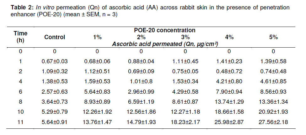
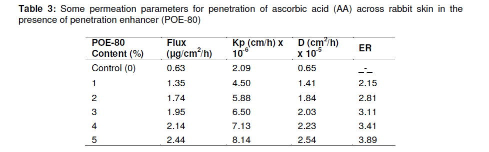
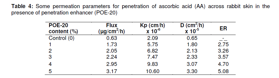
Tropical Journal of Pharmaceutical Research, Vol. 10, No. 3, June, 2011, pp. 281-288 Research Article Penetration Enhancing Effect of Polysorbate 20 and 80 on the In Vitro Percutaneous Absorption of L-Ascorbic Acid N Akhtar1, MU Rehman1, HMS Khan1, F Rasool2, T Saeed2 and G Murtaza3* 1Faculty of Pharmacy and Alternative Medicine, The Islamia University of Bahawalpur, Bahawalpur 63100; 2University College of Pharmacy, University of the Punjab, Lahore, Pakistan; 3Department of Pharmaceutical Sciences, COMSATS Institute of Information Technology, Abbottabad, Pakistan Received: 8 August 2010 Revised accepted: 2 April 2011 Code Number: pr11037 DOI: 10.4314/tjpr.v10i3.1 Abstract Purpose: To investigate the penetration enhancing effect of two polysorbates -polyoxyethylene 20 (POE-20) and polyoxyethylene 80 (POE-80) -on the in vitro percutaneous absorption of ascorbic acid (AA). Keywords: L-Ascorbic acid, Skin permeation, Polyoxyethylene (Polysorbate), Flux, Penetration enhancers When a drug system is applied topically, the drug diffuses passively out of its carrier or vehicle. The two principal absorption routes are transepidermal (diffusion directly across the stratum corneum) and transfollicular (diffusion through the follicular pore). The stratum corneum is a source of high diffusional resistance to most compounds and thus constitutes the skin’s foremost barrier layer [1]. Percutaneous penetration enhancement techniques fall primarily into two categories: physical and chemical. Physical penetration enhancement involves an externally applied force to augment the delivery of the target agent across the skin [2] while in chemical penetration enhancement, penetration enhancers, which are compounds that alter the barrier properties of the stratum corneum by reversibly affecting the lipid bilayers or the protein structures in corneocytes, are used. Penetration enhancing compounds can induce skin irritation and may significantly alter stratum corneum lipids, in some cases, irreversibly. Non-ionic surfactants have not been reported to cause skin irritation [3]. Ascorbic acid (AA) is a vitamin for humans and other primates, guinea pig, bats, passeriform birds, and most fishes and invertebrates. Other animals synthesize it as an intermediate in the uronic acid pathway of glucose metabolism [4]. It is a colourless and odourless crystalline substance, slightly sour in taste and optically active. It is soluble in water and alcohol but practically insoluble in chloroform, solvent ether and light petroleum. It is readily oxidized, particularly in the presence of copper and iron. AA is also rapidly destroyed by alkalis. It is a powerful reducing agent [5]. Topical ascorbic acid (AA) has been shown to reduce chronic ultraviolet radiation damage in murine skin and to increase the minimal erythema dose (MED) value in human skin, indicating its value in providing UV barrier. Topical application of a 5 % AA cream has been shown to improve the clinical appearance of photo-damage as well as lead to dermal production of collagen and elastin discernible at the ultra structural level [6]. There are now data confirming the benefits of topically applied AA and supporting its use as a cosmeceutical [5]. A significant increase in AA levels after topical application of different AA derivatives, including magnesium ascorbyl phosphate, ascorbyl-6-palmitate and dehydroascorbic acid, to porcine skin could not be shown [6]. Polysorbate 80 and polysorbate 20 are nonionic surfactants used in the manufacture of a variety of pharmaceutical pr oducts. They are known to enhance the permeability of phospholipid membranes, causing leakage of low molecular mass compounds. Interaction between biomembranes and polysorbate 80 and polysorbate 20 has been shown in a 2 to 4fold increase in the synthesis rates of phospholipids, indicating that the polysorbates cause some damage to epidermal membranes. Furthermore, polysorbate 20 and 80 also induce alteration in the physicochemical properties of biomembranes, specifically, increase in the permeability of sarcoplasmic reticulum [7]. The aim of the present study was to examine the permeation enhancing effect of two polysorbates on the in vitro percutaneous absorption of AA via hairless rabbit skin. EXPERIMENTAL Materials Ascorbic acid (Merck, Germany), Tween (polysorbate) 80 (Merck, Germany), Tween (polysorbate) 20 (Fisher Scientific, UK), glycerin (Sarfraz Pharma, Pakistan), and a depilatory cream (Anne French, Pakistan) were used in this study. Solubility study To determine the solubility of AA in 50 % aqueous glycerin, a large amount of AA was added to 2 ml of 50 % aqueous glycerin in a 5 ml vial capped with a rubber stopper. The mixture was then blended slowly using magnetic stirrer coated with teflon. The equilibrated mixture was centrifuged for 15 min at 5,000 rpm to get rid of the undissolved AA. A sample withdrawn from close to the surface of the supernatant was passed through a 0.45 µm membrane filter with the aid of a Millipore syringe filter unit, diluted appropriately with 50 % aqueous-glycerin mixture and the concentration of AA determined by UV spectrophotometry (Shimadzu 1601, Japan) at 264 nm [8] after suitable dilution with 50 % aqueous glycerin. Blank was 50 % aqueous glycerin. This determination was carried out in triplicate. Preparation of calibration curve for ascorbic acid An accurately weighed amount (100 mg) of AA was dissolved in 50 % aqueous glycerin and the volume was made up to 10 ml in a volumetric flask. An aliquot (0.1 ml) of this solution was transferred to a 10 ml volumetric flask and made up with 50 % aqueous glycerin to obtain 100 µg/ml stock solution. Aliquots of 0.2, 0.4, 0.6, 0.8, 1.2, and 1.6 ml, respectively, of the stock solution were separately diluted to 10 ml with aqueous glycerin to produce 2, 4, 6, 8, 12 and 16 µg/ml solutions of AA, respectively. The absorbance of these solutions was determined spectrophotometrically against blank (50 % aqueous glycerin) at 264 nm. This test was performed in triplicate. Preparation of rabbit skin Rabbit skin was used to evaluate the percutaneous delivery of AA [6]. Male white rabbits, weighing 1 -1.25 kg, were acquired from the animal house of the Department of Pharmacy, the Islamia University of Bahawalpur. The animal study was approved by The Islamia University of Bahawalpur Ethical Committee for In vivo Studies (ref no. 45-IUB/M.Phil.), and was conducted according to the international guidelines of Helsinki Declaration [9]. The hair on the dorsal area of the rabbit was carefully shaved off with an electric hair clipper, avoiding any damage to the skin. A depilatory (hair removing cream) was applied to the shaved area to completely remove any residual hair and then cleaned with a wet cotton cloth. This procedure was carried out a day prior to excising the skin in order to allow the skin to condition itself to the environment. Thus, the same circular area on the back of each rabbit, marking out the specific skin segment to be placed between the two half cells, was used [10]. The rabbit was sacrificed by cervical dislocation and the hairless skin was excised from the animal with surgical scissors. Since the skin was not firmly attached to the viscera, it was easily lifted from the animal after the incision was made. The subcutaneous fat and other extraneous tissues were removed with a scalpel [11]. Full thickness skin sections were prepared, rinsed with distilled water, wrapped in aluminum foil and stored at -50 oC (ultra-low temperature freezer, Sanyo, Japan) until used [7]. Examination of skin barrier integrity Before the experiment, the barrier integrity of the skin was checked by physical methods,. First, the integrity of the skin was assessed qualitatively by visual inspection and then by a transepidermal water loss (TEWL) apparatus, Tewameter™ (Courage + Khazaka, Germany). TEWL values before skin removal and after storage were obtained. Normal TEWL value of rabbit skin is 4 – 5 g/m2/h and so only specimens with TEWL levels of < 15 g/m2/h were used for permeability test [12]. Preparation of formulations Test formulations were prepared by dissolving 400 mg of the drug (AA) in 2 ml of 50 % aqueous glycerin solution in a small vial with the aid of a small stirrer coated with teflon. This formulation was a saturated solution based on the outcome of the solubility test carried out as described above. The solvent/medium used, i.e., 50 % aqueous glycerin, was selected due to the stability of AA in it [13]. Varying concentrations of the penetration enhancers (POE-20 and POE-80) in the formulations, ranging from 1 -5 %, were achieved by dissolving amounts of the enhancers ranging from 20 -100 mg of the enhancer in the AA solution. All the test formulations were freshly prepared and used immediately in the permeation study. The water used for all the solutions was distilled, de-ionized and degassed. Control formulation was similarly prepared except that enhancer was not added. Permeation studies The donor chamber solution chosen was 50 % glycerin. It has previously been demonstrated that glycerin, when used as a donor solution, does not alter skin penetration characteristics [12]. The skin was saturated with the receptor solution for 12 h at 4 °C prior to the permeation test to equilibrate it [5]. The apparatus used was vertical Franz diffusion cell (PermeGear, Bethlehem, USA) with an effective diffusional surface area of 1.767 cm 2. The volume of the receptor chamber was approximately 12 ml. The receptor medium was poured into the lower (reveptor) chamber to fill it and the skin was sandwiched between the lower (receptor) and upper (donor) compartments, with the superficial horny layer facing the upper chamber. Since AA is sensitive to light, the whole set-up was wrapped in aluminum foil to minimize photodegradation [13]. Stretching of the skin, as evidenced by distortion or expansion of the circular outline, was corrected for and the half cells were held tight together by a stainless steel holder. Immense care was taken to keep air from being trapped beneath the skin [14]. If air bubble was noticed, the Franz cell was tilted to remove the bubble from the assembly via the side arm of the sampling port. The receptor compartment solution, which was stirred at 600 rpm throughout the duration of the test with a teflon-coated small magnetic stirrer, was maintained at 37.0 ± 0.2 °C with the aid of a water bath equipped with a peristaltic pump. The donor compartment contained 1.5 ml of the test formulation (corresponding to infinite dose conditions) and was also occluded with aluminum foil [4]. Samples (0.14 ml) were withdrawn from the receptor compartment using a syringe fitted with a long needle at regular intervals for up to 11 h, diluted with 50 % aqueous glycerin solution and assayed spectrophotometrically at 264 nm (after appropriate dilution) against a blank comprising the test formulation but without the drug [9]. On each sampling occasion, the same volume of fresh receptor medium at 37 0C as was removed was added to the receptor compartment to replenish the receptor medium [12]. The test was carried out in duplicate. Computation of permeation data As a result of the sampling of large volumes from the receptor medium (and replenishment with equal volumes of the fresh medium), the receptor medium was constantly being diluted. Consequently, the receptor compartment concentration of AA was corrected for sample removal and replenishment using Eq 1 [15]. C'n = Cn (Vt / Vt-Vs) (C'n-1 / Cn-1)…….. (1) where C'n = corrected drug concentration in the nth sample, Cn = measured drug concentration in the nth sample, C'n-1 = corrected drug concentration in the (n-1)th sample, Cn-1 = measured drug concentration in the (n-1)th sample, Vt = total volume of receptor solution, Vs = volume of the sample, and C'1 = C1. Permeation rate The corrected data were expressed as the cumulative drug permeation per unit of skin surface area using Eq 2. Qn = C'n/A …………………………….... (2) where A = 1.767 cm2 Steady-state flux (Jss) The cumulative amount of drug per unit area (Qn, µg) in the receiver chamber was plotted as a function of time (t, h), and steady-state flux (Jss, µg/h/cm2) was calculated from the slope of the linear portion (5 -10 h) of the curve [9]. Permeability coefficient (Kp) Apparent permeability coefficient (Kp, cm/h) was calculated according to Eq 3. Kp = Jss/Cd ……………………….….. (3) where Cd = drug concentration in the donor compartment, i.e., 20.0 % w/v (20.0×10⁴ µg/ml). It was assumed that under sink conditions, drug concentration in the receptor compartment was negligible compared to that in the donor compartment [11]. Enhancement ratio Enhancement ratio (ER), which measures the penetration enhancing activity of the enhancers, was calculated as in Eq 4. ER = Kp1 /Kp0 ……………………… (4) where Kp0 is the permeability coefficient of control (i.e., without enhancer) and Kp1 is the permeability coefficient in the presence of enhancer. Diffusion coefficient (D) Diffusion coefficient (D) was calculated using Eq 5 [13]. Kp = D/A2 ……………………….…. (5) Thus, D = Kp x A2 …………………………… (6) where D and Kp are as defined above and A2 = square of effective absorption area. Statistical analysis Statistical analysis of the experimental data was carried out by one way analysis of variance (ANOVA) at a significance level of p < 0.05 using SPSS 12.0 software (IBM). RESULTS Solubility of ascorbic acid (AA) The solubility of AA in 50 % glycerin aqueous solution was 1 g/100 ml. The coefficient of regression (R2) of the standard solubility curve was 0.9997 while the regression equation was y = 0.0563x + 0.0047. In vitro permeation of AA Tables 1 and 2 show the effect of the penetration enhancers, POE-80 and POE-20, respectively, on the amount of ascorbic acid (AA) permeated across rabbit skin. The results show that as the concentration of penetration enhancer increased, AA permeation also increased. Other permeation data The other AA permeation data derived from this study, namely, steady state flux (Jss), permeability coefficient, diffusion coefficient and enhancement ratio are given in Tables 3 and 4. Without the enhancer, AA flux was 0.626 µg/cm2/h while mean permeability coefficient (Kp) was 2.09 × 10⁻⁶ cm/h. AA flux (µg/cm2/h) was 3.17 and 2.44 for POE-20 and POE-80, respectively. Mean permeability coefficient (Kp, cm/h) was 10.6 × 10⁻⁶ and 8.14 × 10⁻⁶ for POE-20 and POE-80, respectively. Maximum flux (3.17 µg/cm2/h) at POE-20 concentration of 5 % was observed with an enhancement ratio (ER) of 5.07 in relation to control (i.e., AA without enhancer) while maximum flux (2.44 µg/cm2/h) at POE-80 concentration of 5 % was observed with an ER value of 3.89. The values of diffusion coefficient (cm2/h) were 3.30 × 10⁻⁵ and 2.54 × 10⁻⁵ for POE-20 and POE-80, respectively. The magnitude of the various permeation paraneters varied with the content of penetration enhancer, with the magnitude increasing as enhancer concentration was raised. DISCUSSION Percutaneous drug absorption is thought to occur via various skin auxilae including hair follicles, sweat ducts and sebaceous glands at an early stage as well as at a steady state. It is believed that nonionic surfactants, as penetration enhancers, seep into the intercellular lipid bilayers of skin thereby decreasing the crystallinity of these lipid bilayers and amplifying their permeability characteristics [16]. Another mechanism of action of non-ionic surfactants is that they cause the emulsification of the sebum and hence increase the thermodynamic activity of the drug [17]. Nonionic surfactants are widely used in topical formulations. Among these surfactants are the polysorbates (Tweens), including the ones used in this study. It is evident from the permeability coefficient (Kp) data that the penetration enhancers used in this study, polysorbate 20 (POE-20) and 80 (POE-80), increased the permeation of ascorbic acid (AA) through hairless rabbit skin in vitro, and that the effect of the enhancers was concentration-dependent. It would seem that the enhancers increased AA permeation by decreasing skin resistance to the diffusion of the drug. The faster penetration of the drug through skin due to the presence of POE-20 and POE-80 can be explained by following mechanisms. First, adsorption and fusion of drug molecules onto the surface of skin, resulting in the high thermodynamic activity gradient of drug at the interface, which is the driving force for drug permeation. Second, the influence of penetration enhancer decreases stratum corneum hindrancebarrier characteristics. Third, modification in the structure of stratum corneum may result in more loose intracellular lipid barrier in the stratum corneum and thus become more permeable to permeation enhancers [19,20]. The enhancement effect of POE20, a polysorbate containing C12 saturated hydrophobic group, can be attributed to an enhancement in thermodynamic activity due to micellar complexation. In contrast, POE80 effect may be due to change in the barrier properties of the skin and in the vehiclestratum corneum partition coefficient. Sarpotdar and Tatz [22] observed an enhancement in the transdermal flux of hydrocortisone in the presence of nonionic surfactants, including POE20 and POE80. In another work, POE80 was reported to improve the skin permeation of hydrocortisone and lidocaine [23]. It has also been shown that POE20 accelerated the percutaneous absorption of 5flourouracil and captopril, and particularly increased the transdermal flux of captopril [1923]. These findings are largely in agreement with the results obtained in the present work. CONCLUSION This work further supports the earlier claims of polysorbate 20 and 80 as penetration enhancers for transdermal delivery of drugs. Furthermore, the higher the concentration of the penetration enhancer, the higher the permeability of ascorbic acid (AA). Increase in AA permeation was achieved with enhancer concentrations as low as 1 %. This is important because these surfactants, being non-ionic, are much less damaging to the skin than other classes of surfactants and enhancers. REFERENCES
Copyright © 2011 - Pharmacotherapy Group, Faculty of Pharmacy, University of Benin, Benin City, 300001 Nigeria The following images related to this document are available:Photo images[pr11037t4.jpg] [pr11037t2.jpg] [pr11037t1.jpg] [pr11037t3.jpg] |
| |||||||||
