 |
| About Bioline | All Journals | Testimonials | Membership | News |
|
||||||
|
||||||
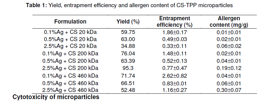
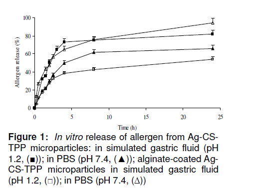
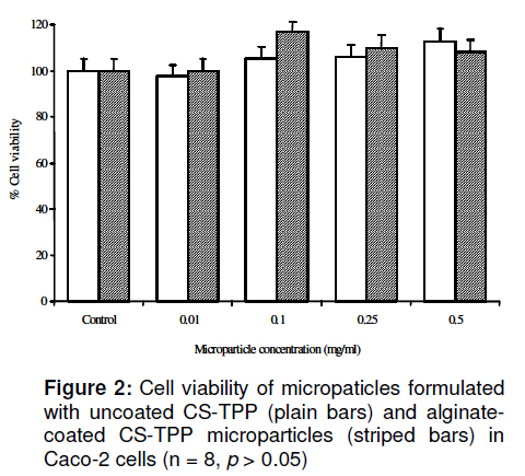
Tropical Journal of Pharmaceutical Research, Vol. 10, No. 3, June, 2011, pp. 317-324 Research Article Development of Alginate/Chitosan Microparticles for Dust Mite Allergen Tittaya Suksamran, Praneet Opanasopit *, Theerasak Rojanarata and Tanasait Ngawhirunpat Faculty of Pharmacy, Silpakorn University, Nakhon Pathom 73000, Thailand Received: 17 November 2010 Revised accepted: 11 April 2011 Code Number: pr11041 DOI: 10.4314/tjpr.v10i3.8 Abstract Purpose: To develop chitosan/alginate microparticles for the mucosal delivery of allergen from dust mite (Dermatophagoides pteronyssinus). Keywords: Alginate, Chitosan, Microparticle, Allergen delivery, Dust mite, Dermatophagoides pteronyssinus INTRODUCTION Chitosan, as a cationic polysaccharide, has gained increasing attention in pharmaceutical research and development due to its favorable biological properties, such as nontoxicity, biodegradability and mucoadhesive properties. Additionally, chitosan micro/nanoparticles can be easily prepared by ionic gelation method using tripolyphosphate (TPP) as precipitating agent [1,2]. In spite of its unique properties, chitosan has an apparent pKa of 5.6 -6.2 and is only soluble in acidic solutions. When incubated in physiological fluid environment, chitosan loses its mucoadhesive capacity and permeation enhancing effect due to deprotonation of chitosan, which causes chitosan to lose its advantage over other carriers for mucosal vaccine delivery. However, chitosan has limited ability for controlling release in acid medium [3]. One approach to overcome this and other obstacles is by coating an acid-resistant polymer, such as sodium alginate, onto the surface of chitosan microparticles [4]. Usually, chitosan–alginate microparticles are obtained by two principally different procedures. The first is a one-step method, where a complex coacervate membrane is formed at the interface between the alginate and chitosan solutions when the alginate solution is dropped directly into a solution of calcium chloride mixed with chitosan, while the other method is a two-step method, where chitosan-TPP microparticles are recovered and subsequently coated with calcium alginate. Recently, we successfully prepared bovine serum albumin (BSA)-loaded alginate microparticles by crosslinking alginate with calcium chloride solution using an electrohydrodynamic spraying technique [5]. The advantage of this method was attributed to the mild condition, which excluded the application of harmful organic solvent, at room temperature, and also efficiently retained the bioactivity of macromolecules (protein, DNA, etc) during encapsulation. The main objective of this study, therefore, was to prepare a microparticulate system (coated with sodium alginate and incorporating dust mite allergen) by cross-linking alginate with calcium, and chitosan with TPP, and characterize them for their release and other physicochemical properties. EXPERIMENTAL Materials High viscosity sodium alginate, derived from brown algae (AHV, 40000 cps) and 3-(4,5dimethylthiazol-2-yl)-2,5-diphenyl tetrazolium bromide (MTT) were purchased from Sigma-Chemical Co., St. Louis, MO, USA. Chitosan (mol wt: 20, 200 and 460 KDa) was purchased from Seafresh Chitosan Lab Bangkok, Thailand). Allergenic extract standard mite (dust mite allergen, Ag) was obtained from ALK-Abelló, Inc., Round Rock, TX, USA. Penta-sodium triphosphate (TPP) was purchased from Ajax Chemicals, New South Wales, Australia. Caco-2 cells were obtained from American Type Culture Collection (Rockville, MD,USA). Dulbecco’s modified Eagle’s medium (DMEM), trypsin-EDTA, penicillin-streptomycin antibiotics and fetal bovine serum (FBS) were supplied by Gibco-Invitrogen, Grand Island, NY, USA. Preparation of chitosan salt (CS) Chitosan hydrochloride salt (CS) was prepared as previously described [1]. Briefly, 1 %w/w CS solution was prepared by dissolving chitosan in distilled water containing hydrochloric acid in a 1:0.8 molar ratio and then stirred for 12 h at room temperature. The solution (10 ml) was spraydried (Minispray Dryer, Büchi 190, Postfach, Switzerland) at an inlet temperature of 125 ± 2 0C at spraying rate of 8 ml/min. Preparation of alginate-coated, allergenloaded chitosan microparticles Chitosan microparticles were prepared by ionotropic gelation [1]. Briefly, CS and TPP were dissolved in water with magnetic stirring to prepare solutions of 0.1% w/v CS and 0.2 % w/v TPP. Allergen-loaded chitosan microparticles (Ag-CS-TPP) were prepared by drop-wise addition of 10 ml TPP solution containing dust mite allergen (0.1, 0.5, 2.5 %w/v) into a 40 ml CS solution while stirring at 700 rpm for 20 min. The resultant particles were collected, washed with distilled water and separated by centrifugation at 3000 rpm for 15 min. They were then lyophilized (Labconco, Freezone 2.5, Kansas City, MO, USA) and stored at 4 °C. Blank CS-TPP microparticles (without allergen) were similarly prepared. To prepare alginate-coated microparticles (Ag-CS-TPP), 2.5 % of allergen (Ag)-CS-TPP microparticles were selected. Briefly, the powders (sodium alginate and CaCl2) were dissolved separately in water using a magnetic stirred to obtain solutions of 0.025, 0.05, 0.1, 0.25, 0.5 %w/v and CaCl2 (4 or 6 % w/v), respectively. Fifty microliters of the alginate solution were mixed well with 13 mg of CS-TPP microparticles and extruded dropwise by electrohydrodynamic spraying techniques as previously described [5] through a glass syringe fitted with a 20-gauge blunt needle into 150 mL of 4 or 6 %w/v CaCl2 solution under gentle stirring. This setup used the applied voltage for 18 kV (Protek® DC power supply) with the distance from the needle tip to the receptor solution (CaCl2) and extrusion rate fixed at 30 cm and 1 ml/h, respectively. After extrusion, the microparticles were collected in CaCl2 with gentle stirring for 30 min, separated from the CaCl2 solution by centrifugation at 5862 g for 10 min, washed twice and re-suspended in distilled water. This suspension was lyophilized and then stored at 4 °C. Blank microspheres (without allergen) were similarly prepared. Morphology and particle size distribution Morphological characterization of the microparticles was performed by inverted microscopy (Eclipse TE 2000-U, Japan) at a magnification of x 20 as well as by transmission electron microscope (Jeol Jem1230, Tokyo, Japan). Particle size analysis was measured using Zetasizer Nano ZS (Malvern Instruments, Malvern, UK). Reverse-phase HPLC analysis of allergen The reverse-phase high performance liquid chromatography (HPLC) system for the determination of allergen consisted of an Agilent HPLC system (Agilent 1100 series, USA) at a flow rate of 1.0 ml/min. For separation, a C18 reverse-phase column (5 µm, 150 mm x 0.5 mm, Waters, USA) was used. The following gradient elution profiles were used: solvent A, 0.1%v/v trifluoroacetic acid (TFA) in water; and solvent B, 0.085 %v/v TFA in acetonitrile [6]. The injection volume was 20 µl. The detection was performed using a fluorescence detector with an excitation wavelength of 280 nm and emission wavelength of 304 nm. Determination of allergen (Ag) entrapment efficiency One hundred milligrams of Ag-CS-TPP microparticles were dissolved in 2 ml of 5M HCl (pH 1.2) while the same amount of alginate-coated Ag-CS-TPP microparticles was dissolved in phosphate buffer saline (PBS, pH 7.4). The mixture, in each case, was stirred at room temperature until completely dissolved. The amount of allergen in the microparticles was determined by a reverse-phase HPLC. The yield, entrapment efficiency (EE), and loading capacity (LC) were calculated as in Eqs 1 -3, respectively [7]. Yield (%) = (Wm/Wt ) x 100 ………… (1) where Wm is the weight of microparticles and Wt is the theoretical weight of microparticles. EE = (Pt/ Lt) x 100 ……………..……. (2) where Pt is the amount of allergen embedded in microparticles. Lt is the theoretical amount of allergen (obtained from feeding condition) incorporated into microparticles. LC = Pt/Mt ……………………………… (3) where Pt is the amount of allergen embedded in microparticles (in mg), and Mt is the total amount of microparticles harvested (in g). Assessment of in vitro allergen release In vitro release studies were performed by suspending 10 mg of microparticles in 1.5 ml microcentrifuge tubes with 1 ml of PBS pH 7.4 or simulated gastric fluid without pepsin (0.2% w/v NaCl and 0.7% v/v HCl) pepsin (pH 1.2). All the tubes were then incubated at 37 °C in a shaker-incubator shaking at 200 rpm to maintain the particles in suspension (sink conditions). To determine the amount of allergen released after a given time (1, 2, 4, 8, 12 and 24 h), the sample was centrifuged for 15 min at 1077 x g. The supernatant was measured in triplicate by reverse-phase HPLC using fluorescence detector at an excitation wavelength of 280 nm and emission wavelength of 304 nm. Cell-culture experiments The colonic adenocarcinoma cell line, Caco 2 cells, were grown in Dulbecco's modified Eagle's medium (DMEM) at pH 7.4, supplemented with 10 % fetal bovine serum, 2 mM L-glutamine, 1 % non-essential amino acid solution and 0.1 % penicillinstreptomycin solution in a humidified atmosphere (5 % CO2, 95 % air, 37 °C). The cells were grown under standard conditions until 60 – 70 % confluency. Cells from passages 20 -40 were used for the experiments. Evaluation of cytotoxicity The cytotoxic effects of the microparticles were investigated in Caco-2 cells by an 3(4,5-dimethylthiazol-2-yl)-2.5diphenyltetrazolium bromide (MTT) cytotoxicity assay [8]. The cells were seeded at a density 2 × 10⁴ cells/well in 96-well cell culture plates and pre-incubated for 24 h prior to microparticle treatment. The cells were then treated with the microparticles at various concentrations ranging from 0.01 to 0.5 mg/ml in serum-free medium (pH 7.4) for 24 h. After treatment, the microparticle suspensions were removed and fresh cell culture medium was added, and then incubated for 4 h to stabilize the cells. Finally, the cells were incubated with 100 µl MTT containing medium (0.1 mg/ml MTT in serumfree medium) for 4 h; the medium was removed, and the formazan crystal formed in the living cells was dissolved in 100 µl DMSO per well. Relative cell viability (RCV) was calculated based on absorbance at 550 nm using a microplate reader (Universal Microplate Analyzer, model AOPUS01 and AI53601, Packard BioScience, CT, USA) using Eq 4. Viability of non-treated control cells was arbitrarily defined as 100 %. RCV = (OD550,sample – OD550,blank )/(OD550,control – OD550,blank ) x 100 ……………….……… (4) Statistical analysis All experimental measurements were performed in triplicate and the data expressed as mean ± standard deviation (S.D.). Statistically significant differences were examined using one-way analysis of variance (ANOVA) followed by LSD post-hoc test. The level of significance was set at p < 0.05. RESULTS Entrapment efficiency and allergen content of microparticles Table 1 shows the yield, entrapment efficiency and allergen content of Ag-CS-TPP microparticles. The yield obtained with this technique was in the range 34 -76 % As the molecular weight (MW) of chitosan (CS) increased, allergen entrapment efficiency and content also increased. The highest allergen content (0.3 mg/g) was obtained for the microparticles containing 2.5 % Ag and CS MW of 460 kDa. Hence, this microparticle formulation was selected for alginate coating. Alginate-coated Ag-CS-TPP microparticles The lower the alginate solution concentration, the smaller the particles obtained. CaCl2 concentration of 4 %w/v yielded microparticles with smaller size and more spherical shapes. Hence, the microparticles containing 0.025 %w/v alginate and 4 %w/v and the allergen content of alginate-coated Ag-CS-TPP microparticles were 26±8.7 % and 0.3 mg/g particles, respectively. In vitro allergen release The release profile of allergen from the coated and uncoated Ag-CS-TPP microparticles is shown in Figure 1. Allergen release at pH 7.4 from alginate-coated Ag-CS-TPP microparticles was approximately 95 % over a period of 24 h, compared with about 40 – 50 % at 24 h at pH 1.2. On the other hand, allergen release over a period of 24 h from the uncoated microparticles at pH 1.2 was approx. 80 % which was higher than at pH 7.4 (65 %). In the case of alginate-coated CS-TPP microparticles, the higher allergen release was observed in PBS buffer (pH 7.4). Cytotoxicity of microparticles The cytotoxicity results, shown in Fig 2, indicate that after 24 h, there was no significant difference (p > 0.05) in viability between the cells treated with microparticles and untreated cells (control) at concentrations up to 0.5 mg/mL. DISCUSSION Allergen entrapment Allergen entrapment in CS-TPP microparticles was most likely facilitated by electrostatic interaction of the positivelycharged (due to the amino group) chitosan and the negatively-charged allergen. The increase in entrapment efficiency with chitosan (CS) molecular weight (MW) has previously been reported [2,11,12] and may be due to the increase in CS solution viscosity with increase in MW which hinders the diffusion of the allergen from the droplet. The increase in the solution viscosity when higher chitosan concentration was used could be a major factor that influenced the rise in entrapment efficiency. Bazzo et al [12] who studied poly (3-hydroxybutyrate)/ chitosan/piroxicam and ketoprofen composite microparticles found that encapsulation efficiency decreased when chitosan concentration decreased. Increase in solution viscosity as chitosan concentration rose may account for the increase in entrapment efficiency of the microparticles. It has been claimed that enhancement of polymer solution viscosity elevated entrapment efficiency because it is more difficult for the drug to diffuse into the outer phase of the system [11]. The spread length of chitosan chain in solution usually varies with MW, which could affect protein interaction and encapsulation. It has been found that bovine serum albumin (BSA) encapsulation efficiency increased from 61.1 to 69.9 and 78.2 % when the MW of chitosan was changed from low to medium and high, respectively [2]. This probably explains why the CS-TPP microparticles incorporating high MW CS showed the highest allergen content. The diameter of the beads formed using the electrostatic droplet technique would be dependent on the bore size of the needle used and the viscosity of the alginate solution. However, since needle bore size was not varied in this study, the viscosity of sodium alginate could also have influenced the diameter and shape of the beads produced. The beads became more spherical as the concentration of sodium alginate solution increased [9]. Allergen release The allergen release from the microparticles can be partly attributed to the peculiar characteristics of chitosan. Chitosan dissolves easily at low pH while it is insoluble at higher pH ranges. The mechanism of the polymer’s pH-sensitive swelling involves the protonation of the amine groups of chitosan at low pH. This protonation leads to chain repulsion, diffusion of proton and counter ions along with water inside the gel and dissociation of secondary interactions. This produces the matrix that dissolves in the stomach. However, the higher allergen release from alginate-coated CS-TPP microparticles may be due to the fact that alginate, with its carboxylate groups, shrinks at low pH but dissolves at high pH, and this explains the retardation of allergen release pH 1.2. Upon mixing, the carboxylate groups of alginate and the ammonium groups of chitosan ionically interact to form a polyelectrolyte complex. Complexation of alginate with chitosan reduces the porosity of the matrix and thus should decrease decrease the diffusion of the encapsulated material [14]. However, we found that higher allergen release from the alginate-coated microparticles actually occurred. And this might be due to the high affinity of phosphate ions to calcium ions. Therefore, when phosphate is present in the dissolution medium used, probable destabilizing of calcium-crosslinked alginate matrix by phosphate ions has to be taken into account as it may cause a faster opening or breakdown of crosslinking than in 0.1M HCl medium (pH 1.2) where such a phenomenon does not occur. Loss of calcium ions from the alginate coating resulted in increase of the permeability of the coating and consequently, increased diffusion of the allergen into the release medium [15]. Cytotoxicity of microparticles The findings of the present work correlate with results of another study which indicate that both alginate-coated and uncoated chitosan microparticles did not induce spleen cell death [15]. However, a recent study showed that CaCl2-crosslinked alginate microparticles caused a slight reduction in cell viability in human umbilical vein endothelial cells (HUVEC) cells except supersaturated particle concentrations were employed [16]. CONCLUSION A suitable chitosan microparticles delivery system with sodium alginate coating yielded a formulation in the microsize range, and showing superior controlled allergen release property to the uncoated microparticles. The particles were non-toxic, thus indicating that alginate-coated chitosan microparticles were safe and can be further developed for mucosal allergen delivery. ACKNOWLEDGEMENT The authors wish to thank the Silpakorn University Research and Development Institute for financial support. REFERENCES
Copyright © 2011 - Pharmacotherapy Group, Faculty of Pharmacy, University of Benin, Benin City, 300001 Nigeria The following images related to this document are available:Photo images[pr11041t1.jpg] [pr11041f2.jpg] [pr11041f1.jpg] |
| |||||||||
