 |
| About Bioline | All Journals | Testimonials | Membership | News |
|
||||||
|
||||||
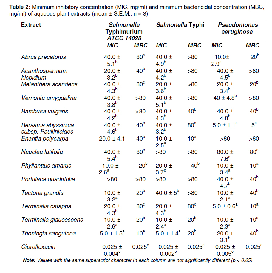
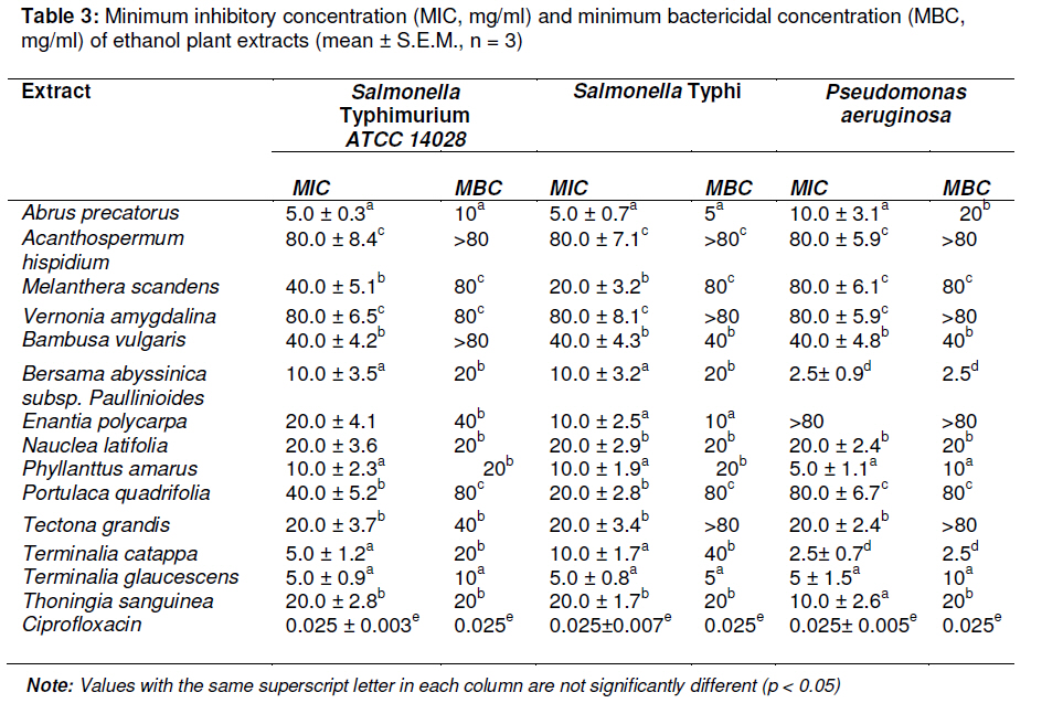
Tropical Journal of Pharmaceutical Research, Vol. 10, No. 3, June, 2011, pp. 335-340 Research Article Evaluation of the Antibacterial Activity of 14 Medicinal Plants in Côte d’Ivoire GEK Bolou1, I Bagré1*, K Ouattara1 and AJ Djaman1,2 1Biochemical Pharmacodynamy Laboratory, Biosciences Department, Cocody University, Abidgan; Received: 10 July 2010 Revised accepted: 24 April 2011 Code Number: pr11043 DOI: 10.4314/tjpr.v10i3.3 Abstract Purpose: To evaluate the antibacterial potentials of fourteen ethnobotanically selected plants traditionally used in different parts of Côte d’Ivoire for the treatment of typhoid fever and gastrointestinal disorders. Keywords: Antimicrobial activity, Ivorian medicinal plants, MIC, MBC. INTRODUCTION Medicinal plants have been known for their healing and/or disease-curing qualities for centuries. Some drugs of plant origin used in conventional medical practice are direct plant extracts or plant materials that have been suitably prepared and standardized [1]. Many readily available plants in Côte d’Ivoire are used in traditional folklore medicine for the treatment of typhoid fever and gastrointestinal disorders such as cholera, diarrheoa and dysentery [2,3]. However, several of them have not been investigated from a pharmacological point of view to demonstrate their antibacterial properties, which could support their use as antityphoid, anticholera or antidiarrhoeal remedies in traditional medicine. The objective of the present study was to evaluate the antibacterial activity of the extracts of some plants used in Côte d’Ivoire traditional healthcare system, against a group of pathogenic bacteria. These plants are used by the indigenous people in different parts of Côte d’Ivoire for the treatment of infectious diseases such as cholera, diarrhoea, dysentery and other gastrointestinal disorders [4,5]. A total number of fourteen species of plants belonging to eleven different families were tested on S. typhimurium, S. typhi and P. aeruginosa. EXPERIMENTAL Plant materials Fourteen medicinal plants -[Abrus precatorius Linn. (Fabaceae), Acanthospermum hispidium Schrank. (Asteraceae), Melanthera scandens Schum. & Thonn. (Asteraceae), Vernonia amygdalina Del. (Poaceae), Bambusa vulgaris Schrad. (Poaceae), Bersama abyssinica subsp. paullinioides Planch. (Melianthaceae), Enantia polycarpa Engl. & Diels (Annonaceae), Nauclea latifolia SM. (Rubiaceae), Phyllanthus amarus Schum. and Thonn. (Euphorbiaceae), Portulaca quadric-folia Linn. (Portulacaceae), Tectona grandis Linn. (Verbenaceae), Terminalia catappa Linn. (Combretaceae), Terminalia glauce-scens Planch. (Combretaceae) and Thoningia sanguinea Vahl. (Balanophoraceae) -were ethnobotanically selected for antimicrobial screening in the present study. The fresh plants were collected from Daloa in Central West Region of Ivory Coast in June 2009. The botanical identification of the plant samples was carried out by Pr Ake Assi, of the Department of Botany, University of Cocody-Abidjan. Voucher specimens of the plant materials were conserved in the herbarium of Centre National de Floristique (CNF) of Abidjan. The plant species, parts used, local name, voucher specimen numbers and the traditional uses of the plants are listed in Table 1. Bacterial strains The organisms used were clinical isolates of Salmonella typhi, Pseudomonas aeruginosa and a collection strain of Salmonella typhimurium ATCC 14028 (provided by Laboratoire de Bactériologie and Virologie of Institut Pasteur de Côte d’Ivoire). Extraction procedure The freshly collected parts of the plants were air-dried at room temperature for 7 days and powdered. Briefly, 20 g of the powder of each of the plant materials was separately soaked in 500 ml distilled water for 24 h with constant stirring. The suspension was filtered through Whatman filter paper no. 1. The filtrate was concentrated by vacuum using a rotary evaporator to obtain the dry aqueous extract. In the same way, the ethanol (70 %) extract was also obtained. Antibacterial tests Isolates were considered susceptible, less susceptible, or resistant to a particular antimicrobial agent on the basis of the diameters of the inhibitory zones that matched the criteria of the manufacturer's interpretation table, which is based on the guidelines of the National Committee for Clinical Laboratory Standards [6,7]. Minimal inhibitory concentration (MIC) was determined according to Wilkinson and Gentry [8]. Two-fold dilutions of the extract were made in the concentration range of 0.625 to 80.0 mg/ml. The tubes were inoculated with a microorganism suspension at a final density of 106 ufc/ml prepared according to the procedure of Société Française de Microbiologie (SFM) [9]. The tubes were incubated at 37 °C for 24 h. The lowest concentration of the tube which did not show any visible growth after macroscopic examination was considered as the MIC. Minimal bactericidal concentration (MBC) is defined as the concentration producing a 99.9 % reduction in colony forming units (CFU) number in the initial inoculum. It was determined by subculture on nutrient agar as previously described [10]. Briefly, the tubes without growth after 24 h of incubation were subcultured on Mueller Hinton agar in Petri dishes for 24 h. MBC was determined as the lowest concentration that showed no bacterial growth in the subcultures [11,12]. The tests were performed in triplicate. Dilutions of ciprofloxacin served as positive control. Statistical analysis The data are presented as mean ± SEM. All the data were analyzed by one-way ANOVA and differences between the means were assessed with Neuman-Keuls’s multiple comparison tests. Differences were considered significant at p < 0.05. All analyses were carried out using Graph Pad software, version 5.01 (USA). RESULTS The results of MIC and MBC tests are presented in Tables 2 and 3, respectively. Generally, each plant studied exhibited antibacteral activity against at least one tested bacterial strain. The aqueous and ethanol extracts of B. abyssinica, P. amarus, T. catappa and T. glaucescens presented MIC and MBC values ranging from 2.5 to 80 mg/ml against S. typhimurium, S. typhi and P. aeruginosa. The ethanol extracts of B. abyssinica and T. catappa exhibited strong MIC and MBC values of 2.5 mg/ml against P. aeruginosa. The ethanol extracts of T. glaucescens and A. precatorius showed strong bactericidal activities with MIC and MBC values of 5 mg/ml against S. typhi while the aqueous extract of T. sanguinea and the ethanol extract of T. glaucescens exhibited strong bactericidal activities with MIC and MBC values of 5 and 10 mg/ml, respectively. The ethanol extract of A. hispidium, and aqueous extracts of V. amygdalina and B. vulgaris were inactive against the three strains. Also, the aqueous extract of A. hispidium, and ethanol extracts of V. amygdalina and T. grandis were inactive against S. typhi and P. aeruginosa strains. DISCUSSION Phytoconstituents present in plants, namely, flavonoids, alkaloids, tannins and triterpenoids, have demonstrated exciting potentials for the expansion of the range of modern chemotherapies against a wide spectrum of microorganisms [13,14]. The antimicrobial activities of various plants have been reported by several researchers [15,16]. The results of this study show that all the selected plants exhibited varied antimicrobial activities against the tested organisms. The extracts tested were effective antibacterial agents against a group of microorganisms that are implicated in either typhoid fever and/or other gastrointestinal disorders/ infectious diseases such as diarrheoa and dysentery. The ethanol extracts showed the better bactericidal activity. Thus, ethanol is the more suitable extraction solvent for the plants assessed. Previous studies also indicate that ethanol was the best solvent for extracting antimicrobial substances from some plants [17,18]. Among the fourteen plants evaluated, the extracts of A. precatorius, B. abyssinica and T. glaucescens were the most effective against the selected strains, followed by T. catappa and T. sanguinea in that order. This study has not only shown the scientific basis for some of the therapeutic uses of traditional plants, but has also confirmed the ethnomedicinal claims for the selected plants. CONCLUSION The results of this study lend some scientific credence to the indigenous uses of the Ivorian medicinal plants evaluated for the treatment of infectious diseases and gastrointestinal disorders such as cholera and diarrhoeal diseases. There is, however, a need for further investigation of the plants that exhibited the highest bactericidal activity with a view to identifying and isolating their active principles. ACKNOWLEDGEMENT The authors wish to thank Pr. Aké Assi Laurent of the Department of Botany, University of Cocody-Abidjan, for the botanical identification and collection of the plants used in this study. REFERENCES
Copyright © 2011 - Pharmacotherapy Group, Faculty of Pharmacy, University of Benin, Benin City, 300001 Nigeria The following images related to this document are available:Photo images[pr11043t1.jpg] [pr11043t2.jpg] [pr11043t3.jpg] |
| |||||||||
