 |
| About Bioline | All Journals | Testimonials | Membership | News |
|
||||||
|
||||||
Iranian Journal of Pharmacology & Therapeutics, Vol. 2, No. 1, 2003, pp. 12-14 An Improved Method for Injection of Bolus Doses of Drugs into the Perfusion Circuit of Isolated Perfused Rat Kidney Utilizing a Six-port Injection Valve Soltan Ahmed Ebrahimi and Ali Rouzrokh Razi Institute for Drug Research,
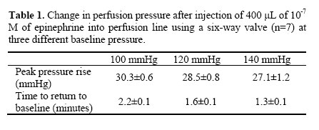
Iran University of Medical Sciences, Tehran, Iran. Received May 6, 2003; Code Number: pt03002 ABSTRACT The isolated perfused rat kidney experiment was introduced in 1959 for studying the regulation of renal blood flow and is recognized as a valuable preparation for studying physiological and biochemical aspects of renal function such as hemodynamics, glomerular filtration rate (GFR) and overall handling of fluids. Dose-response curves are obtained by injection of bolus doses of drugs into the perfusion line. However current injection methods can cause several problems such as low reproducibility and altered baseline pressure. The aim of the present work is to develop a simple method of introducing the drug into the perfusion circuit which is free from these aberrations. This was achieved using a six-way injection valve placed in the perfusion circuit, just before the kidney. To assess the reproducibility of this method, 400 μL epinephrine (10-7 M) was injected seven times into an isolated perfused rat kidney. The mean peak pressure rise (mmHg) was 30.3±0.6, 28.5±0.8 and 27.1±0.6 at 100, 120 and 140 mmHg base perfusion pressures respectively. Base pressure returned to pre-injection levels under all conditions tested. Low standard deviation of pressure maxima indicates the high reproducibility of this method while multiple injections can be made in a relatively shorter time. This method can be applied to all organ perfusion setups such as isolated hind limb, tail, arteries and arterioles. Keywords: Kidney, Perfusion, Pressure The isolated perfused rat kidney was introduced by Weiss et al. in 1959 for studying the regulation of renal blood flow [1] and it has been recognized as a suitable preparation for studying many physiological and biochemical aspects of renal function such as hemodynamics, glomerular filtration rate (GFR) and overall handling of fluids [2]. The methodology and perfusion techniques are explained in detail by Maack [2]. Perfused kidney is exposed to experimental substances by two major routes: addition of substances to perfusion medium reservoir for continuous perfusion or direct injection of bolus doses of drugs into the perfusion circuit before the kidney. Injection of bolus doses into the perfusion line is most appropriate for obtaining dose-response curves and is usually performed by either of the following methods:
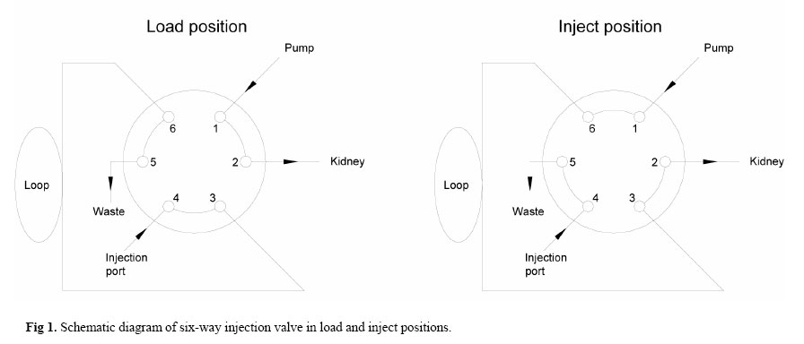
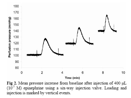
The first method while fast and easy to perform has several drawbacks, including low reproducibility due to alterations in injection speed and improper change of baseline pressure as a result of the force applied to the syringe piston. Moreover, since the perfusion pressure varies from one injection to next, variation in drug-organ contact time occurs leading to loss of reproducibility. Using a second pump to inject the drug, although more precise, requires multiple calculation of main flow rate and subsequent adjustment of injection flow rate. On the other hand, washing and loading the pump tubing prior to injection of each dose is time consuming while extra care must be taken in order to inject a precisely defined volume. A computer controlled module can be used to calculate the required pump speed in order to maintain the constancy of main perfusion pressure while the bolus dose is injected at a fix ratio of perfusate. Such modules and required software would make the system quite complicated and costly. In theory, a perfect injection must be made as fast as possible to avoid perturbation of the dynamic regimen that is already established in the kidney and pressure transducer. In perfusion experiments, the main difficulty is injecting a precise amount of the drug in the perfusion line without stopping or altering the flow of perfusate [4]. We have developed a practical and inexpensive method with high reproducibility that does not alter the baseline pressure regardless of the main perfusion flow. Materials and Methods Male Wistar rats (300-350 g) having free access to commercial pellet chow and tap water were anaesthetized with pentobarbital (50 mg/kg). The abdominal cavity was exposed by a ventricular incision, heparin was injected into the vena cava (500 U/kg) and renal artery was cannulated using a number 20 hypodermic needle with a polished tip via the superior mesenteric artery without disruption of flow. Perfusion was initiated in situ and continued with Tyrode's solution of the following composition (mM): KCl 2.68, NaCl 136.9, MgCl2 1.05, NaHCO3 11.90, NaH2PO4 0.42, CaCl2 1.8, and glucose 5.55 and equilibrated with 95% O2 and 5% CO2. Perfusion medium was fed to the kidney by means of a peristaltic pump (LKB, Varioperpex II) through PTFE tubings (Pharmacia Biotech, 18-8207-01) with a constant flow at 85-95 mmHg. The ligatures around the cannula were tied and kidney was removed and placed in a thermostated glass chamber. Alterations in perfusion pressure, arising from changes in renal vascular resistance, were recorded on a Beckman polygraph (R-612) by means of a pressure transducer (Beckman, 4-327) situated parallel to perfusion cannula. The injection technique we adapted has already been practiced in chromatography for many years. The injecting mechanism is known as six-way injection valve as shown in Fig 1. This is done using a precisely machined manual or motorized valve located just before the kidney (Valco C22). The valve, which is mounted in the path of the perfusate, provides two flow paths. In the load position, the valve connects the pump directly to the kidney. Using a syringe, the sample contained the perfusion medium is injected into a loop which has a small defined volume. In the inject position, the sample which is in the loop at atmospheric pressure, is directly inserted into the flow path of the perfusate. In manual injectors, this is done using a handle that allows the valve to rotate 60 degrees thus connecting sample loop to the perfusion line. Highly reproducible injections are achieved when the loop is completely filled with the sample. To assess the method, the renal vasculature was constricted by injection of 400 μL (10-7 M) epinephrine to the perfusion line at three different baseline pressures (100 mmHg, 120 mmHg and 140 mmHg) (Fig 2). Vasoconstriction was measured as mean peak increase in perfusion pressure after seven sequential injections as shown in Table 1. Standard deviation of mean shows the reproducibility of injections. Results and Discussion The loading and injection, marked by small vertical events, do not alter the baseline pressure resulting in an identical baseline regardless of the main perfusion pressure (Fig 2). The variability in peak pressure and time required to return to baseline is demonstrated in Table 1. The mean peak pressure rise was almost unchanged in the three different baseline pressures, which was due to the exact amount of drug injected each time. The time required for pressure to return to baseline decreased as the baseline pressure was raised. This is probably due to higher flow rates and shorter exposure of renal vessels to the agonist. This process does not depend on the main flow rate since the content of the loop is placed in the path of the perfusion line and the main flow is not altered at all. Low standard deviation of pressure change indicates the high reproducibility of this method after seven sequential injections. Our method has several other advantages. Multiple injections can be made in a relatively shorter time. Minor changes can be detected in the perfusion pressure resulting in higher sensitivity of the detection method. This method can be applied to all organ perfusion experiments such as isolated hind limb, tail, arteries and arterioles. References
Copyright © 2003 by Razi Institute for Drug Research (RIDR) The following images related to this document are available:Photo images[pt03002t1.jpg] [pt03002f2.jpg] [pt03002f1.jpg] |
| |||||||||