 |
| About Bioline | All Journals | Testimonials | Membership | News |
|
||||||
|
||||||
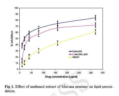
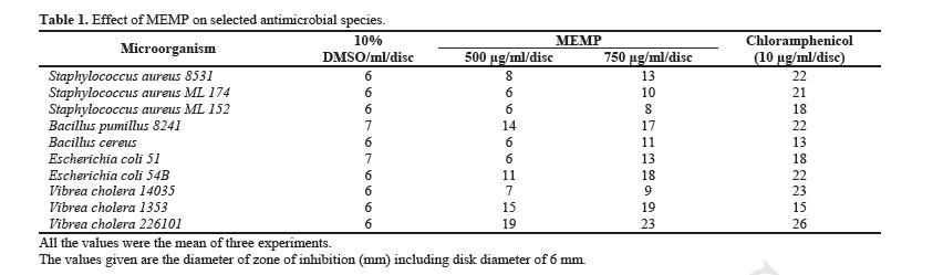
Iranian Journal of Pharmacology & Therapeutics, Vol. 4, No. 1, 2005, pp. 32-35 In Vitro Lipid Peroxidation and Antimicrobial Activity of Mucuna pruriens Seeds YERRA RAJESHWAR, MALAYA GUPTA and UPAL KANTI MAZUMDER Division of Pharmaceutical Chemistry and Pharmacology, Department of Pharmaceutical Technology, Jadavpur Uni-versity, Kolkata, India. Address correspondence to: Prof. Upal Kanti Mazumder, Department of Pharmaceutical Technology, Jadavpur Univer-sity, Kolkata, India. E-mail: yrajeshwar@yahoo.co.in Received May 5, 2005; Code Number: pt05008 ABSTRACT The present investigation is aimed to carry out the in vitro lipid peroxidation and antimicrobial activities of the methanol extract of Mucuna pruriens (MEMP) (Family: Fabaceae) seeds. Lipid peroxidation was monitored by the change in optical density of the prepared concentrations (10-320 µg/ml) and the % inhi-bition was calculated. Ascorbate/FeSO4-induced peroxidation was inhibited by standard antioxidants such as quercetin, L-ascorbic acid and MEMP. Moreover, the % inhibition of the methanol extract increased with increase in concentration. IC50 value for the MEMP, L-ascorbic acid and quercetin for lipid peroxida-tion was found to be 217.25 µg/ml, 41 µg/ml and 19.75 µg/ml respectively. The antimicrobial activity of MEMP was determined by disc diffusion method with various Gram positive and Gram-negative microor-ganisms. MEMP showed broad-spectrum antimicrobial activity against all the tested microorganisms ex-cept Staphylococcus aureus ML 152 and Vibrae cholera 14035. The results obtained in the present study indicate that MEMP can be a potential source of natural antioxidant and antimicrobial agent. Keywords: Mucuna pruriens seeds, In vitro lipid peroxidation, Antimicrobial activity There has been growing interest in the investigation of the natural products from plants for the discovery of new antimicrobial and antioxidant agents as well as an alternative route for the substitution of synthetic chemi-cals, side effects of which are always in question. For this, the essential oils and the extracts of many plants have been prepared and screened for their antimicrobial and antioxidant activities leading to the accumulation of a large number of reports in the literature concerning the above mentioned properties of plants [ 1 - 5 ]. Because of the side effects and the resistance that pathogenic mi-croorganisms build against antibiotics, much recent at-tention has been paid to extracts and biologically active compounds isolated from plant species used in herbal medicine [ 6 ]. Plant based antimicrobials represent a vast untapped source for medicines and further exploration of plant antimicrobials needs to occur. Antimicrobials of plant origin have enormous therapeutic potential. They are effective in the treatment of infectious diseases while simultaneously mitigating many of the side ef-fects that are often associated with synthetic antimicro-bials [ 7 ]. Mucuna pruriens is a twinning herb found all over tropical parts of India. It, an Indian indigenous legumi-nous plant, is well known for producing itch. This prop-erty is attributed to the trichomes (hair) present on the pods. It has been established that this unique property is accounted by the presence of 5-hydroxy tryptamine (5-HT) in the hair. [ 8 ]. It is also likely that histamine and kinin like substances may also be responsible [ 9 ]. Some reports show that anti-histaminics afford protection against the itch [ 9 ]. It has been reported to be anti-diabetic [ 10 ]. Mucuna pruriens seeds are herbaceous forage and food legumes that have for a long time found widespread usage as rotation crops for management of various pests and pathogens, as well as in soil improve-ment and weed control [ 13 , 14 ]. Seeds of Mucuna pru-riens are known to produce the unusual non-protein amino acid 3-(3,4-dihydroxyphenyl)-l-alanine (L-DOPA), a potent neurotransmitter precursor that is, at least in part, believed to be responsible for the toxicity of Mucuna seed [ 15 ]. L-DOPA, a potentially neurotoxic agent used in the treatment of Parkinson’s Disease, is found in relatively large amounts in Mucuna pruriens seeds [ 16 , 17 ] to the point where the seeds have been suggested as a medical source of L-DOPA [ 16 ] and even in the treatment of Parkinson’s Disease [ 18 ]. Previously, we reported the antiepileptic and anti-neoplastic activity of methanol extract of Mucuna pru-riens root [ 19 ] from our laboratory. Our recent findings revealed that the methanol extract of Mucuna pruriens seeds showed significant in vitro antioxidant activity [ 20 ]. In this work, we have tested the in vitro lipid per-oxidation and antimicrobial activity (against Gram posi-tive and Gram negative bacteria) of the methanol extract of Mucuna pruriens. MATERIALS AND METHODS Chemicals Quercetin, L-ascorbic acid and TBA were purchased from Sigma Chemical Co. (St. Louis, MO, USA). All other chemicals and reagent used were of analytical grade. Plant Extract The seeds of Mucuna pruriens (MP) were purchased from the United Chemicals and Allied Products, Kol-kata, India. They were identified by the Botanical Sur-vey of India (BSI), Kolkata, India. For the extract, the seeds were dried in shade and powdered in a mechanical grinder. The powder of MP seeds was initially defatted with petroleum benzine (60-80°C) followed by 1000 ml of methanol by using a Soxhlet extractor for 72 h at a temperature not exceeding the boiling point of the sol-vent [ 21 ]. The extract was filtered using Whatman filter paper (No. 1) and then concentrated in vacuum and dried. The extract thus obtained was directly used in the assay of lipid peroxidation and antimicrobial activity. Previously isolated classes of compounds The phytochemical study revealed that the methanol extract of Mucuna pruriens (MEMP) seeds contained alkaloids, flavones, saponins, aminoacids, and fatty ac-ids [ 22 ]. Bacterial Strains Employed Microorganisms (Staphylococcus aureus 8531, Staphylococcus aureus ML 174, Staphylococcus aureus ML 152, Bacillus pumillus 8241, Bacillus cereus, Es-cherichia coli 51, Escherichia coli 54B, Vibrea cholera 14035, Vibrea cholera 1353, and Vibrea cholera 226101) were obtained from the stock culture of Central Drugs Laboratory, Kolkata; Indian Institute of Chemical Biology, Kolkata and Mycology and Plant Pathology Laboratory, Calcutta University, Kolkata, India. Lipid Peroxidation Lipid peroxidation induced by Fe2+-ascorbate system in rat liver homogenate by the method of Bishayee and Balasubramaniyam [ 23 ] was estimated as thiobarbituric acid reacting substances (TBARS) by the method of Ohkawa et al. [ 24 ]. The reaction mixture contained rat liver homogenate 0.1 ml (25% w/v) in Tris-HCl buffer (20 mM, pH 7.0); KCl (30 mM); FeSO4 (NH4)2SO4.7H2O (0.06 mM); and various concentra-tions of Mucuna pruriens extract in a final volume of 0.5ml. The reaction mixture was incubated at 37°C for 1 h. After the incubation period, 0.4ml was removed and treated with 0.2ml sodium dodecyl sulphate (SDS) (8.1%); 1.5 ml thiobarbituric acid (TBA) (0.8%); and 1.5 ml acetic acid (20%, pH 3.5). The total volume was made up to 4.0 ml with distilled water and then kept in a water bath at 95 to 100°C for 1 h. After cooling, 1.0 ml of distilled water and 5.0 ml of n-butanol and pyridine mixture (15:1 v/v) were added to the reaction mixture, shaken vigorously and centrifuged at 4000 rpm for 10 min. The butanol-pyridine layer was removed and its absorbance at 532 nm was measured to quantify TBARS. Inhibition of lipid peroxidation was deter-mined by comparing the optical density (OD) of treat-ments with that of the control. Quercetin and L-ascorbic acid were used as standard. Determination of Antimicrobial Activity Antimicrobial activity was measured using the stan-dard method of diffusion disc plates on agar [ 25 ]. 0.1 ml of each culture of bacteria was spread on agar plate sur-faces. For antibacterial assays, all bacterial strains were grown in Mueller Hinton Broth medium (Merck) for 24 h at 37°C. The concentration of bacterial suspensions was adjusted to 108 colony forming units (108 cfu/ml) in Mueller Hinton Agar. Paper discs (6 mm in diameter) were impregnated on the agar to load 10 µl of each sam-ple. The impregnated disks were placed on the medium suitably spaced apart and the plates were incubated at 5°C for 1 h to permit good diffusion and then trans-ferred to an incubator at 37°C for 24 h. The results were recorded by measuring the zones of growth inhibition surrounding the disc. Clear inhibition zones around the discs indicated the presence of antimicrobial activity. All data on antimicrobial activity are the average of triplicate analyses. In order to determine the antibacte-rial effect of the MEMP, chloramphenicol (10 µg/ml/disc) were used as positive control. Inhibition diameters were measured after incubation for 24 h at 37°C. RESULTS Effect of MEMP on Lipid Peroxidation The effect of MEMP and commercially available an-tioxidants namely quercetin and L-ascorbic acid on the in vitro inhibition of lipid peroxidation is showed in Fig 1 . The generation of lipid peroxidase by Fe2+-ascorbate in rat liver homogenate seems to be inhibited by MEMP with IC50 value of 217.25 µg/ml. A similar effect was produced by L-ascorbic acid (IC50 =41 µg/ml) and quercetin (IC50 =19.75 µg/ml), indicating that the effect of MEMP on the inhibition of lipid peroxide production is significant (p < 0.05). The inhibition percentage of lipid peroxidation in the presence of extract was found to be 61.41% of the corresponding controlsm. The val-ues for L-ascorbic acid and quercetin were found to be 72.11% and 84.09%, respectively at 320 µg/ml. Effect of MEMP on Antimicrobial Activity The data presented in Table 1 indicate that the methanol extract of Mucuna pruriens (MEMP) inhibit the growth of some of the tested microorganisms (Gram positive and Gram negative) to various degrees. The MEMP at a concentration of 500 μg/ml and 750 μg/ml exhibited significant (p < 0.05) antimicrobial effect against all the tested microorganisms. The extract showed strong antibacterial activity against Bacillus pumillus 8241, Escherichia. Coli 5B and Vibrae Chol-era 1353 and 226101. However, their activity against Staphylococcus aureus ML 152 and Vibrae cholera 14035 was found to be significantly (p > 0.05) less then the control. The antimicrobial activity was compared with the standard Chloramphenicol at a concentration of 10 μg/ml. Statistical Analysis All treatments were performed in triplicate and each data point in the results is the mean of two or three rep-licate tests. All experiments were repeated at least once. The statistical significance of a treatment effect was evaluated by student’s t-test and the values were ex-pressed as mean ± SEM. Probability limit was set at p < 0.05. Discussion Unsaturated lipids in liver tissue are very susceptible to peroxidation when they are exposed to reactive oxy-gen species (ROS). In the present investigation we have incubated the liver tissue in presence of a ROS generat-ing system, ascorbate/FeSO4, and examined the effect on tissue homogenate by measuring the optical density (OD) at 532 nm. The results of the investigations re-vealed that MEMP had potent lipid peroxidation inhibition activity. The antimicrobial activity of the MEMP was studied by the disc diffusion method against various microor-ganisms. Disc diffusion methods are used extensively to investigate the antibacterial activity of natural sub-stances and plant extracts. These assays are based on the use of discs as reservoirs containing solutions of the substances to be examined. In the case of solutions with a low activity, however, a large concentration or volume is needed. Because of the limited capacity of discs, holes or cylinders are preferably used [ 26 ]. MEMP showed a broad spectrum of activity against all the bac-terial strains as shown in Table 1 . Chloramphenicol (10 µg/ml/disc) was used as a positive control. On the basis of the results obtained in the present study, we conclude that the methanol extract of Mucuna pruriens had significant in vitro lipid peroxidation and antimicrobial activity. The components responsible for the inhibition of lipid peroxidation of MEMP are cur-rently unclear. Further studies are needed to isolate the active components, responsible for the lipid peroxida-tion and antimicrobial activities. ACKNOWLEDGMENTS One of the authors, Yerra Rajeshwar is grateful to All India Council for Technical Education and Training (AICTE), New Delhi, India, for providing financial support for this investigation. REFERENCES
Copyright © 2005 by Razi Institute for Drug Research (RIDR) The following images related to this document are available:Photo images[pt05008t1.jpg] [pt05008f1.jpg] |
| |||||||||