 |
| About Bioline | All Journals | Testimonials | Membership | News |
|
||||||
|
||||||
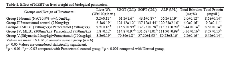
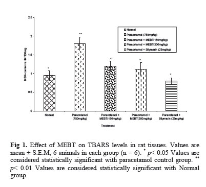
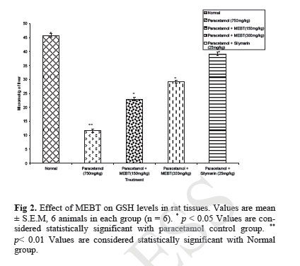
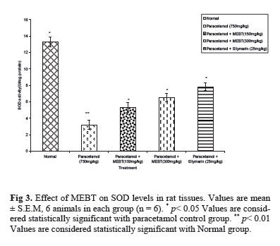
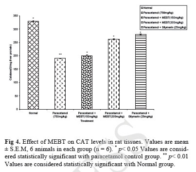
Iranian Journal of Pharmacology & Therapeutics, Vol. 4, No. 1, 2005, pp. 64-69 Hepato Protective and Antioxidant Role of Berberis tinctoria Lesch Leaves on Paracetamol Induced Hepatic Damage in Rats KANDA SAMY MURUGESH, VEERENDRA CHANNABASAPPA YELIGAR, BHIM CHARAN MAITI and TAPAN KUMAR MAITY Department of Pharmaceutical Technology, Division of Pharmaceutical Chemistry, Jadavpur University (K.S.M., V.C.Y., T.K.M.); Indian Institute of Chemical Biology (B.C.M.), Kolkata, India. Address correspondence to: Dr.Tapan K. Maity. Division of Pharmaceutical Chemistry, Department of Pharmaceutical Technology, Jadavpur University, Kolkata-700032, India. E-mail: tapanmaya2002@yahoo.com Received May 1, 2005; Code Number: pt05014 ABSTRACT The scientific evaluation of medicinal plants used in the preparation of folk remedies has provided modern medicine with effective pharmaceuticals for the treatment of diseases. The methanol extract of Berberis tinctoria Lesch (Berberidaceae) leaves was investigated for its hepatoprotective and antioxidant effects on paracetamol (750 mg/kg) induced acute liver damage in Wistar albino rats. Hepatoprotection activity was measured by using biochemical parameters such as serum glutamate oxalate transaminase and serum Glutamate Pyruvate Transaminase (SGOT and SGPT), alkaline phosphatase (ALP), bilirubin and total protein. The methanol extract of Berberis tinctoria (MEBT) at the doses of (150 mg/kg and 300 mg/Kg) produced significant hepatoprotective effect by decreasing the activity of serum enzymes, bilirubin and lipid peroxidation while it significantly increased the levels of glutathione (GSH), catalyse (CAT) and super oxide dismutase (SOD) in a dose dependant manner. The effects of MEBT were comparable to that of standard drug silymarin. These results suggest that MEBT may have potential therapeutic value in the treatment of some liver disorders, probably by its antioxidative effect on hepatocytes. Keywords: Berberis tinctoria, Hepatoprotective effect, Antioxidants, Paracetamol In recent years many researchers have examined the effects of plants used traditionally by indigenous healers and herbalists to support liver function and treat diseases of the liver. In most cases, research has confirmed traditional experience and wisdom by discovering the mechanisms and mode of action of these plants as well as reaffirming the therapeutic effectiveness of certain plants or plant extracts in clinical studies. Several hundred plants have been examined for use in a wide variety of liver disorders. Just a handful has been fairly well researched [ 1 ]. The plant Berberis tinctoria Lesch (berberidaceae) is a shrub, very variable in size and form, in the open often 2 to 3 feet high, but in the forest sometimes reaching a height of 15 feet with thick stem and long scandent branches bearing numerous slender leafy twigs [ 2 ]. It is locally called as Oosikala and medicinally used by the Kurumbas, the Nilgiri tribe, for stomach-ache (root paste). The aqueous root paste along with honey is used as an antimicrobial agent against skin diseases. The wood, root bark and extract have been used in skin dis-eases, menorrhagia, diarrhoea, jaundice and infections of the eyes [ 3 ]. The present study was undertaken to study the possible hepatoprotective and antioxidant role of methanol extract of leaves of Berberis tinctoria Lesch. Paracetamol (acetaminophen) is a widely used anti-pyretic and analgesic which produces acute liver damage if overdoses are consumed. Paracetamol is mainly metabolized in liver to excretable glucuronide and sulphate conjugates [ 4 , 5 ]. However, the hepatotoxicity of paracetamol has been attributed to the formation of toxic metabolites when a part of paracetamol is activated by hepatic cytochrome P-450 [ 6 ], to a highly re-active metabolite N-acetyl-P-benzoquinone imine (NAPQI) [ 7 ]. NAPQI is initially detoxified by conjuga-tion with reduced glutathione (GSH) to form mercapturic acid [ 8 ]. However, when the rate of NAPQI formation exceeds the rate of detoxification by GSH, it oxidizes tissue macromolecules such as lipid or SH group of protein and alters the homeostasis of calcium after depleting GSH. Plant derived natural products such as flavonoids, terpenoids and steroids etc. have received considerable attention in recent years due to their diverse pharmacological properties including antioxidant and hepatopro-tective activity [ 9 - 11 ]. There has been a growing interest in the analysis of certain flavonoids, triterpenoids and steroids stimulated by intense research into their potential benefits to human health. One of their main properties in this regard is their antioxidant activity, which enables them to attenuate the development of tumor and inflammatory diseases. Antioxidants play an important role in inhibiting and scavenging radicals, thus providing protection to humans against infection and degenerative diseases. Realizing the fact, this research was carried out to evaluate the antioxidant and hepatoprotective activity of methanol extract of Berberis tinctoria leaves (MEBT) against paracetamol induced hepatic damage in rats. Silymarin is marketed as one of the standard hepato-protective herbal formulation. Silymarin may be collectively called as silybin, silydianin and silychristine which is the active constituents of milk thistle of Sily-bum marinum. The hepatoprotective effects of silymarin in humans after ingestion of Amanita toxins have been repeatedly demonstrated. In one series of 18 patients treated with silymarin, all patients survived except one particularly high dose suicide. Many studies have demonstrated the beneficial hepatoprotective effects of treatment with silymarin. MATERIALS AND METHODS Plant Material The plant Berberis tinctoria was collected from Doddabetta forest area, Udhagamandalam, Nilgiri district, Tamil Nadu in the month of February 2004. It was identified by the Botanical Survey of India, Coimbatore and a voucher specimen (BSI/SC/5/21/03-04/Tech. 1674.) was kept for future reference. The underground root was separated from the aerial parts and the leaves were then shade dried and mechanically powdered separately to obtain a course powder, which was then subjected to successive extraction in a soxhlet apparatus using petroleum ether (60-80°C),chloroform and methnol. Solvent elimination under reduced pressure afforded the chloroform extract (2.5% yield) and methanol extract (12% yield) respectively. The resulting Methanol extract was then used for hepatoprotective activity and antioxidant studies. Animals Studies were carried out using Male Wistar albino rats (150-180 g). They were obtained from the animal house, Indian Institute of Chemical Biology (IICB), Kolkata, India. The animals were grouped and housed in polyacrylic cages (38 × 23 × 10 cm) with not more than six animals per cage and maintained under standard laboratory conditions (temperature 25 + 2°C) with dark and light cycle (14/10 h).They were allowed free access to standard dry pellet diet (Hindustan Lever, Kolkata, India) and water ad libitum. The mice were acclimatized to laboratory condition for 10 days before commencement of experiment. All procedures described were reviewed and approved by the University Animals Ethical Committee. Drugs and Chemicals Silymarin was purchased from Micro labs Tamil-nadu India, 1-Chloro-2, 4-dinitrobenzene [CDNB], Bovine serum albumin (Sigma chemical St. Louis, MO, USA), Thiobarbituric acid, Nitro blue tetrazolium chloride (NBT) (Loba Chemie, Bombay, India), 5,5'-dithio bis-2-nitrobenzoic acid (DTNB),The solvents and/or reagents obtained were used of analytical grade and obtained from Sicco research laboratory, Mumbai, India. Paracetamol-Induced Liver Damage in Rats (Acute Model) [12] Five groups (I-V) each comprising of six male Wis-tar albino rats weighing in the range of 150-180 g were selected. Group I served as control and was fed orally with normal saline 5 ml/Kg daily for seven days. Groups II rats were similarly treated as group I .Group III and IV were treated with 150 mg and 300 mg/kg methanolic leaf extract leaves/day/orally respectively for seven days. While group V were fed silymarin 25 mg/k daily [ 13 ] as standard for seven days. On the seventh day, 750 mg/kg paracetamol suspension was given by oral route in a dose of to all rats except rats in group I. The biochemical parameters were determined 18 hours after the last dose. Biochemical Studies Blood was obtained from all animals by puncturing retro-orbital plexus. The blood samples were allowed to clot for 45 min at room temperature. Serum was separated by centrifugation at 2500 rpm at 30ºC for 15 min and used for the estimation of various biochemical parameters namely SGOT, SGPT [ 14 ], SALP [ 15 ], serum bilirubin was estimated by Malloy et al method [ 16 ] and the protein content was measured by Lowry et al method [ 17 ]. After collection of blood samples the rats were killed and their livers were excised, rinsed in ice cold normal saline, followed by 0.15 M Tris-HCl (pH 7.4) blotted dry and weighed. A 10 % w/v of homogenate was prepared in 0.15 M Tris-HCl buffer and processed for the estimation of lipid peroxidation [ 18 ]. A part of homogenate after precipitating proteins with Trichloro no space acetic acid (TCA) was used for estimation of glutathione [ 19 ]. The rest of the homogenate was centri-fuged at 1500 rpm for 15 min at 4°C. The supernatant thus obtained was used for the estimation of SOD [ 20 ] and CAT activity [ 21 ]. Determination of Thiobarbituric Acid Reactive Sub-stances (TBARS) TBARS in tissues was estimated by the method of Ohkawa et al [ 18 ]. To 0.5 ml tissue homogenate, 0.5 ml saline and 1.0 ml 10% TCA were added, mixed well and centrifuged at 3000 rpm for 20 min. To 1.0 ml of the protein-free supernatant, 0.25 ml of thiobarbituric acid (TBA) reagent was added; the contents were mixed and heated for 1 h at 95ºC. The tubes were cooled to room temperature under running water and absorption measured at 532 nm. The levels of lipid peroxides were expressed as no space moles of thiobarbatituric acid reactive substances (TBARS)/mg protein. Determination of Reduced Glutathione (GSH) To measure the reduced glutathione (GSH) 0.2 ml of tissue homogenate was mixed with 1.8 ml of EDTA solution. To this 3.0 ml precipitating reagent (1.67 g of met phosphoric acid, 0.2 g of EDTA disodium salt, 30 g sodium chloride in 1 L of distilled water) was added, mixed thoroughly and kept for 5 min before centrifugation. To 2.0 ml of the supernatant, 4.0 ml of 0.3-M diso-dium hydrogen phosphate solution and 1.0 ml of DTNB (5, 5-dithio bis 2-nitro benzoic acid) reagent were added and was read at 412 nm. Absorbance values were com-pared with a standard curve generated from known GSH. Assay of Super Oxide Dismutase (SOD) The activity of SOD in tissue was assayed by the method of Kakkar et al [ 20 ]. The assay mixture con-tained 1.2 ml sodium pyrophosphate buffer (pH 8.3, 0.025 mol/L), 0.1ml phenazine methosulphate (186 mM), 0.3 ml NBT (300 mM), 0.2 ml NADH (780 mM) and approximately diluted enzyme preparation and wa-ter in a total volume of 3 ml. After incubation at 30ºC for 90 sec, the reaction was terminated by the addition of 1.0 ml glacial acetic acid. The reaction mixture was stirred vigorously and shaken with 4.0 ml n-butanol. The color intensity of the chromogen in the butanol layer was measured at 560 nm against n-butanol and concentration of SOD was expressed as units/mg pro-tein. Absorbance values were compared with a standard curve generated from known SOD. Assay of Catalase (CAT) Catalase was assayed according to the method of Aebi et al [ 21 ]. The estimation was done spectropho-tometrically following the decrease in absorbance at 230 nm. The tissue was homogenized in M/150 phosphate buffer (pH 7.0) at 1–4ºC and centrifuged at 5000 rpm. The reaction mixture contained 0.01 M phosphate buffer (pH 7.0), 2 mM H2O2 and the enzyme extract. The spe-cific activity of catalase was expressed in terms of units/mg protein. Absorbance values were compared with a standard curve generated from known CAT. Statistical Analysis Data were expressed as Mean ± SEM for 6 rats in each group. The biochemical parameters were analysed statistically using one-way ANOVA, followed by Dun-nett’s multiple comparison test (DMRT).The minimum level of significance was fixed at p < 0.05. RESULTS Biochemical parameters are presented in the table. Administration of paracetamol 750 mg/kg; p.o) after, 18 hours intoxication, caused a marked elevation of en-zyme levels in paracetamol treated group, in comparison with the control. There were significant restoration of enzyme levels on administration of MEBT (Group III and IV, Table 1 ) and silymarin at the dose of 200 mg/kg (Group V, Table 1 ). It was observed that the size of the liver was enlarged in paracetamol intoxicated rats but it was nor-mal in drug treated group ( Table 1 ). A significant reduc-tion (p < 0.05) in liver weight supports this finding. The effect of MEBT on rat liver lipid peroxidation, glutathione, and antioxidant enzyme (SOD and CAT) levels are shown in Fig 1, 2, 3, 4. Thiobarbituric acid reactive substance levels were significantly (p < 0.05) increased in the paracetamol control rats when com-pared with the normal rats. Treatment with MEBT (150 mg and 300 mg/kg) significantly (p < 0.05) prevented the increase in TBARS levels and brought them near to normal level (Fig 1). Glutathione, SOD, and CAT levels were significantly (p < 0.05) increased in MEBT treated groups (Fig 2, 3, 4). The effects of MEBT were com-pared with that of standard reference drug silymarin. DISCUSSION Paracetamol (acetaminophen), a widely used antipy-retic and analgesic drug produces acute liver damage if accidental overdoses are consumed. The covalent bind-ing of N-acetyl-p-benzoquinoneimine, an oxidation product of paracetamol, to sulphydryl groups of protein resulting in cell necrosis and lipid peroxidation induced by decrease in glutathione in the liver as the cause of hepatotoxicity have been reported earlier [ 22 , 23 ]. In the assessment of liver damage by paracetamol the determination of enzyme levels such as SGPT and SGOT is largely used. Necrosis or membrane damage releases the enzyme into circulation; therefore, it can be measured in serum. High levels of SGOT indicates liver damage, such as that due to viral hepatitis as well as cardiac infarction and muscle injury. SGPT catalyses the conversion of alanine to pyruvate and glutamate, and is released in a similar manner. Therefore, SGPT is more specific to the liver, and is thus a better parameter for detecting liver injury [ 24 ]. Elevated levels of serum enzymes are indicative of cellular leakage and loss of functional integrity of cell membrane in liver [ 25 ]. Se-rum ALP and bilirubin level on other hand are related to the function of hepatic cell. Increase in serum level of ALP is due to increased synthesis, in presence of in-creasing biliary pressure [ 26 ]. This present study evaluated the hepatoprotective ef-fects of MEBT in paracetamol induced liver toxicity. Acute administration of paracetamol produced a marked elevation of the serum levels of SGOT, SGPT, ALP, serum bilirubin and total proteins in treated animals (Group II to V) when compared with that of the l con-trol group (Group I). Treatment with MEBT at a dose of 150 mg/kg and 300 mg/kg significantly reduced the elevated levels of the enzymes. Treatment with MEBT decreased the serum levels of SGOT, SGPT towards the respective normal value that is an indication of stabilization of plasma membrane as well as repair of hepatic tissue damage caused by paracetamol. The above changes can be considered as an expression of the functional improvement of hepato-cytes, which may be caused by an accelerated regenera-tion of parenchyma cells. Effective control of alkaline phosphatase (ALP) and bilirubin levels points towards an early improvement in the secretary mechanism of the hepatic cell. Lipid peroxidation has been postulated as being the destructive process in liver injury due to paracetamol administration [ 26 ]. In our study, elevations in the lev-els of TBARS in liver of rats treated with paracetamol were observed. The increase in TBARS levels in liver suggests enhanced lipid peroxidation leading to tissue damage and failure of antioxidant defense mechanisms to prevent formation of excessive free radicals. Treat-ment with MEBT significantly reversed these changes. Hence it may be possible that the mechanism of hepato-protection of MEBT is due to its antioxidant effect. Glutathione is one of the most abundant tripeptide, non-enzymatic biological antioxidants present in the liver. Its functions are concerned with the removal of free radical species such as hydrogen peroxide, superox-ide radicals, alkoxy radicals, and maintenance of mem-brane protein thiols and as a substrate for glutathione peroxidase (GPx) and GST [ 27 ]. In our present study the decreased level of GSH has been associated with an enhanced lipid peroxidation in paracetamol treated rats. Administration of MEBT significantly increased the level of glutathione in a dose dependent manner. Increase in serum activity of superoxide dismutase (SOD) is a sensitive index in hepatocellular damage and is the most sensitive enzymatic index in liver injury [ 28 ]. SOD has been reported as one of the most impor-tant enzymes in the enzymatic antioxidant defense sys-tem. It scavenges the superoxide anion to form hydro-gen peroxide, hence diminishing the toxic effect caused by this radical. In the present study, it was observed that the MEBT caused a significantly increased in the hepatic SOD activity of the paracetamol induced liver damage in rats. This shows MEBT can reduce reactive free radicals that might lessen oxidative damage to the tissues and improve the activities of the hepatic antioxidant enzyme. Catalase (CAT) is an enzymatic antioxidant widely distributed in all animal tissue and the highest activity is found in the red cells and in liver. CAT decomposes hydrogen peroxide and protects the tissue from highly reactive hydroxyl radicals [ 29 ]. Therefore, the reduction in the activity of these enzymes may result in a number of deleterious effects due to the accumulation of super-oxide radicals and hydrogen peroxide. Administration of MEBT increases the activities of SOD and CAT in paracetamol induced liver damage in rats to prevent the accumulation of excessive free radicals and protects the liver from paracetamol intoxication. So results of this study demonstrated that the MEBT has significant action on paracetamol induced hepato-toxicity. Detailed studies on the mechanism of action and phytochemical analyses are in progress at our labo-ratory. ACKNOWLEDGEMENT One of the authors K. Murugesh, Senior Research Fellow, grateful to Jadavpur University, for providing all facilities. REFERENCES
Copyright © 2005 by Razi Institute for Drug Research (RIDR) The following images related to this document are available:Photo images[pt05014t1.jpg] [pt05014f9.jpg] [pt05014f2.jpg] [pt05014f7.jpg] [pt05014t5.jpg] [pt05014f3.jpg] [pt05014f1.jpg] [pt05014f4.jpg] |
| |||||||||