 |
| About Bioline | All Journals | Testimonials | Membership | News |
|
||||||
|
||||||
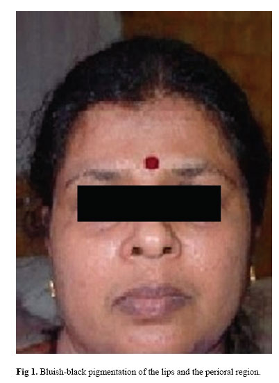
Iranian Journal of Pharmacology & Therapeutics, Vol. 4, No. 1, 2005, pp. 70-71 Hyperpigmentation of the Skin Following Chloroquine Treatment – Case Series Report PANAMBOOR SABITHA, MATTI PRABHA ADHIKARI and MARIA KURUVILLA Department of Pharmacology (P.S.); Department of Medicine (M.P.A.); Department of Dermatology (M.K.); Kasturba Medical College, Mangalore, Karnataka State, India. Address correspondence to: Dr. P. Sabitha, Department of Pharmacology, Kasturba Medical College, Mangalore, Karna-taka State, India. E-mail: sabita_rao1@rediffmail.com Received May 1, 2005; Code Number: pt05015 ABSTRACT In this article, we are collectively presenting case reports of 15 patients who developed pigmentary changes of the skin and mucus membrane during the course of chloroquine therapy for connective tissue disorders. These female patients developed hyper pigmentation of the skin, largely on the exposed parts of the body. The pigmentary changes varied from brownish/ grayish/ bluish-black in color, patchy or dif-fuse, intense or mild. The shortest time lag between onset of chloroquine therapy and development of pigmentary changes was 3 months. The patients should be informed about this cosmetically important toxic effect of chloroquine and advised to avoid direct exposure to sunlight. Keywords: Chloroquine, Hyperpigmentation of skin, Connective tissue disorder Chloroquine has affinity for melanin and it gets con-centrated in pigmented (melanin-containing) structures which may explain its toxic effects on the eye (kerato-pathy and retinopathy) and that on the skin when used over a prolonged period for treating rheumatoid arthri-tis, systemic/discoid lupus erythematosis and other con-nective tissue disorders. Dermatological toxic effects include photosensitivity, photo allergic dermatitis, rashes, pruritus, bluish-black, grayish or brown pigmen-tation of skin and mucous membrane, discoloration of nail beds, bleaching of hair and eyebrows [ 1 - 9 ]. These dermatological manifestations are of cosmetic concern and warrant warning the patient at the outset. In this article, we are collectively presenting a series of case reports of 15 patients suffering from connective tissue disorders who developed cutaneous and mucosal pig-mentary changes after starting chloroquine therapy. SUBJECTS AND DESCRIPTION OF PIGMENTARY CHANGES These 15 patients visited our hospital once every three months between the years 1999-2001, when hy-droxychloroquine was not freely available in India. All the 15 were females, nine being treated with chloro-quine for SLE, five for rheumatoid arthritis, one for multiple connective tissue disorder. All these patients were between age group 22-50 years, average age being 36 years. Patients were receiving chloroquine 150 mg/day of base (250 mg salt) by oral route. The patients developed pigmentary changes at dif-ferent sites on the body ( Fig 1 and Fig 2 ) – periorbital / infraorbital (fifteen), cheeks (four), bridge of the nose (two), perioral / side of the mouth (fifteen), tongue (one), lips (two), sclera (one), sides of the neck/ back of the neck (ten), arms (four), palms (five), nails (four), trunk (upper abdomen / upper chest / upper back (six), legs (one), sole (one). The pigmentary changes varied from brownish / grayish / and bluish-black in color, patchy or diffuse, rippled (neck of five patients), mild as well as intense. Four patients started developing these changes within three-six months, four patients in six -nine months, three patients in nine-twelve months, and four patients in 12-16 months, after starting chloro-quine, at one site or the other and the changes were pro-gressive in nature. The skin complexion of these pa-tients varied from dark (seven), moderately fair (eight). No other toxic effects of chloroquine were reported in these patients. Oral Prednisolone (20-60 mg/day) was the concomitant medication in all the 15 patients and oral methotrexate (5 mg once a week) was received by two of the rheumatoid arthritis patients. Two of the patients who were concerned about their beauty, were switched over to hydroxychloroquine and the pigmenta-tion resolved within a period of six months. Attempts were not made to stop or reduce the dose of chloroquine in the remaining patients. DISCUSSION AND CONCLUSION The aforesaid pigmentary changes of the skin al-though, could be explained by the underlying disorder or other concomitantly administered drug (predniso-lone), have followed reasonable temporal sequence to administration of chloroquine and expected response pattern to chloroquine, hence, substantiating their defi-nite causal relationship with chloroquine use. Chloro-quine when used at a dose of 150 mg/day for more than three months duration in the treatment of connective tissue disorders can produce pigmentary changes of the skin and mucus membrane, irrespective of the disease for which it was being used, age or skin complexion of the patients. The pigmentation on the face and the ex-posed parts of the body, although not deleterious to health, may concern women especially those with fair complexion. Patients should be well informed about this potential toxic effect while starting therapy and advised to take proper measures to protect themselves from sunlight. The pigmentation resolved in two patients following chloroquine withdrawal despite being substituted by hydroxychloroquine. Hence, hydroxychloroquine may score over chloroquine not only with reference to ocular toxicity [ 9 ], but also with reference to cutaneous toxic-ity, for its long term use in patients suffering from con-nective tissue disorders. However, further studies are warranted to corroborate the above data. REFERENCES
Copyright © 2005 by Razi Institute for Drug Research (RIDR) The following images related to this document are available:Photo images[pt05015f2.jpg] [pt05015f1.jpg] |
| |||||||||