 |
| About Bioline | All Journals | Testimonials | Membership | News |
|
||||||
|
||||||
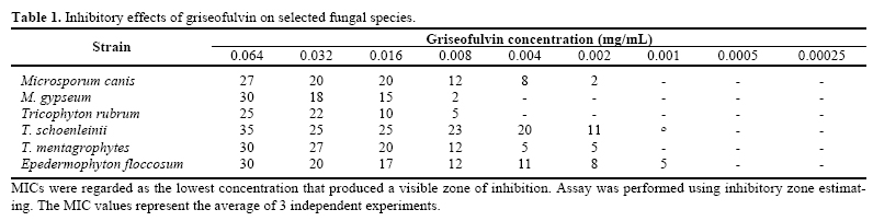
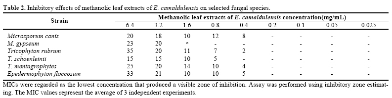
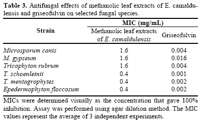
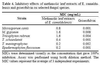
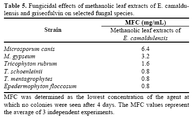
Iranian Journal of Pharmacology & Therapeutics, Vol. 4, No. 2, 2005, pp. 80-83 Anti Dermatophyte Activities of Eucalyptus camaldulensis in Comparison with Griseofulvin MEHRABAN FALAHATI, NASIM OMIDI TABRIZIB and FERESHTEH JAHANIANI Department of Parasitology, Iran University of Medical Sciences (M.F., N.O.T.); Razi Institute for Drug Research, Iran University of Medical Sciences (F.J.), Tehran, Iran. Address correspondence to: Dr. Fereshteh Jahaniani, Razi Institute for Drug Research, Iran University of Medical Sci-ences, Tehran, Iran E-mail: fereshteh_j_2000@yahoo.com Received June 16, 2005; Revised August 25, 2005; Accepted September 4, 2005 Code Number: pt05018 ABSTRACT Methanolic leaf extracts of Eucalyptus camaldulensis were investigated for in vitro antifungal activities against Microsporum canis, Microsporum gypseum, Tricophyton rubrum, Tricophyton schoenleinii, Trico-phyton mentagrophytes and Epedermophyton floccosum. The studies were carried out using broth dilu-tion method, agar dilution method and inhibitory zone estimation. The effects of the plant extract were compared with those of griseofulvin. Eucalyptus camaldulensis showed antifungal activity against all the dermatophytes tested with MIC values ranging from 0.4 to 1.6 mg/mL using inhibitory zone estimation, 0.4-1.6 mg/mL using agar dilution method and 0.2 to 1.6 mg/mL using broth dilution method. The mini-mum fungicidal concentration (MFC) of the extracts ranged from 0.8 to 6.4 mg/mL. The results obtained suggest that E. Camaldulensis has anti-dermatophyte activity. Keywords: Antifungal activities; Eucalyptus camaldulensis; Griseofulvin; Dermatophytes Human infections, particularly those involving the skin and mucosal surface constitute a serious problem, especially in tropical and subtropical developing coun-tries; dermatophytes and Candida spp. being the most frequent pathogen. Herbal medicines have been impor-tant sources of products for the developing countries in treating common infections including fungal diseases. Some studies have demonstrated that the oil and leaf extracts of Eucalyptus spp. have antifungal and repellent activity [ 1 - 2 ]. Crude methanolic extract of E. Camaldu-lensis has been reported to inhibit the growth of Can-dida albicans [ 5 ]. Also, it has been shown that ethanolic leaf extract of Eucalyptus camaldulensis had marked fungicidal effect against clinical dermatophytic fungal isolates; Microsporium gypseum and Trichophyton men-tagrophytes [ 6 ]. The phytochemical analysis of the crude extracts of the Eucalyptus spp. revealed the presence of saponin, saponin glycosides, steroid, cardiac glycoside, tannins, volatile oils, phenols and balsam (gum) [ 5 ]. Qualitative phytochemical tests, thin layer chromatography (TLC) and TLC-autography of certain active extracts demon-strated the presence of common phytocompounds in the plant extracts including phenols, tannins and flavonoids as major active constituents. Some compounds in the leaves of E. Camaldulensis include: essential oil (1 to over 2%), betulinic acid, eucalyptic acid, eucalyptolic acid, oleanolic acid and ursolic acid [ 7 ]. In this study, the antifungal effect of E. Camaldulensis growing in Iran has been studied against some dermatophyte spp including Microsporum canis, M. gypseum, Tricophyton rubrum, T. schoenleinii, T. mentagrophytes and Eped-ermophyton floccosum, which have not been studied previously. MATERIALS AND METHODS Plant Material The leaves of E. Camaldulensis were collected from national herbarium garden Tehran, Iran in 2001. Extraction and Isolation The leaves of E. Camaldulensis were dried at room temperature (20-23ºC) and ground into a powder using a blender. The dried leaf powder was (200 g) extracted with 80% methanol (800 mL) by refluxing for 3 hours. The solution was filtered and evaporated to dryness with a rotary evaporator. A 256 mg/mL stock solution of extract was prepared in DMSO and kept at -20ºC for future use. Microorganisms Dermatophyte strains included PTCC (Persian Type Culture Collection) (5070) Microsporum gypseum, PTCC (5060) Microsporum canis, PTCC (5069) Trichophyton rubrum PTCC (5143) and Trichophyton schoenleinii were obtained from Iranian scientific and industrial institute. Trichophyton mentagrophyte and Epedermophyton floccosum were isolated from patients at Mycology department of Iran Medical Science Uni-versity. Dermatophyte strains identity were confirmed by slide culture and urease test [ 8 ]. All strains were maintained in 20% glycerol and 10% lactose at -190ºC in liquid nitrogen. Inucolum Preparation All the fungi spp. was maintained on Sabouraud dextrose agar. Sterile distilled water containing 0.05% Tween 80 was added to the surface growth and spores and hypae were scraped off with a sterile wire loop. A spectrophotometer set at 530 nm used to adjust the sus-pension to 90% transmittance. This resulted in a con-centration of about 1×10⁶ CFU/mL. Inhibitory Zone Estimating Qualitative antifungal screening was carried out us-ing the agar-well diffusion assay [ 9 ]. Twenty mL of sterilized sabouraud dextrose agar medium were poured into a 15 cm Petri dish. Twenty µL of inoculums sus-pension of each test organism was distributed evenly over the surface. A 6mm well was cut in the centre of each plate using the wide-end of a sterilized Pasteur pipette. Fifty µl of serial dilution of methanolic leaf extracts of Eucalyptus camaldulensis or giseofolvin were placed into the wells. The plates were incubated for 5 days at 30ºC. Results of the qualitative screening were recorded as the average diameter of the inhibition zone surrounding the wells containing the test solution. Results were compared with griseofulvin. The MIC was regarded as the lowest concentration that produced a visible zone of inhibition. Agar Dilution Method Two mL of melted sabouraud was added to each of 10 sterile universal containers (numbered 1-10). 2 mL of the methnaolic leaf extract solution of E. Camaldu-lensis (12.8 mg/mL) was added to container 1. The con-tents were mixed and 2 mL transferred to container 2. This serial dilution was repeated through to container 9. 2mL was discarded from container 9. Eighteen mL of melted sabouraud dextrose agar was added to each con-tainer; the contents were mixed and poured into a 9 cm diameter Petri dish (numbered 1-10). After solidifica-tion of the medium, 20 µl of inoculum were spread onto each plate. The plates were incubated at 30ºC for 5-7 days. The final concentrations of E. Camaldulensis ex-tract ranged from 6.4 to 0.025 mg/mL. For griseofulvin, the final concentration ranged from 0.064 to 0.00025 mg/mL. The MIC was the lowest drug concentration at which there was no visible fungal growth after incuba-tion [ 10 ]. Broth Dilution Method One mL of sterile liquid sabouraud medium was added to 11 sterile capped tubes, each. 1mL of E. Camaldulensis methanolic leaf extract suspension (12.8 mg/mL in medium) was added to tube 1. The contents were mixed and 1mL was transferred to tube 2. This serial dilution was repeated through to tube 9. 1 mL was discarded from tube 9. Fifty µL of inoculum was added to tubes 1-10 and the contents were mixed. Medium control (no inoculum and no drug) and inoculum control (no drug) tubes were prepared. The final concentrations of E. Camaldulensis extract ranged from 6.4 to 0.025 mg/mL. For griseofulvin, the final concentration ranged from 0.064 to 0.00025 mg/mL. The tubes were incu-bated at 30ºC for 72 h. The fungal growth in each tube was detected turbidometrically at 530 nm. MIC was defined as the drug concentration at which the turbidity of the medium was the same as the medium control. Ten µl aliquot of cell suspension from the tube without observed growth of fungi was inoculated on to sabouraud dextrose agar, and Minimum fungicidal con-centration (MFC) of test compound was determined as the lowest concentration of the agent at which no colo-nies were seen after 4 days at 30ºC [ 11 ]. RESULTS Inhibitory Zone Estimating All tested fungi were affected by griseofulvin and E. Camaldulensis methanolic leaf extract. The greatest inhibitory effect of griseofulvin was recorded with Tri-cophyton schoenleinii (35 mm; inhibition zone). Trico-phyton mentagrophytes showed the most susceptibility against E. Camaldulensis methanolic leaf extract (35 mm). The results are presented in Table 1 and Table 2 . Agar Dilution Method The MICs of griseofulvin and E. Camaldulensis methanolic leaf extract obtained by the agar dilution assay are presented in Table 3 . Griseofulvin showed the greatest and the least antifungal activity against Trico-phyton schoenleinii and Microsporum gypseum, respec-tively. The MICs of E. Camaldulensis methanolic leaf extract ranged from 0.4 to 1.6 mg/mL. Epedermophyton floccosum, Tricophyton schoenleinii and Tricophyton mentagrophytes were the most susceptible strains against E. Camaldulensis methanolic leaf extract. Broth Dilution Method The MICs of griseofulvin and E. Camaldulensis methanolic leaf extract obtained by the broth dilution assay are presented in Table 4 . The MICs of griseo-fulvin and E. Camaldulensis methanolic leaf extract ranged from 0.001 to 0.008 mg/mL and 0.2 to 1.6 mg/mL, respectively. The least sensitive strain to griseofulvin was Microsporum gypseum. The most sen-sitive dermatophyte strains to E. Camaldulensis metha-nolic leaf extract were Tricophyton mentagrophytes and Epedermophyton floccosum. The MFCs of E. Camaldu-lensis methanolic leaf extract ranged from 0.8 to 6.4 mg/mL ( Table 5 ). DISCUSSION The emergence of anti fungal resistant strain of vari-ous fungi such as Candida, dermatophyte and crypto-coccus neoformans has prompted research into develop-ing new strategies for fighting fungal infections [ 12 ] which may be less toxic to man. Some studies have demonstrated the inhibitory effects of essential oil and leaf extract of Eucalyptus spp. against some fungi strains [ 1 - 4 ]. In this study, the inhibitory effects of methanolic leaf extracts of Eucalyptus camaldulensis were studied against six species of pathogenic dermato-phytes including Microsporum canis, Microsporum gypseum, Tricophyton rubrum, Tricophyton schoenleinii, Tricophyton mentagrophytes and Epeder-mophyton floccosum. Inhibitory zone estimating, agar dilution method and broth dilution method were used in this study, and the results were compared with each other and griseofulvin. Our results demonstrated that methanolic leaf extract of E. Camaldulensis has concentration dependent anti-fungal activity against all tested strains. All the methods used, showed that Tricophyton mentagrophytes was the most susceptible strain to methanolic leaf extracts of E. Camaldulensis. As expected from a crude extract with many compo-nents, the methanolic leaf extract of E. Camaldulensis had a much larger MICs value than griseofulvin. How-ever as the methanolic leaf extract of E. Camaldulensis, is known to have a very low toxicity [ 13 ], one might conclude that the mixture would probably produce less side-effects and toxicity compared with conventional chemotherapeutic agents. The results obtained suggest that E. Camaldulensis can be used in treating diseases caused by the test organisms. ACKNOWLEDGMENTS We thank Dr. L. Akhlaghi and Ms. S. Farahyar for their consultations. Also, the authors would like to thank Dr. S.A. Ebrahimi for his help. REFERENCES
Copyright © 2005 by Razi Institute for Drug Research (RIDR) |