 |
| About Bioline | All Journals | Testimonials | Membership | News |
|
||||||
|
||||||
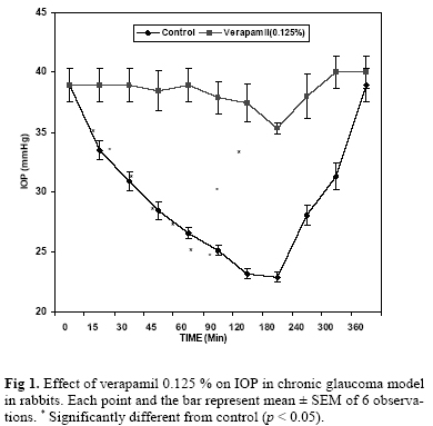
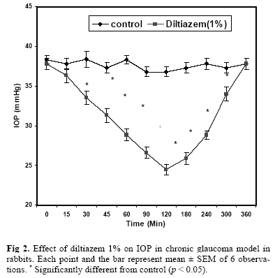
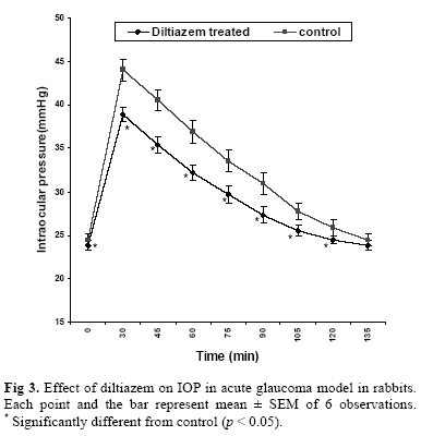
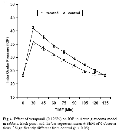
Iranian Journal of Pharmacology & Therapeutics, Vol. 4, No. 2, 2005, pp. 95-99 Effect of Calcium Channel Blockers on Intraocular Pressure in Rabbits ASHUTOSH JANI, RAMESH K GOYAL, GAURANG B SHAH and ANITA A MEHTA Department of Pharmacology, L.M. College of Pharmacology, Ahmedabad (R.K.G., A.A.M.); Department of Pharma-cology, K.B. Institute of Pharmaceutical Education & Research, Gandhinagar (A.J., G.B.S.), India. Address correspondence to: Dr. Anita A. Mehta, Department of Pharmacology, L.M. College of Pharmacology, Ahmeda-bad, India. E-mail: dranitalmcp@rediffmail.com Received November 14, 2005; Revised December 24, 2005; Accepted December 27, 2005 Code Number: pt05021 ABSTRACT The objective of the study was to evaluate the antiglaucoma effect of calcium channel blockers diltiazem and verapamil. Albino rabbits were used and chronic glaucoma was induced in them using freshly pre-pared 150 units of alpha–chymotrypsin. 0.1 mL of drug solution was administered topically into the left eye whereas the right served as control. The pressure recording was carried out at 15, 30, 45, 60, 90, 120, 180, 240, 300, 360 and if required at 420 and 480 min after drug instillation. Acute glaucoma was induced using 5% glucose solution administered intravenously, through the marginal ear vein, at a dose of 15 mL/kg body weight. The Intraocular Pressure (IOP) was recorded with a Schiotz-type indentation tonometer, which was previously calibrated by an open manometric calibration procedure. Topical ad-ministration of diltiazem (1%) (37.8 ± 0.632456 to 24.48 ± 0.6531) and verapamil (0.125%) (38.95 ± 1.40 to 22.85 ± 0.43) significantly reduced the elevated IOP (>30 mmHg) in alpha chymotrypsin induced chronic glaucoma model and diltiazem (1%) and verapamil (0.125%) prevented acute rise in the intraocu-lar pressure induced by intravenous administration of 5% glucose. Verapamil and diltiazem have IOP low-ering effect and can be utilized as potential investigative antiglaucoma drugs. Keywords: Antiglaucoma drugs, Calcium channel blockers, Diltiazem, Verapamil Calcium is an important intracellular messenger, of-ten interacting, with cyclic nucleotides to control a broad spectrum of physiological actions [ 1 ]. The topical administration of calcium ionophores A23187 and X573A has been shown to increase the Intraocular Pres-sure (IOP) [ 2 ]. Calcium channel blockers may plaeid an important role in clinical management of cardiovascular disorders over the past three decades. These drugs block membrane bound calcium channels and inhibits the calcium influx, cause relaxation of smooth muscle cells in vascular walls, a decrease in vascular tone, and an im-provement in blood flow [ 3 ]. Ferrante et al. [ 4 ] showed that nitrendipine and D600, a verapamil analogue, caused relaxation of pros-taglandin induced constriction of isolated calf retinal vessels. Both verapamil and diltiazem showed relaxation of cat opthalmocilliary artery ring segments in vi-tro. In normal human subjects studies using color Dop-pler ultrasonography, topical verapamil was found to reduce the resistive index in the central retinal artery. When verapamil was administered, the episodes of transient visual dimming ceased immediately. In addition, soon thereafter, visual acuity improved, the rubeosis partially regressed, and the hypotony reversed. This indicates that verapamil may be effective in treating cases of ocular ischemic syndrome, when vasospasm is a contributing cause [ 5 ]. Intraocular pressure is maintained as a steady state between aqueous humor formation and outflow. Calcium influx could have several effects on aqueous humor dynamics, including a hydrostatic component caused by an effect on arterial blood pressure and ciliary body perfusion, and an osmotic component caused by and effect on the active secretion of sodium, calcium and other ions by ciliary epithelium [ 6 ]. Effect of calcium channel blockers (CCB) on aqueous humour dynamics and intraocular pressure remains controversial since wide ranges of results are obtained. After systemic administration, CCBs failed to reduce the IOP in both rabbits [ 7 ] and humans, [ 8 , 9 ] although several labs have reported ocular Hypertensive [ 10 ] and ocular Hypotensive [ 11 - 14 ] response after oral or intravenous administration of these drugs. Results from the topical applied CCBs on IOP are conflicting. Beatty et al. [ 10 ] found that these drugs produced dose related increase in IOP in albino rabbits and humans, whereas Payne et al. [ 13 ] noted that vera-pamil, diltiazem and nifedipine had no effect on IOP in rabbits. On the other hand it has seen that [ 8 , 15 , 16 ] topical administration of verapamil and nifedipine effec-tively reduced the IOP in rabbits in a dose related fash-ion. In humans, Abelson et al. [ 17 ] and Mooshian et al. [ 17 ] reported a decrease in IOP after a single topical dose of verapamil in ocular hypertensive subjects. Recently Netland et al. [ 18 ] also found that verapamil sig-nificantly lowers the IOP in normal human volunteers. Despite the fact that no conclusion has been reached about in the effect of these drugs on IOP, evidence sug-gest that topical application of CCBs could be effective in management of ocular hypertension [ 17 , 18 , 20 ] and low tension glaucoma [ 19 ] However, such a potential role in the treatment of glaucoma is largely based on the circumstantial evidence and has not undergone an ade-quate preclinical and clinical evaluation. So present in-vestigation was undertaken to study effect of calcium channel blockers on intraocular pressure in the rabbit eye. MATERIALS AND METHODS The experiments were initiated after seeking approval from the institutional Animal Ethics committee of L.M. College of Pharmacy. Animals Albino rabbits of either sex weighing, 2.0-2.5 kg were used in the study. They were individually housed in metallic cages in well-ventilated rooms, under hygi-enic conditions. Animals were given water ad libitum and fed with green leafy vegetable available form the local market. Reagents and Drugs Verapamil, Pure powder was as a gift sample from Prof. G.B. Shah, Gandhinagar. Diltiazem was a gift sample from Cadila Pharma, Ahmedabad. Sterile water for Injection was obtained from Claris Life sciences Ltd. Phosphate buffer was prepared from Chemicals obtained from Rankem (Ranbaxy India). Preparation of Drugs Verapamil was prepared in phosphate buffer and di-luted to required strength and diltiazem injection was diluted with sterile water for injection. Experimental Procedure Acute ocular hypertension model in rabbits [20, 22 ]. Rabbits weighing 2.0-2.5 kg were used for the study. The basal IOP was measured using a Schiotz indenta-tion tonometer. In our preliminary studies on rabbits lower concentration of verapamil and diltiazem were found effective in lowering the IOP. Hence 0.1 mL of 0.1% verapamil and 1% diltiazem were used in further studies. The drug solution of verapamil was prepared in phosphate buffer and diltiazem injection was diluted with sterile water for injection. The drug solutions pre-pared was instilled topically into the left eye and right eye received the vehicle and served as a control. After 30 min of drug administration, the IOP was measured, and 5% glucose solution was administered intrave-nously, through the marginal ear vein, at a dose of 15 mL/kg body weight. The IOP recording was carried out every 15 min for 75 min in both eyes. The IOP was re-corded with a Schiotz-type indentation tonometer, which was previously calibrated by an open manometric calibration procedure. Studies on the chronic ocular hypertension Model [ 23]. Albino rabbits weighing 2.0-2.5kg were lightly anesthe-tized with ketamine (50 mg/kg i.v.). A cannula attached to a reservoir was inserted into the anterior chamber with the help of a 30-gauge needle, to provide a hydro-static pressure of 25mmHg during injection of alpha-chymotrypsin. Then a second appropriately shaped, 30-gauge needle was introduced near the pupil. Freshly prepared 150 units of Alpha-chymotrypsin (Sigma, St.Louis, MO, U.S.A.) prepared in 0.5 mL of sterile saline was irrigated through the cannula into the posterior chamber. Care was taken to prevent the contact of Alpha-chymotrypsin with corneal stroma. Both cannulas were carefully removed without significant loss of aqueous humor. Initially and after 2 days, the IOP was measured daily with a Schiontz type indentation tono-meter using 5.5-7.5 and 10 g weights. By drawing a graph of days versus IOP, the maximum period required to achieve a stable increase in IOP was determined. It was found that 15 days were sufficient to achieve a sta-ble increase in IOP. IOP was measured after 15 days for 3 consecutive days, every morning, to assure stable IOP. The rejection criterion in our study was the removal of those rabbits from the study that showed IOP < 30 mmHg. However, none of the eyes treated with Alpha-chymotrypsin showed IOP values < 30 mmHg. After achieving the steady elevated IOP, 0.1 mL of drug solu-tion was administered topically into the left eye whereas the right served as control. The pressure recording was carried out at 15, 30, 45, 60, 90, 120, 180, 240, 300, 360 and if required at 420 and 480 min after drug instillation. Statistical method. Results were expressed as mean ± S.E.M. Statistical analyses were compared statistically. With untreated controls using ANOVA followed by Student's t-test. Values of p < 0.05 were considered statistically significant. RESULTS Effect of Verapamil on Alpha-Chymotrypsin Induced Chronic Glaucoma Model in Rabbits Introduction of alpha-chymotrypsin (50 Units) in 0.1mL sterile saline in the posterior chamber of the eye produced a sustained elevation in IOP (>30 mmHg) after 15 days. Topical administration of verapamil (0.125%) to these animals produced a significant fall in intraocular pressure (38.95 ± 1.40 to 22.85 ± 0.43) (Fig 1). Effect of Diltiazem (1%) on Alpha-Chymotrypsin Induced Chronic Glaucoma Model in Rabbits Topical administration of diltiazem (1%) (37.8 ± 0.632456 to 24.48 ± 0.6531) significantly reduced the elevated IOP (>30 mmHg) in alpha chymotrypsin induced chronic glaucoma model (Fig 2). Effect of Verapamil (0.125%) on Acute Glaucoma Model in Rabbits A transient elevation in IOP upto 35-45 mmHg was observed when 5% glucose (15 mL/kg) was adminis-tered intravenously. Pretreatment with verapamil (0.125%) prevented the acute rise in intraocular pressure (IOP) due to infusion of 5% glucose intravenously (Fig 3). Effect of Diltiazem (1%) on Acute Glaucoma Model in Rabbits Diltiazem (1%) prevented acute rise in the intraocular pressure induced by intravenous administration of 5% glucose (Fig 4). DISCUSSION We have studied the ocular hypotensive effect of verapamil in experimentally induced acute and chronic models of gaulcoma in rabbits. verpamil (0.125%) and diltiazem (1%) prevented the acute rise in IOP due to 5% glucose infusion. Chronic and stable elevation of IOP was achieved by administering alpha chymotrypsin into the posterior chamber of rabbit eye. The sustained increase in IOP was caused by an inflammatory reaction in the trabecular meshwork. Data from our study show that duration of action of diltiazem and verapamil were almost the same and comparable to pilocarpine. Vera-pamil (38.95 ± 1.40 to 22.85 ± 0.428) and diltiazem (37.8 ± 0.63 to 24.48 ± 0.5) produced significant reduc-tion in IOP in alpha chymotrypsin induced chronic model. These results conflict with those of Beatty et al. [ 10], who found an increase in IOP after intravenous and topical application of verapamil, nifedipine and diltiazem in rabbits and after topical verapamil in hu-mans. Because the doses of verapamil used by Beatty et al. [ 10] were higher than those applied in most of the aforementioned studies, this may be one of the reason for failure of CCB in reducing IOP reported by Beatty et al. [ 10]. There are however several studies that support our finding. Abelson et al. [ 16 ] proposed that CCBs may have a biphasic effect on IOP, with an ocular hy-potensive action at low and an ocular hypertensive ac-tion at high concentrations. Matsuo [19] studied various cell lines from ocular tissues that were exposed to sustained levels of hydro-static pressure; he observed that that there was reorgani-zation of cell cytoskeleton and changes in the shape. It was also reported that human trabecular cell showed transient rise or oscillation of calcium when hydrostatic pressure was applied. Despite the shift of importance to cup disc optic disc and optic nerve, still the primary aim of treatment still deals with lowering of IOP. Segarra et al. [ 8] also reported that an IOP reduction after unilateral topical application of verapamil and nifedipine in albino rabbits. Abelson et al. [ 16 ] and Mooshian et al. [18] noted a contralateral effect of topi-cally applied verapamil in ocular hypertensive subjects, whereas Netland et al. [19] reported no effect of verapamil on IOP in the contralateral eye after topical ad-ministration in normal subjects. In our study we did not find a clear bilateral effect of verapamil or diltiazem when administered to only one eye, suggesting that these CCB lacks a contralateral effect in these animal models for glaucoma. Calcium channel blockers cause vasodilatations and reduce vascular resistance, increase the capillary blood speed in the optic nerve head [ 12, 16, 19], this make them to be possible drugs useful in the treatment of low-tension glaucoma. L-type (and T-type) calcium channels seem to have a role in cellular growth and several calcium antagonists, and possibly all can inhibit the growth and proliferation of vascular smooth muscle and fibroblasts. Calcium antagonists may also inhibit the synthesis of extra cellular-matrix collagen proteins [ 24], suggesting beneficial effect in glaucoma. Lowering of IOP by verapamil and diltiazem may be due to inhibition of the intracellular uptake of calcium by inactivating the inner phosphorylation-dependent calcium gate of the cellular membrane [ 25]. Its known that trabecular meshwork cells have contractile properties, which may be influenced by Ca2+ influx through voltage-dependent L-type Ca2+ channels, thus the relaxation by these agents can increase the outflow facility. The perfusion studies in dissected human eyes showed dosse-related increases in outlfow facility after verapamil administration [27, 28]. Further Santafé et al., [ 15] reported that CCBs de-crease aqueous humor secretion, although they also cause a slight, although significant, reduction of tono-graphic outflow facility. Also the outflow of aqueous humour influenced by episcleral venous pressure may be directly affected by calcium inhibition [6]. Further Gap junctions, possibly regulated by calcium, exist between nonpigmented and pigmented ciliary epithelial cells, verapamil may interfere with these Gap junctions, altering cellular permeability of the ciliary epithelium and thus inhibiting normal aqueous humour formation [ 11]. Verapamil may also alter the cyclic adenosine monophosphate content in ciliary epithelium cells, thereby affecting intraocular pressure through a de-crease in aqueous humour formation, or an increase in outflow facility [ 28]. Thus in conclusion verapamil and diltiazem have IOP lowering effect and can be utilized as potential In-vestigative antiglaucoma drugs. REFERENCES
Copyright © 2005 by Razi Institute for Drug Research (RIDR) |