 |
| About Bioline | All Journals | Testimonials | Membership | News |
|
||||||
|
||||||
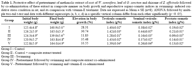
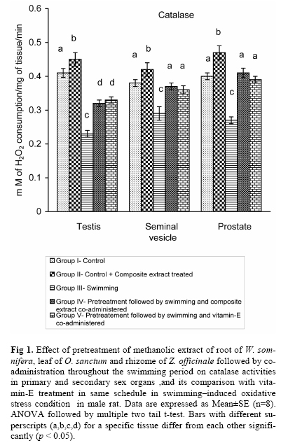
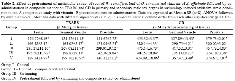
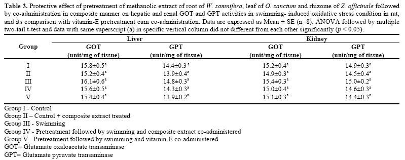
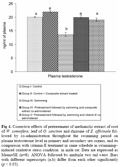
Iranian Journal of Pharmacology & Therapeutics, Vol. 4, No. 2, 2005, pp. 110-117 Protective Effect of Composite Extract of Withania somnifera, Ocimum sanctum and Zingiber officinale on Swimming-Induced Reproductive Endocrine Dysfunctions in Male Rat DEBANKA SEKHAR MISRA, RAJKUMAR MAITI, SARADINDU BERA, KOUSHIK DAS and DEBIDAS GHOSH Reproductive Endocrinology and Family Welfare Research Unit, Department of Human Physiology with Community Health, Vidyasagar University, Midnapore, West Bengal, India. Address correspondence to: Dr. Debidas Ghosh, Reproductive Endocrinology and Family Welfare Research Unit, Department of Human Physiology with Community Health, Vidyasagar Uni-versity, Midnapore, West Bengal, India. E-mail: debidas_ghosh@yahoo.co.in Received July 27, 2005; Revised October 18, 2005; Accepted December 23, 2005 Code Number: pt05024 ABSTRACT The present study has been designed to find out the effects of forced swimming-induced oxidative stress imposition on primary and secondary sex organs and its protection by plant extract in male Wistar strain rats. This work has been designed to find out the scientific basis of the local use of the composite extract of root of Withania somnifera, leaf of Ocimum sanctum and rhizome of Zingiber officinale by village Ayurvedic doctors to protect the health disorders in connection to strenuous physical exercise, and also to search out the potentiality of above mentioned plant products on swimming-induced oxidative damage. Forced intermittent swimming for 8 hours /day for 28 days resulted a significant elevation in the level of products of free radical i.e., thiobarbituric acid reactive substances and conjugated dienes along with sig-nificant diminution in the activities of catalase, superoxide dismutase and glutathione–S–transferase in testis, prostate and seminal vesicle which were protected significantly after co-administration of methanolic extract of said plant parts in composite manner. Testicular steroidogenesis was assessed in this condition by measuring plasma levels of testosterone, which was diminished significantly in swimming group and was protected significantly by the composite extract of the plants parts. The antioxidative potency of this composite extract was compared with potent and standard antioxidant i.e., vitamin-E in forced swim-ming state. This herbal extract has no toxic effect on metabolic organs that has denoted here by the measurement of glutamate oxaloacetate transaminase and glutamate pyruvate transaminase activities in liver and kidney. The results suggest that swimming-induced oxidative stress on male sex organs may be protected by using above mentioned medicinal plants extract. Keywords: Swimming; Oxidative stress; Medicinal plants; Vitamin-E; Reproductive organs In 21st century, people are very much conscious for health promotion following non-chemotherapeutic strat-egy like diet modification, regular exercise etc. Regular physical exercise like swimming may be the most effec-tive strategy to maintain or promote health status. In our country, there is a long tradition of the use of medicinal plants for health recovery [1, 2 ]. Moreover, Government of United States of America also established the Centre of Complementary and Alternative Medicine, and de-fined several ways of such treatment where herbal me-dicinal treatment shows a promising effect [ 3 ]. At a pi-lot project, we screened several locally available medicinal plants having antioxidative reputation and noted that the composite extract of the three plants selected in this work have promising antioxidant potency which is equal to established antioxidant i.e., vitamin-E. Forced swimming, a good physical exercise model, is considered as physical stressor also [ 4 ]. During physical exercise, oxygen utilization increases 10-15 folds [ 5 ] and it is well established that reactive oxygen species (ROS) generation is a direct function of the rate of oxygen utilization [ 6 ]. Oxygen reperfusion is another process of ROS imposition due to physical exercise like swimming though there is also a controversy about oxi-dative stress development due to exercise that focuses on the elevation in antioxidant defense system due to regular exercise [ 7 ]. From literature searching, it has been indicated that remarkable dysfunctions are noted in male reproductive system due to intensive exercise [ 7 , 8 ]. There is a plethora of information about the oxida-tive stress imposition in sex organs due to chronic swimming [ 4 ]. Moreover no information is available about the herbal management of oxidative damage induced by forced swimming on male reproductive or-gans. From trial and error, the effective dose of the composite extract and the ratio of the extract of these three plant parts have been determined for maximal management of oxidative injury caused by forceful swimming on male reproductive organs. On that back-ground, the present experimental design has been for-mulated to find out the level of endogenous anti-oxidative defensive derangement in male sex organs and its protection by co-administration of composite extract of above mentioned medicinal plants in comparison to potent and established antioxidant i.e., vitamin-E. Withania somnifera (W. somnifera) Dunal (Ashwa-gandha) is widely used in Ayurvedic medicine, the tra-ditional medical system of India. Its height is 3-4 feet and grows into a bush and is a member of the family Solanaceae. In India, its growth is maximum and at pre-sent this plant is cultivated for medicinal purpose. Therapeutic importance of the different parts of this plant has a long history and is mentioned in Charak Sanghita. It is an ingredient in many formulations prescribed for a variety of musculoskeletal condition (e.g., arthritis, rheumatism) and as a general tonic to increase energy, improve overall health and longevity and pre-vent disease in athletes, the elderly and during preg-nancy [ 9 ]. It is also used as an antistressor and antioxi-dant agent [ 10 ]. Ocimum sanctum (O. sanctum) is considered as sa-cred plant in the Hindu culture and known as Tulsi or Tulasi in Hindi or Holy Basil in English. It is a tropical annual herb, up to 18 inches tall and grows into a low bush and is a member of the family Lamiaceae (Labi-atae). Its therapeutic importance has been mentioned in Charak Sanghita, the ancient textbook of Ayurveda. Leaves of this plant are used in a variety of pathophysi-ological states like asthma, dysentery, dyspepsia, chronic fever, skin disease, helminthiasis and for ring worms [ 11 ]. It is also used as antistressor [ 12 ]. It is available throughout India. Zingiber officinale (Z. officinale) (Ginger) has been used for the treatment of several diseases since Baydic age. It is used as antioxidant [ 13 ], as well as used in antimotion sickness [ 14 ]. It belongs to the family Zingiberaceae. Its common names are calamus, sweet ginger etc. and cultivated throughout India. MATERIALS AND METHODS Selection of Animals and Care The study was conducted on forty healthy, adult, male albino rats of Wistar strain having a body weight of 120 ± 5 g. They were acclimatized to laboratory con-dition for 2 weeks prior to experimentation. Animals were housed two per cage in a temperature-controlled room (22 ± 2ºC) with 12-12 h dark-light cycle (8.00-20.00 h light: 20.00-8.00 h dark) at a humidity of 50 ± 10 %. They were provided with standard food and water ad libitum. Animal care was provided according to the Guiding Principle for the Care and Use of Animals [ 15 ]. Our University Ethics Committee approved the experi-mental protocol. Plant Materials The root of W. somnifera, leaf of O. sanctum and rhizome of Z. officinale were collected from Gopali, Indian Institute of Technology, Kharagpur, Paschim Medinipur district of West Bengal in the month of May and the material was identified by taxonomist of Botany Department, Vidyasagar University, Midnapore. The voucher specimens were deposited in the Department of Botany, Vidyasagar University, and the voucher speci-men numbers were HPCH No-3, 4, 5, respectively. Preparation of Methanolic Extract of Root of W. somnifera, Leaf of O. sanctum and Rhizome of Z. officinale The plant parts were dried in an incubator for 2 days at 40ºC, crushed in an electrical grinder and then pow-dered separately. 50 g powder of each plant material was extracted in 250 mL of methanol for 18 h in a sox-hlet apparatus. The deep brown of Z. officinale, yellow-ish brown of W. somnifera and deep green of O. sanc-tum extracts in methanol were collected. The extracts were dried at reduced pressure, stored at (0-4)ºC and used for next 7 days of the experiment. As per demand, extracts were prepared further throughout the experi-mental period. When needed, the extracts were sus-pended in olive oil and used in the study. Experimental Design Forty, adult healthy, male albino rats of Wistar strain were divided equally into 5 groups on the basis of the matching of body weights of the animals. The treatment schedule of each group was as follows. Group I (Control Group). Control rats were kept in rat’s cage. Rats of this group received olive oil (0.5 mL/100 g body weight/ day) for 15 days prior to ex-perimentation followed by 28 days of experimental period through oral route at 8.00 h. Group II (Control + Composite Extract Treated Group). Animals were subjected to forceful oral ad-ministration of methanolic extract of these plant parts at the ratio of 1: 2: 2 (W. somnifera: O. sanctum: Z. offici-nale) at the dose of 40 mg /100 g body weight / day / rat in 0.5 mL olive oil for 15 days prior to starting of ex-periment followed by next 28 days of experimentation with out swimming. The herbal mixture was adminis-tered at 8.00 h of each day by gavage. Group III (Swimming Group). Rats were sub-jected to swimming for 8 h/day including rest. The dura-tion of this exercise was fixed for 30 min at a stretch followed by 10 min rest as followed by previous work-ers [ 16 ]. This swimming was continued for 28 days without break. Olive oil was administered through ga-vage as in group I. Group IV (Pretreatment Followed By Swimming, and Composite Extract Co-Administered Group). Rats were subjected to preconditioning by oral admini-stration of methanolic extract of these plant parts for 15 days prior to the starting of swimming at the same ratio as of group II. From 16th day, animals were subjected to swimming for 8 h/ day (including rest) at the same pro-tocol like group III for 28 days. Before 2 h of starting the swimming in each day, all the animals of this group were subjected to oral administration of the methanolic extract of these plant parts in composite way at the same dose as per preconditioning period and as of group II. Food was provided to the animals at least 1 h before swimming and the swimming was started from 10.00 h to 18.00 h including rest everyday. Group V (Pretreatment Followed by Swimming and Vitamin-E Co-Administered Group). Rats were subjected to preconditioning by oral administration of vitamin-E (Alpha- tocopherol succinate) at the dose of 6 mg/100 g body weight/day/rat in 0.5 mL olive oil [ 17 ] for 15 days prior to starting of swimming. From 16th day, animals were subjected to swimming for 8 h/day (including rest) at the same protocol like group III for 28 days. Two hours before starting the swimming in each day, all the animals of this group were subjected to oral administration of the vitamin at the same dose as per preconditioning period. Food was provided to the animals at least 1 h before swimming. Including rest period, swimming was continued from 10.00 h to 18.00 h daily throughout the experimentation. To maintain the same physical stress due to handling of animals and forceful ingestion of extract, all the ani-mals of Group I, II and III were subjected to olive oil treatment by gavage throughout the experimental period at the same time in relation to pre exercise oral treat-ment of herbal mixture to group IV and vitamin-E to group V. Forced Swimming Programme The forced swimming of rats was performed in acrylic plastic pool (90 cm × 45 cm × 45 cm) filled with water to a depth of 37 cm as per design of previous workers [ 18 , 19 ]. The temperature of the water was maintained at 34 ± 1ºC. The rats were loaded with a steel washer weighing approximately 4% of their body weight attached to the tail. This arrangement forced the rat to maintain continuous rapid leg movement [ 20 ]. The fur of the rats was washed with liquid soap prior to swimming and air bubbles trapped in the fur were re-moved periodically to reduce buoyancy and ensure the imposed workload [ 21 ]. After completion of 28 days swimming, all the ani-mals, one after another, were killed within 5 min of post-exercise period. Liver, kidney, testis, seminal vesicle, prostate and blood were collected from each animal. All tissues were refrigerated at –20ºC and within 2h of refrigeration, the tissues were processed for biochemical assay. Biochemical Assay of Catalase (CAT) Catalase activity was measured biochemically [ 22 ]. For the evaluation of CAT activity testis, seminal vesi-cle, prostate, liver and skeletal muscle from each animal were homogenized separately in 0.05 M Tris-hydrochloric acid (HCl) buffer solution (pH-7.0) at the tissue concentration of 50 mg/mL. These homogenates were centrifuged separately at 10,000 g at 4ºC for 10 min. In spectrophotometric cuvette, 0.5 mL of hydrogen peroxide (H2O2) and 2.5 mL of distilled water were mixed and reading of absorbance was noted at 240 nm. Tissue supernatants were added at a volume of 40 µl and the subsequent six readings were noted at 30 sec interval. Biochemical Assay of Superoxide Dismutase (SOD) Testis, seminal vesicle and prostate were homogenized in ice-cold 100 mM Tris-cacodylate buffer to give a tissue concentration of 50 mg/mL and centrifuged at 10,000 g for 20 min at 4 0C. The SOD activities of these supernatants were estimated by measuring the percent-age inhibition of the pyragallol autooxidation by SOD [ 23 ]. The buffer was 50 mM Tris (pH-8.2) containing, 50 mM cacodylic acid (pH-8.2), 1 mM ethylene diamine tetra acetic acid (EDTA) and 10 mM hydrochloric acid (HCl). In a spectrophotometric cuvette, 2 mL of buffer, 100 µl of 2 mM pyragallol and 10 µl of supernatant were poured and the absorbance was noted in a spectro-photometer at 420 nm for 3 min. One unit of SOD was defined as the enzyme activity that inhibited the autooxidation of pyragallol by 50 percent. Estimation of Glutathione-S-Transferase (GST) Activities of GST in the tissue samples were also in-dividually measured spectrophotometrically [ 24 ] using 1-chloro-2,4-dinitrobenzene (CDNB) as substrate. The assay mixture of 3 mL contained 0.1 mL of 1 mM CDNB in ethanol, 0.1 mL of 1 M reduced glutathione (GSH), 2.7 mL of 100 mM potassium phosphate buffer (pH-6.5) and 0.1 mL of supernatant of the tissue ho-mogenate. The formation of the adduct of CDNB, S-2,4-dinitrophenylglutathione was monitored by measur-ing the net increase in absorbance at 340 nm against blank. Enzyme activity was calculated using the extinc-tion coefficient of 6.9/M/cm and expressed in unit/mg of tissue. Estimation of Lipid Peroxidation from the Levels of Thiobarbituric Acid Reactive Substances (TBARS) and Conjugated Dienes (CD) The testis, seminal vesicle and prostate were ho-mogenized separately at the tissue concentration of 50 mg/mL in 0.1 M of ice-cold phosphate buffer (pH-7.4) and the homogenates were centrifuged at 10,000 g at 4ºC for 5 min individually. Each supernatant was used for the estimation of TBARS and CD. For the measurement of TBARS, 0.5 mL homogenate was mixed with 0.5 mL of normal saline (0.9 g% NaCl) and 2 mL of thiobarbituric acid-trichloro acetic acid (TBA-TCA) mixture (0.392g of TBA in 75 mL of 0.25 N HCl with 15 g TCA, the volume of the mixture was made up to 100 mL by 95% ethanol) and boiled at 100ºC for 10 min. This mixture was then cooled at room temperature and centrifuged at 4000 g for 10 min. The whole super-natant was transferred in spectrophotometer cuvette and read at 535 nm [ 25 ]. Quantification of the CD was performed by a stan-dard method [ 26 ]. The lipids were extracted with chloroform-methanol (2:1) mixture followed by centrifuga-tion at 1000 g for 5 min. The chloroform layer was evaporated to dryness under a stream of nitrogen. The lipid residue was dissolved in 1.5 mL of cyclohexane and the absorbance was noted at 233 nm to measure the amount of hydro peroxide formed. Assay of Plasma Testosterone The collected blood was centrifuged and plasma fraction was separated. Plasma level of testosterone was measured following the immuno-enzymatic method by using enzyme linked immuno sorbent assay (ELISA) reader (Merck, Japan) according to the standard proto-col of National Institute of Health and Family Welfare [ 27 ]. Here, we followed commercially available com-petitive solid phase enzyme immunoassay where horse-radish peroxidase was used as enzyme-labelled antigen supplied by EQUIPAR, USA, competes with unlabelled antigen for binding with limited number of site of anti-body on the micro plates (solid phase). After incubation, bound/ free enzyme labeled antigen separations were performed by simple washing. The substrate of enzyme, H2O2 and the chromogen (TMB-3/3’/5/5’ـtetra Methyl Benzedine) were added and after the schedule time en-zyme reaction was terminated by the addition of stop solution, supplied by EQUIPAR, USA. Testosterone concentration in the sample was calculated based on five standards supplied by the EQUIPAR, USA. The absorbance’s of standard and sample were monitored against the blank at 450 nm. The cross-reaction of the testosterone antibody to dihydrotestosterone was 10% and intra-assay precision had a coefficient of variation of 6.2%. As all the samples were run at the same time so there was no inter-assay precision. The assay validated in respect to correctness of the data in our laboratory was 98%. Glutamate Oxaloacetate Transaminase (GOT) and Glutamate Pyruvate Transaminase (GPT) Activities in Liver and Kidney For the assessment of metabolic toxicity, we meas-ured GOT and GPT activities in liver and kidney ac-cording to the method of Goel [ 28 ]. Statistical Analysis Analysis of variance (ANOVA) followed by a mul-tiple two-tail ‘t’ test with Bonferroni modification was used for statistical analysis of the collected data [ 29 ]. Difference were considered significant when p < 0.05. RESULTS Body Weight and Somatic Indices of Sex Organs Body weight was increased at the end of experiment in group I, II, IV and V in respect to their initial body weight ( Table 1 ). In group III, the percentage of eleva-tion in body growth was dramatically less than the other groups due to swimming in non trained rats ( Table 1 ). Testiculo-somatic, seminal vesiculo-somatic and prostato -somatic indices were decreased significantly in group III in comparison to group I and group II. After co-administration of composite extract or vitamin-E in group IV or V respectively these parameters were reset-tled either to or towards the control level ( Table 1 ). Catalase Activity In group II, CAT activities in testis, prostate and seminal vesicle were elevated in respect to group I, IV and V ( Fig 1 ). After swimming (Group III), the activi-ties of this enzyme in above mentioned tissues were decreased significantly in respect to group I ( Fig 1 ). There was a significant protection in this parameter after pretreatment with composite extract followed by its co-administration (Group IV) or vitamin-E pretreatment cum co-administration (Group V) to the animals of the swimming group. Activities of GST and SOD Administration of herbal mixtures to non-swimming animals (Group II) resulted a significant elevation in the activities of GST and SOD in testicular, seminal vesicu-lar and prostatic tissues in respect to the animals of group I, IV and V ( Fig 2 and Fig 3 ). The activities of both the above enzymes were decreased significantly in aforesaid tissues in group III when compared to group I ( Fig 2 and Fig 3 ). Pretreatment followed by co-administration of composite extract to the animals of group IV resulted a significant correction of the above enzyme activities in respect to group III and the values were resettled to the control level ( Fig 2 and Fig 3 ). The protective effect of vitamin-E pretreatment followed by co-administration on SOD and GST activities in above tissues showed an insignificant variation between group IV and V. Quantification of TBARS and CD Quantity of TBARS and CD both were decreased in testis, seminal vesicle and prostate of group II when com-pared to group I, IV and V ( Table 2 ). In non trained rats, 28 days swimming resulted a significant elevation in the values of both the parameters in all of the above mentioned tissues when we compared the data in respect to group I ( Table 2 ). In group IV and V, the values of both these pa-rameters in all of the above mentioned tissue samples were significantly decreased than the group III ( Table 2 ) and the values were resettled to the control level ( Table 2 ). Activities of GOT and GPT In liver and kidney, the activities of GOT and GPT were not altered significantly among the groups ( Table 3 ). Plasma Level of Testosterone After 28 days of swimming (Group III), there was a significant diminution in the plasma level of testosterone in respect to control (Group I). The plasma testosterone level was resettled to the control value in group IV and group V animals after pretreatment cum co-administration of herbal mixture or vitamin-E respectively ( Fig 4 ). DISCUSSION Despite the paradox that exercise results in benefi-cial effects for promotion of good health and prevention of various diseases, there are several reports that strenu-ous exercise causes oxidative stress imposition in sev-eral vital organs by derangement of endogenous anti-oxidant regulators [ 30 ]. Swimming-induced oxidative stress in testis and male secondary sex organs has been established here by noting the low activities of SOD, CAT, GST- important antioxidant enzymes, which is consistent with the ob-servation of others [ 31 ]. The decrease in antioxidant enzyme activities due to swimming might be due to their use against the free radicals destruction and or their inhibition by free radical species [ 32 ]. It is well established that SOD activity is inhibited by hydrogen peroxide that reduced Cu+2 to Cu+1 in SOD [ 23 ]. The reduced Cu+1 can act as promoter of hydroxyl by Haber-Weis reaction [ 23 ]. Low-antioxidant enzyme activities further facilitate the increased susceptibility to lipid peroxidation [ 5 ]. The reduction of hydrogen peroxide is catalyzed by CAT that protects the tissues from highly reactive hydroxyl radicals [ 33 ]. Reduction of hydrogen peroxide and hydro peroxides to non-toxic products are catalyzed by GST and peroxidase. Another possibility for the low levels in the activities of above mentioned antioxidant enzymes in sex organs may be due to low level of testosterone as testosterone promotes the syn-thesis of antioxidant enzymes in sex organs [ 34 ]. This low plasma level of testosterone due to swimming has been noted here. Elevations in the levels of products of free radicals like TBARS and CD in sex organs in swimming group again support the low antioxidant enzyme activity that elevate the lipid peroxidation while TBARS and CD are the products of lipid peroxidation. Another possibility for such elevation in TBARS and CD may be due to ischemia- reperfusion phenomenon [ 35 , 36 ] or due to high rate of catecholamine secretion that generate free radicals either through auto oxidation or through metal ion or superoxide-catalyzed oxidation [ 37 ]. In this experiment, the composite extract of above mentioned plants has been used in relation to the tradi-tional use of herbal mixture by our village Ayurvedic doctors as well as by other workers who reported that composite plant extract give a better result than single plant extract treatment [ 1, 38], and by the outcome of pilot experiments conducted in our laboratory. The composite extract of these plants results significant ele-vation in the level of antioxidant status, and its potency is equivalent to potent established antioxidant known as vitamin-E as shown here. Vitamin-E protects biological membrane against the damaging effects of reactive oxy-gen species, as this vitamin is effective inhibitor of lipid peroxidation because of its association with membrane lipids. Moreover vitamin-E itself elevates scavenger enzymes activity [ 39] as well as gonadal steroidogenesis [ 40]. Our results also support the above view that anti-oxidant status is elevated in vitamin-E co-administeredswimming group. From literature review it has been indicated that W. somnifera and O. sanctum have some spermicidal effect in vitro [ 41]. Moreover, O. sanctum also inhibits sexual behavior [ 42]. These discrepancies may be explained from the angle of dose, solvent used for extract prepara-tion and design of the experiment. On the other hand there is also report that Z. officinale has testicular stimu-lating effects [ 43] which is consistent with our results. The composite extract pre-exposed cum co-administered group shows a significant protection of the sex organs against swimming –induced oxidative stress by diminution in the level of products of free radicals and elevation in the activities of antioxidant enzymes along with level of plasma testosterone .The actual mechanism for such protection would not be explained clearly but following hypothesis may be proposed. The active ingredient(s) present in this extract may destroy the free radical generation or elevate the level of anti-oxidant enzymes along with testosterone biosynthesis. Diminution in sex-organ somatic indices after exercise followed by recovery in extract treatment or vitamin-E pretreatment cum co-administered group suggest the effects of this extract on gonadotrophin and testicular steroid synthesis as testicular growth is indicator of gonadotrophin [44] and the growth of accessory sex organs is indicator of plasma tetosterone [45]. As the composite extract has no general and metabolic toxic effect reflected here from significant veriation in body growth and activities of liver and kidney GOT and GPT, so it may be proposed that the results of this work may be disseminated to the sports community for beneficial outcome from physical exercise-induced oxidative stress imposition that significantly interfere the physical performance and endurance. ACKNOWLEDGEMENTS The financial assistance from University GranCommission, New Delhi in the form of Faculty Improvement Programme scheme to the author D. Misra is gratefully acknowledged. REFERENCES
Copyright © 2005 by Razi Institute for Drug Research (RIDR) |