 |
| About Bioline | All Journals | Testimonials | Membership | News |
|
||||||
|
||||||
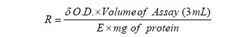
Iranian Journal of Pharmacology & Therapeutics, Vol. 5, No. 1, 2006, pp. 15-20 Research Article Nootropic Activity of Calyces of Hibiscus sabdariffa LinnHANUMANTHACHAR JOSHI and MILIND PARLE Current Author Addresses: Hanumanthachar Joshi, Dept. of Pharmacognosy, Soniya Education Trust’s College of Pharmacy, Dharwad, Karnataka, India. Email: amanjoshi17@yahoo.com (Corresponding Author). Received September 9, 2005; Revised March 10, 2006; Accepted March 11, 2006 Code Number: pt06002 ABSTRACT Nootropic acitivity of calyces of Hibiscus sabdariffa Linn. was studied in mice. Elevated plus maze and passive avoidance paradigm were employed to evaluate learning and memory parameters. Scopolamine (0.4 mg/kg, i.p.) was used to induce amnesia in mice. The aqueous extracts of calyces of Hibiscus sabdariffa (HS-100 and 200 mg/kg, p.o.) significantly attenuated amnestic deficits induced by scopolamine (0.4 mg/kg, i.p.) and natural aging. HS (100 and 200 mg/kg) decreased the transfer latencies and increased step down latencies significantly in the aged mice and scopolamine induced amnesic mice as compared with Piracetam (200 mg/kg, i.p.). To delineate the possible mechanism through which H. sabdariffa elicits the anti-amnesic effects, we studied its influence on central cholinergic activity by estimating the whole brain acetylcholinesterase activity. H. sabdariffa significantly decreased acetyl cholinesterase activity in mice. The results indicate that, the aqueous extract of calyces of H. sabdariffa might prove to be a useful memory restorative agent in the treatment of dementia seen in elderly. The underlying mechanism of action can be attributed to its anti acetylcholinesterase property. Keywords: Nootropic activity, Hibiscus sabdariffa, Memory Alzheimer’s disease is a progressive neurodegenerative brain disorder that occurs gradually and results in memory loss, unusual behavior, personality changes and ultimately death [1]. It is a chronic, progressive disabling organic brain disorder characterized by disturbance of multiple cortical functions, including memory, judgment, orientation, comprehension, learning capacity and language[2]. Nootropic agents such as piracetam [3], pramiracetam, aniracetam [4] and choline esterase inhibitors like Donepezil® are presently used for improving memory, mood and behavior. However, the resulting adverse effects associated with these agents have limited their use [5] and it is worthwhile to explore the utility of traditional medicines in the treatment of various cognitive disorders. Hibiscus sabdariffa Linn. (Malvaceae) is commonly known as red sorrel in English, Lal lambari in hindi, pundibija in kannada and the dried calyx is used as a diuretic, sedative, refrigerant [6] and for making a drink by boiling in water and adding a little salt, pepper, asafoetida and molasses to treat mental fatigue due to excessive heating by the Gondas tribal of Karnataka, India. It is native of tropical Africa or Asia and is now found widely cultivated throughout tropics of India. The tender leaves and stalks are eaten as salad, as curries and an infusion of the calyces are used as laxative, in cardiovascular and nervous diseases [7]. H. sabdariffa reported to be antiseptic, aphrodisiac, astringent, resolvent, cholagogue, digestive, diuretic and stomachic [8]. Roselle is a folk remedy for abscesses, heart ailments and hypertension. Hibiscus tea has been shown to lower blood pressure in patients with essential hypertension [9]. A polyherbal drug, Anna Pavala Sindhooram (Sidha formulation) contains flowers of H. sabdariffa as major constituents and is used in the management of atherosclerosis. Calyces are used in West Indies to color and flavor rum, Seeds are used for preparing an aphrodisiac herbal tea, and the fruits are edible [10]. Calyx of Roselle contains various antioxidants such as, anthocyanin, quercetin, L-ascorbic acid and protocatechuic acid [11]. It also contains anisaldehyde, arachidic acid, b-carotene, b-sitosterol, delphinidin, gossypetin and hibiscetin [12]. H. sabdariffa is reported to possess antihypertensive [13], antioxidant [14], anticancer [15], anticlastrogenic [16], hypolipidaemic [17], hepatoprotective, anti-stress [18-19], antispasmodic [20], diuretic [21] and antidiarrheal [22] activities. In the present study, we investigated the nootropic effects of calyces of H. sabdariffa by employing exteroceptive and interoceptive behavioral models in mice. Elevated plus maze is a neutral exteroceptive model used to assess short-term memory whereas, passive avoidance apparatus is a punishment based exteroceptive model used to test long-term memory [23]. Interoceptive behavioral models such scopolamine and natural aging induced amnesia are widely cited as models simulating human dementia in general and Alzheimer’s disease in particular.To understand the possible mechanism of action by which H. sabdariffa exerts nootropic activity, whole brain acetyl cholinesterase activity was determined. Materials and Methods Plant Materials Red calyces of H. sabdariffa were purchased from local herbal market and were identified by taxonomists at Department of Pharmacognosy. Voucher specimens (HKJ/HS-11) of the collected samples were deposited in the Department of Pharm. Sciences, Guru Jambheshwar University, Hisar, Haryana, India. Preparation of Water DecoctionThe shade-dried calyces were powdered and passed through 10-mesh sieve. The coarsely powdered materials (1000 g) were soaked in distilled water in the ratio of 1:18 (w/v) and boiled for 20 min. The extract was filtered, concentrated using rotavapour apparatus, and dried in freeze drier with high vacuum. A suspension was prepared using distilled water containing 1% (w/v) carboxymethyl cellulose (CMC). Drugs and ChemicalsScopolamine hydrobromide (Sigma Aldrich, USA) and piracetam (Nootropil®, UCB India Pvt. Ltd., Vapi, Gujarat) were diluted in normal saline and administered peritoneally. Phenytoin (Dilantin®suspension, Parke Davis) was administered orally. Volume of administration was 1ml/ 100 g/body weight. All the drugs were administered in the morning session i.e. 8 AM- 9 AM on each day. 5, 5’-dithiobis nitrobenzoic acid (DTNB, Ellman’s reagent, Sigma, USA) and acetyl thiocholine (Sigma USA) were used. Acute Toxicity StudiesH. sabdariffa aqueous extract (HS) at different doses (50-2000 mg/kg) was administered orally to mice with the help of a specially designed oral needle connected to a polythene tube. HS was administered at the same time on each day (i.e. 8 AM- 9 AM). During the first four hours after the drug administration, the animals were observed for gross behavioral changes if any, for 7 days. Parameters such as hyperactivity, grooming, convulsions, sedation, hypothermia and mortality were observed. The doses selected were 100 mg/kg and 200 mg/kg/day. AnimalsSwiss mice of either sex weighing around 18 g (younger ones, aged 8 weeks) and 25 g (older ones, aged 28 weeks) were used in the present study. Animals were procured from disease free animal house of CCS Haryana Agriculture University, Hisar (Haryana, India). They were acclimatized to the laboratory conditions for 5 days before behavioral studies. The animals had free access to food and water and were maintained under 12:12 h light and dark cycles. All experiments were carried out during day time from 900 to 1400 h. Institutional Animals Ethics Committee (IAEC) had approved the experimental protocol and care of animals was taken as per guidelines of CPCSEA, Dept. of Animal Welfare, Govt. of India. Exteroceptive Behavioral ModelsElevated plus Maze (EPM). The elevated plus maze served as the exteroceptive behavioral model (wherein the stimulus existed outside the body) to evaluate learning and memory in mice. The apparatus consisted of two open arms (16 cm × 5 cm) and two covered arms (16 cm × 5 cm × 12 cm). The arms extended from a central platform (5 cm × 5 cm), and maze was elevated to a height of 25 cm from the floor. On the first day, each mouse was placed at the end of an open arm, facing away from the central platform. Transfer latency (TL) was taken as the time taken by the mouse to move into any one of the covered arms with all its four legs. TL was recorded on the first day. If the animal did not enter into one of the covered arms within 90 sec., it was gently pushed into one of the two covered arms and the TL was assigned as 90 sec. The mouse was allowed to explore the maze for 10 sec and then returned to its home cage. Memory retention was examined 24 h after the first day trial on the second day [24-26]. Passive shock avoidance paradigm. Passive avoidance behavior based on negative reinforcement was recorded to examine long-term memory. The apparatus consisted of a box (27 × 27 × 27 cm) having three walls of wood and one wall of Plexiglas, featuring a grid floor (3 mm stainless steel rods set 8 mm apart), with a wooden platform (10 × 7 × 1.7 cm) in the center of the grid floor. The box was illuminated with a 15 W bulb during the experimental period. Electric shock (20V AC) was delivered to the grid floor. Training was carried out in two similar sessions. Each mouse was gently placed on the wooden platform set in the center of the grid floor. When the mouse stepped down and placed all its paws on the grid floor, shocks were delivered for 15 sec and the step-down latency (SDL) was recorded. SDL was defined as the time taken by the mouse to step down from wooden platform to grid floor with its entire paw on the grid floor. Animals showing SDL in the range (2-15 sec) during the first test were used for the second session and the retention test. The second-session was carried out 90 min after the first test. When the animals stepped down before 60 sec, electric shocks were delivered for 15 sec. During the second test, animals were removed from shock free zone if they did not step down for a period of 60 sec. Retention was tested after 24 h in a similar manner, except that the electric shocks were not applied to the grid floor. Each mouse was again placed on the platform, and the SDL was recorded, with an upper cut-off time of 300 sec [27-29]. Experimental Protocol The animals were divided into 32 groups and each group consisted of a minimum of five animals. Separate animals were used for each experiment. Group I: Represented Control group for young mice (n=6). Distilled water (DW), was administered orally for 8 days. TL was noted after 45 min of administration on 8th day and again after 24 h i. e on 9th day. Group II & X: Piracetam, 200 mg/kg, i.p. was injected to both young and aged mice respectively. TL was noted after 45 min of injection and again after 24 h. Group III: Scopolamine (0.4 mg/kg, i.p.) was administered to young mice and TL was noted after 45 min of injection on 8th day and again after 24 h i.e. on 9th day. Group IV & V: HS (100 mg/kg & 200 mg/kg) was administered orally to young mice for 8 days. The last dose was given 45 min before subjecting the animals to elevated plus maze test. TL was noted on 8th day and again after 24 h. Group VI & VII: HS (100 mg/kg & 200 mg/kg, p.o.) was administered to young mice for 8 days. After 45min of administration of the last dose on the 8th day, scopolamine hydrobromide (0.4 mg/kg, i.p.) was administered. TL was noted after 45 min of administration of scopolamine and again after 24 h i.e. on the 9th day. Group VIII: Piracetam (200 mg/kg, p.o.) was administered to young mice for 8 days. After 45min of administration of the last dose on the 8th day, scopolamine hydrobromide (0.4 mg/kg, i.p.) was administered. TL was noted after 45 min of administration of scopolamine and again after 24 h i.e. on the 9th day. Group IX: Served as the control group for aged mice. Distilled water (DW), was administered orally for 8 days. TL was noted after 45 min of administration on the 8th day and again after 24 h i. e on the 9th day. Group XI & XII: HS (100 mg/kg & 200 mg/kg) was administered orally to aged mice for 8 days. The last dose was given 45 min before noting TL on the 8th day. GroupXIII: Control group for young mice. Distilled water (1 mL/100g) was administered p.o. for 8 days. After 90 min of administration on the 8th day, SDL was recorded. Retention was examined after 24 h. Group XIV: Piracetam (200 mg/kg, i.p.) was administered for 8 days to young mice. SDL was recorded after 45 min of administration on the 8th day and again after 24 h. Group XV: Scopolamine hydro bromide (0.4 mg/kg) was administered i.p. to young mice after training on the 8th day and SDL was recorded at 45 min after injection. Group XVI:100mg/kg HS was administered orally for 8 days to young mice. SDL was recorded after 90 min of administration on the 8th day and again after 24 h. Group XVII: 200 mg/kg HS was administered orally for & days to young mice. SDL was recorded after 90 min of administration on the 8th day and again after 24h. Group XVIII & XIX: HS (100 mg/kg & 200 mg/kg, p.o.) was administered to young mice for 8 days. After 45 min of administration of the last dose on the 8th day, scopolamine hydrobromide (0.4 mg/kg, i.p.) was administered. SDL was recorded after 90 min of administration on the 8th day and again after 24 h. Group XX: Piracetam (200 mg/kg, i.p.) was administered for 8 days to young mice. After 45 min of administration of the last dose on the 8th day, scopolamine hydrobromide (0.4 mg/kg, i.p.) was administered. SDL was recorded after 60 min of administration on the 8th day and again after 24 h. Group XXI: Served as control group for aged mice. Distilled water (1 mL/100g) was administered p.o. for 8 days. After 90 min of administration on the 8th day, SDL was recorded. Retention was examined after 24 h. Group XXII: Piracetam (200 mg/kg, i.p.) was administered for 8 days to aged mice. SDL was recorded after 45 min of administration on the 8th day and again after 24 h. Group XXIII & XXIV:HS (100 and 200 mg/kg respectively) orally for 8 days to aged mice. SDL was recorded after 90 min of administration on the 8th day and again after 24 h. Group XXV: served as control and treated with saline water, Group XXVI: was treated with phenytoin (12 mg/kg, p.o.) and Group XXVII: was treated with piracetam (200 mg/kg, p.o.). Group XXVIII and Group XXIX: were treated with HS (100 mg/kg and 200 mg/kg, p.o.) respectively for 8 days and acetyl cholinesterase levels were determined. Estimation of Brain Acetyl Cholinesterase (AChE) ActivityThe time frame of cholinesterase activity estimation was similar to behavioral tests i.e. 8 AM- 11 AM on each day. On the 9th day the animals were killed by cervical dislocation carefully to avoid any injuries to the tissue. The whole brain AChE activity was measured using the Ellman method [30]. The end point was the formation of yellow color due to the reaction of thiocholine with dithiobisnitrobenzoate ions. The rate of formation of thiocholine from acetylcholine iodide in the presence of tissue cholinesterase was measured using a Jasco 530 UV VIS spectrophotometer. The sample was first treated with 5, 5’-dithionitrobenzoic acid (DTNB) and the optical density (OD) of the yellow color compound formed during the reaction at 412 nm every minute for a period of three minutes was measured. Protein estimation was done using Folin’s method. AChE activity was calculated using the following formula:
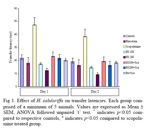
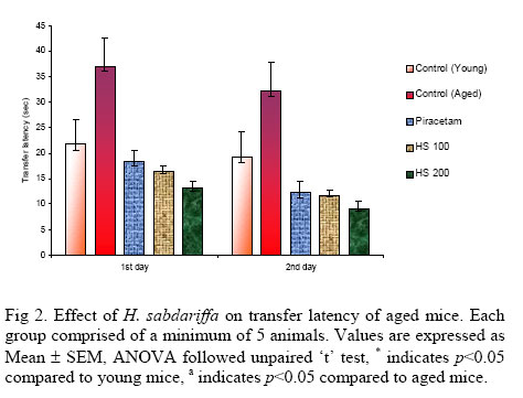
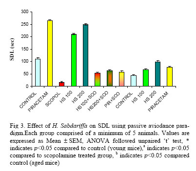
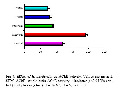
Where R = rate of enzyme activity in ‘n’ mole of acetylcholine iodide hydrolyzed / min / mg protein; d O.D. = Change in absorbance / min; E = Extinction coefficient = 13600 / M / cm. Locomotor FunctionLocomotor activity of control and drug treated animals was measured using a photoactometer (INCO, Ambala, India). Statistical AnalysisAll the results were expressed as mean ± Standard error. The data was analyzed using ANOVA and Student’s (Unpaired) ‘t’ test. Kruskal Wallis one-way ANOVA followed by multiple range tests was used for the analysis of non-normally distributed data. p <0.05 was considered as significant. Results Acute Toxicity Study No mortality was observed following oral administration of HS even with the highest dose (2000 mg/kg). However HS at doses more than 1000 mg/kg produced profuse watery stools in animals. Both the doses of HS had no toxic effect on the normal behavior of the rats. Effect on Locomotor ActivityIn the present study, HS (100 and 200 mg/kg) did not show any significant change in the locomotor function of animals (score: 222.6±8 and 211±15) when tested on photoactometer as compared to control group (score 216.4±12). Effect on Transfer Latency Using EPMAged mice showed higher transfer latency (TL) values on first day and on second day (after 24 h) as compared to young mice, indicating impairment in learning and memory (i.e. ageing-induced amnesia). Piracetam (200 mg/kg, i.p.) pretreatment for 8 days decreased transfer latency of the 8th day 9th days as compared to distilled water treated group, indicating improvement in both learning and memory. Scopolamine (0.4 mg/kg) increased TL significantly (p < 0.05) in young mice on first and second day as compared to control, indicating impairment of both learning and memory (Fig 1 and Fig 2). HS (100 mg/kg, p.o.) decreased TL on the 8th and 9th days in both young and aged mice (p < 0.05) when compared to respective control groups. Higher dose of HS (200 mg/kg, p.o.) improved learning and memory of aged animals rather than the young mice as reflected by marked decrease in TL on the 8th and 9th days, when subjected to elevated plus maze tests (Fig 1 and Fig 2). HS pretreatment for 8 days protected the young as well as the old mice (p < 0.05) against scopolamine induced amnesia. Each group comprised of a minimum of 5 animals. Values are expressed as Mean ± SEM, ANOVA followed unpaired ‘t’ test, * indicates p < 0.05 compared to respective controls, a indicates p < 0.05 compared to scopolamine treated group. Each group comprised of a minimum of 5 animals. Values are expressed as Mean ± SEM, ANOVA followed unpaired ‘t’ test, * indicates p < 0.05 compared to young mice, a indicates p < 0.05 compared to aged mice. Effect on SDL Using Passive Avoidance ParadigmHS (100 and 200 mg/kg, p.o.) treatment profoundly increased step down latency (SDL) as compared to control group on the second day indicating improvement in memory of young mice. Scopolamine hydrobromide (0.4 mg/kg, i.p.) decreased SDL on second day after training, indicating impairment of memory. HS (200 mg/kg, p.o.) administered orally for 8 days significantly (F (7, 32) = 59.312. p < 0.05) reversed amnesia induced by scopolamine and natural ageing (Fig 3). Each group comprised of a minimum of 5 animals. Values are expressed as Mean ± SEM, ANOVA followed unpaired ‘t’ test, * indicates p < 0.05 compared to control (young mice), a indicates p < 0.05 compared to scopolamine treated group, b indicates p < 0.05 compared control (aged mice). Effect on Acetylcholinesterase ActivityThe acetylcholiesterase activity of whole brain was markedly elevated (p < 0.05) after phenytoin (12 mg/kg, p.o.) treatment. Piracetam (200 mg/kg, p.o.) and HS (100 and 200 mg/kg, p.o.) significantly lowered AChE activity (Fig 4). Values are mean ± SEM, AChE- whole brain AChE activity, a indicates p<0.05 Vs control (multiple range test), H = 16.67; df = 5; p < 0.05. DiscussionAlzheimer’s disease is a neurodegenerative disorder associated with a decline in cognitive abilities; patients also frequently have non-cognitive symptoms, such as depression, apathy and psychosis that impair daily living [31]. The most common cause of dementia in the elderly is probably Alzheimer’s disease (AD) [32]. The National Institute of Health predicts, if the current trend continues, there will be more than 8.5 million AD patients by the year 2030 in USA alone [33]. Administration of infusion prepared with 10 g of dry Calyx of H. sabdariffa in 0.5 L water administered orally twice a day before breakfast for 4 weeks had shown beneficial effects on mild to moderate hypertension [34]. Subjects administered with a single oral dose of 150 mL H. sabdariffa aqueous extract exhibited health protecting effects. In Thailand, Roselle tincture (1-3 mL daily) and infusion (1-2 cups) daily is given to treat hyperlipidaemia, bladder stone, gastric ulceration [35]. It has also been used for indigestion, loss of appetite, respiratory problems, as anthelmintic and antibacterial agent [36]. The present study suggests that H. sabdariffa is a potential anti-cholinesterase agent. It also possesses nootropic activity in view of its facilitatory effect on retention of learned task. Central cholinergic system plays an important role in learning and memory [37]. Phenytoin is known to reduce hippocampal ACh concentration and causes cognitive impairment [38]. In our study, phenytoin per se (12 mg/kg, p.o.) significantly elevated brain AChE activity. Piracetam (250 mg/kg, p.o.) and HS (100 and 200 mg/kg, p.o.), on the other hand significantly (p<0.05) lowered this activity indicating the counteracting actions of these drugs on the cholinergic system. HS also reversed the scopolamine-induced impairment in learning and memory, when assessed on passive avoidance paradigm. Piracetam, the first representation of a class of nootropic agents, has been shown to improve memory deficits in geriatric individuals. Repeated injections of piracetam had improved learning abilities and memory capacities of laboratory animals [39]. Passive avoidance behavior is based on negative reinforcement and is used to examine long-term memory [40-41]. Both piracetam and H. sabdariffa meet major criteria for nootropic activity, namely improvement of memory in absence of cognitive deficit [42-43]. In the present study, H. sabdariffa significantly inhibited the AChE activity in the mice whole brain homogenate, indicating its potential in the attenuation of symptoms of cognitive deficits. Hence, the memory improving activity of H. sabdariffa may be attributed to its antioxidant [14], neuroprotective, pro-cholinergic and anti-acetylcholinesterase properties and can be of enormous use in delaying the onset and reducing the severity of Alzheimer’s disease. Further investigations using more experimental paradigms are required for further confirmation of nootropic potential of dried calyces of H. sabdariffa in the treatment of various cognitive disorders. AcknowledgementsAuthors are deeply grateful to Dr. R.P. Bajpai, Honorable Vice-Chancellor, Guru Jambheshwar University, Hisar, for research facilities and motivation. We are thankful to UCB India Pvt. LTD., (Gujarat), for supply of piracetam. We also thank the Principal, Soniya Education Trust’s College of Pharmacy, Dharwad, Karnataka, India, for encouragement. References
Copyright © 2005 by Razi Institute for Drug Research (RIDR) The following images related to this document are available:Photo images[pt06002f2.jpg] [pt06002f4.jpg] [pt06002f3.jpg] [pt06002f1.jpg] |
| |||||||||