 |
| About Bioline | All Journals | Testimonials | Membership | News |
|
||||||
|
||||||
Iranian Journal of Pharmacology & Therapeutics, Vol. 5, No. 1, 2006, pp. 21-25 Research Article Effect of Aqueous Extract of Leaf of Aegle marmelos on Testicular Activities in RatsUttam Kumar Das, Rajkumar Maiti, Debasis Jana and Debidas Ghosh Current Author Addresses: Uttam Kumar Das, Reproductive Endocrinology and Family
Welfare Research Unit, Department of Human Physiology with Community Health. Received April 12, 2005; Revised August 12, 2006; Accepted August 14, 2006 Code Number: pt06003 ABSTRACT The aqueous extract of leaf of Aegle marmelos (Bael) at the dose 50 mg/100 g body weight resulted a significant diminution in the activities of key testicular steroidogenic enzymes along with low levels of plasma testosterone and relative wet weights of sex organs in respect to control without any significant alteration in general body growth. Germ cells numbers in different generation at stage VII of seminiferous epithelial cell cycle were diminished significantly after the treatment of the above extract. The above mentioned dose did not exhibit any toxicity in liver and kidney. Therefore, it may be predicted that the aqueous extract of leaf of Aegle marmelos has a potent antitesticular effect at a specific dose. Keywords: Aegle marmelos, Steroidogenic key enzymes, Plasma testosterone, Germ cells Since population explosion is a leading cause of poverty and pollution in developing countries, the World Health Organization (WHO) has constituted a population control programme, which includes studies having traditional medical practices. At present global attempt has been taken to search out the effect of herbal product for contraceptive purposes [1, 2]. Few herbal contraceptives have been developed but the potentiality of these contraceptives is very minimum and mode of action beyond our knowledge till now. It has been noted that the leaves of Aegle marmelos (A. marmelos) are used for contraceptive purpose in males in different tribal areas. This plant is tree type and grows in almost all districts in West Bengal, India. This is under the family of Rutaceae. The height of this plant is 20-30 ft and leaves are pinnately compound in type. From Ayurvedic medicine, it has been claimed the leaf of A. marmelos posses contraceptive efficacy [3]. Proper scientific research on the contraceptive effects of A. marmelos and its mode of action has not been carried out till now. On this back ground the present study has been conducted to investigate the effects of aqueous extract of leaf of A. marmelos on parameters such as testiculo-somatic, seminal vesiculo-somatic, prostato-somatic, epididymal-somatic indices, activities of testicular steroidogenic key enzymes, plasma testosterone level, seminiferous tubular diameter (STD) and quantification of germ cells at stage-VII of seminiferous epithelial cell cycle. From trial and error the effective dose was selected to search out whether it has any antitesticular effect and to delineate the possible mode of action for such effect. To determine whether the aqueous extract of leaf of A. marmelos at the applied dose has any toxic effects on metabolic organs like liver and kidney, the activities of transaminases and phosphatases were studied in both the organs. Therefore, the aim of the present study is to find out whether the aqueous extract of leaf of the plant has any contraceptive potency or not and to focus a light on the possible ways of its contraceptive activities. Materials and Methods Plant Material Leaves of A. marmelos were collected from Midnapore in the month of May-June. The material was identified by taxonomist of Central National Herbarium (Kolkata), Botanical Survey of India (BSI), Shibpur, Howrah and Department of Botany, Vidyasagar University, Midnapore. The voucher specimen was deposited in the Department of Botany of Vidyasagar University having voucher specimen number HPCH No. 2. Preparation of Aqueous Extract of Leaf of A. MarmelosAqueous extract of leaf of A. marmelos (Bael) was prepared according to the method of National Institute of Health and Family Welfare (NIHFW), New Delhi, India. Fresh leaves were dried at 40ºC for 48 hours and crushed in an electrical grinder. An aqueous extract was prepared by refluxing 200 g of leaf powder with 1.0 litre double distilled water. Extract was dried at reduced pressure and finally lyophilized. Selection of AnimalIn this experiment sixteen healthy and fertile adult male albino Wistar strain rats (Chakraborty Brother, Midnapore, India), 3 months of age, weighing 150±10 g were selected for acclimation for a period of15 days in our laboratory condition (12 h dark: 12 h light, 28±2ºC) prior to experimentation. They were provided with standard food and water ad libitum. Animals were divided into two equal groups according to the mode of treatment. TreatmentEight animals were forcefully fed by gavage at a dose of 50 mg of aqueous extract/0.5 mL of distilled water/100 g body weight/day for 28 days. The remaining eight animals were considered as control and they were treated with vehicle only by forceful feeding (0.5 mL of distilled water/100 g body weight/day for 28 days). The gavage treatment was performed at 6-8 AM and at fasting condition regularly and animal food was provided at 10 AM in each day. On 29th day, body weights of all animals were recorded. For blood collection from dorsal aorta by heparinized syringe, all the animals were anaesthetized by application of light ether exposure. The animals were killed by decapitation. The testis, prostate, seminal vesicle, epididymis, liver and kidney of each animal were dissected out, and their wet weights were recorded. The right testis of each animal was kept at –20ºC for enzymatic studies. The left testis of each animal was placed in Bouin’s fluid for histological studies. Plasma was separated from the collected blood by centrifugation at 3000 g for 5 minutes and was kept at –20ºC for assay of testosterone by ELISA. Assay of Testicular Δ5, 3β-hydroxysteroid Dehydrogenase (Δ5, 3β-HSD) ActivityTesticular Δ5, 3β-hydroxysteroid dehydrogenase activity was measured biochemically [4]. The right testis of each animal was homogenized in 20% spectroscopic grade glycerol (BDH Chemical Division, Bombay, India) containing 5 mM potassium phosphate (Loba Chemical Company, Bombay, India) and 1 mM EDTA (Organon, Kolkata, India) at a tissue concentration of 100 mg/mL homogenizing mixture and it was centrifuged at 10,000 g for 30 minutes at 4°C. The supernatant at a volume of 1 mL was mixed with 1 mL of 100 µM of sodium pyrophosphate buffer, pH=8.9 (Loba Chemical Company, Bombay, India), 40 µL of ethanol containing 30 mg of dehydroepiendosterone (Organon, Kolkata, India) and 960 µL of 25 mg% of bovine serum albumin (BSA) making the incubation mixture a total of 3 mL. Enzyme activity was measured after addition of 100 µL of 0.5 µM nicotinamide adenine dinucleotide (NAD)(SRL, Bombay, India) to the tissue supernatant mixture in a spectrophotometer cuvette at 340 nm against a blank (with out NAD). One unit of enzyme activity was the amount causing a change in absorbance of 0.001/min at 340 nm. Assay of Testicular 17β-hydroxysteroid Dehydrogenase (17β-HSD) ActivityThe activity of testicular 17 β-HSD was measured biochemically [5]. The part of the supernatant prepared for the assay of D5, 3b- HSD (above) was used for assaying 17β -HSD activities. The supernatant (1 mL) was mixed with 1 mL of 440 μM sodium pyrophosphate buffer, pH=10.2 (Loba Chemical Company), 40 mL ethanol containing 0.3 μM testosterone (Sigma Chemical Company, St Louis, MO. USA) and 960 μl of 25% of BSA that make the incubation mixture a total of 3 mL. Enzyme activity was assessed after the addition of 100 μL of 0.5 μM NAD to the incubation mixture in a spectrophotometer cuvette at 340 nm against a blank with out NAD. One unit of enzyme activity was equivalent to a change in absorbance of 0.001/min. at 340 nm. Assay of Plasma TestosteronePlasma level of testosterone was measured following the immuno-enzymatic method by ELISA reader [6]. Horse reddish peroxidase was used as the enzyme labeled antigen that competes with unlabeled antigen for binding to a limited number antibody. After incubation, bound/free enzyme labeled antigen separation was performed by simple solid phase washing. The substrate of enzyme, H2O2 and chromogen were added as per scheduled and the enzyme reaction was stopped by a solution supplied by Equiper (Italy). The color intensity was inversely proportional to the testosterone concentration in the sample. Testosterone concentration in the sample was calculated based on 5 standards supplied by Equiper. The absorbance of standards and sample was monitored against the blank at 450 nm. The cross reaction of the testosterone antibody to dehydrotestosterone is 10% and intra-run precision had co-efficient of variation of 6.2%. The entire sample was run at the same time so there was no inter -run variation. Measurement of Alkaline Phosphatase (ALP), Acid Phosphatase (ACP), Glutamate Oxaloacetate Transaminase (GOT) and Glutamate Pyruvate Transaminase (GPT) Activities in Liver and KidneyAlkaline phosphatase activities of liver and kidney were estimated biochemically [7]. For quantitative estimation of ALP, the above organs were homogenized separately in a Potter Elvijhem homogenizer at a tissue concentration of 20 mg/mL using ice cold homogenizing medium (0.22 M Tris-Hcl buffer pH=7.5). Homogenate at a volume of 0.25mL was added in a centrifuge tube containing 1 mL buffer (1 mM p-nitrophenol phosphate in 1M Tris buffer, pH=8.0). The mixture was incubated at 37ºC for 30 minutes in water bath. The assay was based on the formation of p-nitrophenol (pNP) in the hydrolysis of p-nitrophenol phosphate (pNPP). The activity was measured spectrophtometrically at 420 nm.
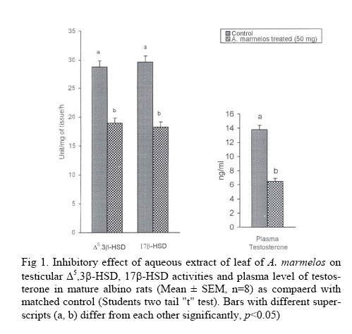
Acid phosphatase activities of liver and kidney were estimated biochemically [8]. For determination of acid posphatase activity, the same homogenizing medium was used at the tissue concentration as in ALP. Activity of ACP was measured in acetate buffer at pH 4.5 using p-nitrophenol phosphate as substrate and OD was noted at 420 nm using spectrophotometer. Glutamate oxaloacetate transaminase (GOT) and glutamate pyruvate transaminase (gpt) are important indicators for the assessment of metabolic toxicity and we also measured GOT and GPT activities in those organs biochemically [9]. The above-mentioned tissues were homogenized separately at a tissue concentration of 50 mg/mL in 0.1M of ice-cold phosphate buffer (pH=7.4). The homogenates were centrifuged at 5000 rpm at 4ºC for 15 min. The substrate GOT used here were 200 mM/L of DL-aspertate and 2 mM/L of α- ketoglutarate where the substrates of GPT were 200 mM/L DL-alanine and 2 mM/L of α- ketoglutarate (pH=7.4). The absorbance was noted at 505nm. The change in absorbance was determined by subtracting the blank reading from corresponding test reading. The enzyme activity was expressed in term of unit/mg of tissue. Quantitative Study of Spermatogenesis at Stage VIIParaffin blocks of testis were serially cut at 5µm thickness and stained in haematoxyline and eosin. The quantitative analysis of spermatogenesis was carried out at stage VII of seminiferous epithelial cell cycle. Characteristics cellular associations of germ cells present at this stage are spermatogonia -A (ASg), preleptotene spermatocytes (pLSc), mid-pachytene spermatocytes (mPSc) and step 7 spermatids (7Sd). The relative number of each variety of germ cells at stage VII of seminiferous epithelial cells, i.e. ASg, pLSc, mPSc and 7Sd were counted. The different nuclei of germ cells (except step 19 spermatids, which cannot be enumerated precisely) were counted in 25 round tubular cross section at stage VII of the cycle in each rat. All the nuclear count (crude count) of the germ cells were corrected using the Abercrombie formula [10]. Histometric StudyThe prepared slides were placed under high power objectives in a phase contrast microscope, and with the help of stage and ocular micrometer, the seminiferous tubular diameters (STD) were measure. For a single measurement of this parameter, 10 round or oval shaped seminiferous tubule were selected and the diameter was noted in each tubule by taking the upward and down ward margin lines of the tubule as well as the left side and right side margin lines of the tubule from each rat [11] Statistical AnalysisStudent's two-tail "t" test was used for statistical analysis of the collected data [12]. Difference were considered significant when p<0.05. Results Organo-Somatic Indices In this experimental profile, there was no significant difference in body weight between two groups (Table 1). Testiculo-somatic, seminal vesiculo-somatic, prostato-somatic and epididymal somatic indices were decreased significantly without any significant alteration in hepato-somatic and reno-somatic indices at the effective dose of aqueous extract treated rats in comparison to control (Table 1). Testicular Δ5, 3β-HSD, 17β-HSD Activities and Plasma Level of TestosteroneTesticular Δ5, 3β-HSD and 17β-HSD activities were decreased significantly in treated group in comparison to control (Fig 1). Plasma testosterone level was decreased significantly in treated group in comparison to control (Fig 2). Quantification of Germ Cells at Stage VIIQuantitative study of spermatogenesis at stage VII of seminiferous epithelial cell cycle revealed that leaf extract treatment resulted in significant reduction in the number of ASg, pLSc, mPSc and 7Sd in respect to the control (Table 2). Histrometric StudyHistometric studies indicated that STD was decreased significantly in leaf extract treated rats compared to the control (Table 2).
Phosphatase and Transaminase Activities in Liver and Kidney Activities of ACP, ALP, GOT and GPT in the liver and kidney were not altered in the leaf extract treated rats in comparison to control (Table 3).
Discussion The present paper provides a number of observations regarding effects of aqueous extract of leaf of A. marmelos on testicular functions in male albino rats. From results, it has been revealed that there was no significant alteration in somatic growth in treated rats compared to the control. This suggests that this leaf extract has no general toxic effect on body growth. Diminution in the weight of testiculo-somatic, seminal vesiculo-somatic, prostato-somatic and epididymal somatic indices in the treated group may be due to low plasma level of pituitary gonadotrophins and testosterone as these growth indices are controlled by plasma level of pituitary gonadotrophins and testosterone [13, 14]. The diminution in the activities Testicular Δ5, 3β-HSD and 17β-HSD, the key enzymes for androgenesis [15, 16], may be due to inhibition in pituitary gonadotrophins secretion [17, 18]. The low plasma level of testosterone reported here also supports this inhibition in steroidogenic enzyme activities. Quantitative study of spermatogenesis was carried out at stage-VII because the cellular associations of germ cells in this stage were composed of elements positioned equidistant in the entire process of spermatogenesis. Therefore, counts of germ cells at this particular stage of the cycle are representative of the condition of spermatogenesis as a hole because all varieties of germ cell are present at this stage [19]. Reduction in the number of all the varieties of germ cells at stage VII of seminiferous epithelial cell cycle in the leaf extract treated rats may be due to low plasma level of pituitary gonadotrophins and testosterone, which are the major regulators of spermatogenesis [20, 21]. Moreover, diminution in STD in leaf extract treated rats also supports the low plasma level of testosterone as STD is one of the indicators of plasma level of testosterone [22]. To determine whether the effective dose of leaf extract of a A. marmelos that induces antitesticular effect have any metabolic toxicity, we measured ACP, ALP, GOT and GPT activities in the liver and kidney because these enzymes are the indicators of metabolic toxicity [23, 24]. Specific enzyme for toxicity study indicated that there was no significant alteration in ACP, ALP, GOT and GPT activities in liver and kidney of the leaf extract treated group. Therefore, it may be claimed that the applied dose of leaf extract of A. marmelos has antigonadal effect without induction of any toxic effects on metabolic organs. In conclusion the data of our study suggest that the aqueous extract of the leaf of A. marmelos results in diminution in the activities of testicular androgenic key enzymes and plasma testosterone along with inhibition in spermatogenesis with out any induction of hepatic and renal toxicity. The actual mode of action of this leaf extract for its hypogonadal activities is not clear from this experiment and more information is necessary to better understand the effect of this leaf extract on the functional physiology of the reproductive system. This study creates a hope to develop a new drug from an herbal product in this new era of herbal drug technology. AcknowledgementAuthors are thankful to the Department of Botany, Vidyasagar University for the preservation of our herbarium specimen. References
The following images related to this document are available:Photo images[pt06003f1.jpg] | ||||||||||||||||||||||||||||||||||||||||||||||||||||||||||||||||||||||||||||||||||||||||||||||||||||||||||||||||||||||||||||||||||
| |||||||||