 |
| About Bioline | All Journals | Testimonials | Membership | News |
|
||||||
|
||||||
Iranian Journal of Pharmacology & Therapeutics, Vol. 5, No. 2, 2006, pp. 159-162 Research Article Ofloxacin ocular inserts: Design, Formulation and EvaluationSreenivas SA, Hiremath SP and Godbole AM Current
Author
Addresses: Sreenivas
SA,
Department
of
Pharmaceutics,
K.L.E.S’s
College
of
Pharmacy,
Hubli-580031,
Karnataka.
India.
E-mail: saiseenu7@rediffmail.com (Corresponding
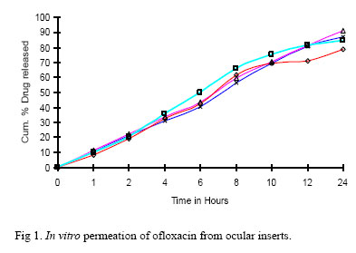
Author). Received January 13, 2006; Revised October 28, 2006; Accepted October 30, 2006 Code Number: pt06027 ABSTRACT In developing a drug delivery strategy, issues of absorption, distribution, metabolism, and elimination must be considered. The eye presents unique opportunities and challenges when it comes to the delivery of pharmaceuticals. While absorption by this route is bungling, there are few side effects with conventional ocular dosage forms. Hence, ocular inserts were prepared with prolonged release of drug and minimum swelling within cul-de-sac using ofloxacinas a model drug; and hydroxy propyl methyl cellulose, methyl cellulose, poly vinyl pyrrolidone and poly vinyl alcohol as polymers. PEG-400 was incorporated as plasticizer. The main purpose of the study was to deliver the drug in zero order kinetics. Solvent casting technique was followed to prepare ofloxacin ocular films. Eight formulations were formulated and subjected to various physicochemical evaluations. Ocular inserts prepared were smooth and passed all the evaluation tests performed. Formulation OF2 shows a maximum cumulative percentage drug release of 91.27 % at the end of 24 hours. Ocuserts formulated also passed the test for sterility. They showed zero-order release of the drug in the in vitro and in vivo release studies. The drug in the films was found to be active against selected microorganisms as was proved by microbial efficacy studies. A high correlation coefficient was found between in vitro and in vivo release rate studies. Shelf-life of the product was found to be more than one year.The results of in vitro, in vivo, kinetic treatment (zero order and Korsemeyer’s regression values) and rate constant ‘k’ value suggest that OF2 was the best formulation among the formulations studied for formulating ofloxacin ocular insert. Keywords: Ocusert, Mmicrobial, Zero order, Korsemeyer, in vivo The eye as a portal for drug delivery is generally used for local therapy against systemic therapy in order to avoid the risk of eye damage from high blood concentrations of the drug, which is not intended. The unique anatomy, physiology and biochemistry of the eye render this organ impervious to foreign substances, thus presenting a constant challenge to the formulator to circumvent the protective barriers of the eye without causing permanent tissue damage [1]. Most ocular treatments like eye drops and suspensions call for the topical administration of ophthalmically active drugs to the tissues around the ocular cavity. These dosage forms are easy to instill but suffer from the inherent drawback that the majority of the medication they contain is immediately diluted in the tear film as soon as the eye drop solution is instilled into the cul-de-sac and is rapidly drained away from the precorneal cavity by constant tear flow and lacrimo-nasal drainage. Therefore, only a very small fraction of the instilled dose is absorbed by the target tissue for this reason, concentrated solutions and frequent dosing are required for the instillation to achieve an adequate level of therapeutic effect. One of the new classes of drug delivery systems, ocular inserts, which are gaining worldwide praise, release drugs at a pre-programmed rate for a longer period by increasing the precorneal residence time [2-4]. Ofloxacin is a pale yellow or bright yellow, crystalline powder. Ofloxacin is antibacterial and is the most commonly used fluroquinolone.It inhibits the enzyme bacterial DNA gyrase, which nicks double stranded DNA, introduces negative supercoils and then reseals the nicked ends. This is necessary to prevent excessive positive supercoiling of the strands when they separate to permit replication or transcription. The bactericidal action probably results from digestion of DNA by exonucleases whose production is signaled by the damaged DNA [5]. Ofloxacin has a broad antimicrobial spectrum against gram-positive and gram-negative microorganisms. This drug is routinely used in the many ocular conditions like infections, inflammations, conjunctivitis, blepharitis, iritis, corneal ulcer etc [6,7]. In the present study, it was aimed to prepare ocular films containing ofloxacin along with hydrophilic and hydrophobic polymers with better solubility and longer duration of action delivering the drug in zero order kinetics. Materials and MethodsThe in vitro release [9] studies were carried out by using biochemical donor-receiver compartment model designed by using commercial semipermeable membrane of transparent cellophane paper.It was tied at one end of the open cylinder, which acted as donor compartment. The ocusert was placed inside the donor compartment.The semipermeable membrane was used to simulate ocular in vivo conditions like corneal epithelial barrier.In order to simulate the tear volume, 0.7 mL of water for injection was placed and maintained at the same level throughout the study. Whole experiment was conducted at room temperature. The entire surface of the membrane was in contact with reservoir compartment which contained 12 mL of water for injection and stirred continuously using a magnetic stirrer. Samples of 1.0 mL were withdrawn at periodic intervals and replaced with equal volume of medium.The drug content was analyzed at 410 nm using Shimadzu UV visible spectrophotometer after suitable dilution.
In vivo studies [10]. The inserts were sterilized using U.V. radiation for 10 minutes at 25 cm height on both sides before the in vivo study. Male rabbits (Orytolagus cumiculus), 10-12 weeks old weighing 1-2 kg were chosen in the present study.They were kept three per cage with husk bedding and were fed with standard diet and water as much as required.A dark and light cycle of 12 hours was maintained. The ocular inserts were placed into the lower conjunctival cul-de-sac of rabbits into eight eyes (after five minutes of sterilization process), each one eye of eight rabbits is served as control. Ocular inserts were removed carefully at 2, 4, 6, 8, 12 and 24 hours and analyzed for residual drug content.The drug remaining was subtracted from the initial drug content of insert; which gave the amount of drug released in the rabbit eye. Kinetic Analysis [11]. The in vitro and in vivo data were analyzed by a zero order kinetics equation as well as Korsemeyer’s equation to understand the release profile and release mechanism. When a graph of the cumulative percentage of the drug released from the matrix against time is plotted, zero order release is linear in such a plot, indicating that the release rate is independent of concentration. The rate of release of the drug can be described mathematically as follows: Rate of release = (dCs/t) = k where Cs = concentration of the drug present in the matrix, k = rate constant and t = time. Since Cs is a constant, and x = amount of drug released described as dx/dt = k integration of the equation yields x = kt + constant. A plot of x versus t results in a straight line with the slope = k. The value of k indicates the amount of the drug released per unit of time and the intercept of the line at time zero is equal to the constant in the equation. The curves plotted may have different slopes, and hence it becomes difficult to exactly pinpoint which curve follows perfect zero order release kinetics. Therefore, to confirm the kinetics of drug release, data’s were also analyzed using Korsemeyer’s equation [11]. Korsemeyer et al. used a simple empirical equation to describe general solute release behavior from controlled release polymer matrices: Mt/M∞ = atn where Mt/M∞ = fraction of drug released, a = kinetic constant, t = release time and n = the diffusional exponent for drug release. The slope of the linear curve gives the ‘n’ value. Peppas stated that the above equation could adequately describe the release of solutes from slabs, spheres, cylinders and discs, regardless of the release mechanism. The value of ‘n’ gives an indication of the release mechanism. When n = 1, the release rate is independent of time (zero order) (case II transport); n = 0.5 for Fickian diffusion; and when 0.5 < n < 1, diffusion and non-Fickian transport are implicated. Lastly, when n > 1.0 super case II transport is apparent. ‘n’ is the slope value of log Mt/M∞ versus log time curve. Further, stability studies were performed for selected formulations as per the International Conference on Harmonization (ICH) guidelines. Result and DiscussionThe prepared films were evaluated for the thickness of each film using a micrometer of sensitivity of 0.001 mm (Mitutoyo, Japan). The average of five readings was taken. The mean thickness, standard deviation and percent coefficient of variation were calculated. All the eight formulations, measured thickness with low standard deviation values ensured the uniformity of the films prepared by solvent casting technique and it was found to be in the range from 0.15 ± 0.01 to 0.20 ± 0.01 millimeters. For the various formulations drug content uniformity was found to vary between 99.01 ± 0.37 to 101.0 ± 0.92 %. The estimation of drug content was found to be almost same with their low standard deviation value. Cumulative percentage drug release of each film in the in vitro release studies and in vivo release studies were based on the mean content of the drug present in the respective films. The weight of all the films was found to be uniform indicating good distribution of drug, polymers and plasticizer. The percentage moisture absorption was calculated for all the eight formulations in triplicate. According to the results obtained, the moisture absorption is more in the formulations where hydrophilic polymers are present. Formulation OF5 has shown the maximum percentage moisture absorption of 12.13 ± 0.516, as the film contains hydroxy propyl methyl cellulose and methyl cellulose as polymers; due to their hydrophilic nature. Formulation OF4 has shown the minimum percentage moisture absorption of 6.28 ± 0.148 as it contains polymers of less hydrophilicity or lipophilic nature. In general, it can be concluded that, the hydroxy propyl methyl cellulose and methyl cellulose have more tendency to absorb moisture as compared to polyvinyl pyrrolidone and polyvinyl alcohol. At humid condition, there was more moisture absorption but there was no change in the integrity; which was observed by its physical appearance. The percentage moisture loss was calculated for all the eight formulations in triplicate. It was observed that when the formulations were kept at very dry condition the maximum moisture loss has been occurred. Formulation OF6 showed the maximum amount of moisture loss 16.52 ± 0.464 and formulation OF3 had shown a minimum loss of 8.40 ± 0.506. Presence of poly vinyl pyrrolidone increases the percentage of moisture loss. The inserts were sterilized by using UV radiation for 10 minutes at 25 cm height on both sides and films were tested for sterility as per Indian Pharmacopoeia. No microbial or fungal growth was seen in any of the formulations which indicated that the films were sterilized completely. Ocular inserts passed the test for sterility. The drug was also found to be active against selected microorganisms as was proved by microbial efficacy studies. In the present study in vitro diffusion were carried out in triplicate. At different time interval sample was withdrawn and cumulative percentage drug released in mg was calculated, on the basis of mean amount of Ofloxacin present in the respective films. Formulation OF2 showed a maximum cumulative percentage drug release of 91.27 % at the end of 24 hours, followed by the formulations OF1 (87.16 %), OF6 (84.49 %), OF5 (78.55 %), OF4 (75.90 %), OF3 (73.05 %), OF8 (67.16 %) and OF7 (63.29 %). The percentage of drug release of all the formulations is presented in Fig 1. The formulations which gave good results with highest percentage were selected for further studies such as in vivo, kinetic treatment and stability studies. Film OF2 containing PVP (600 mg) and HPMC (400 mg) showed a release of 91.27 % at the end of 24 hours which indicated that, the polymer combination with same quantities can be used for the formulation of ocular film for therapeutic drug management in the systemic circulation. OF2 is a combination of hydrophilic polymers. This film has shown good compatible nature in IR studies indicating no drug polymer incompatibility.The programmed release is due to the formation of hydrogen bonds between the drug and polymers which have helped in rate control release of drug. PVP also has good adhesive property which is helpful, when the ocular film is inserted in the cul-de-sac. In vivo release studies were performed using rabbits in triplicate at periodic intervals. Formulations OF1, OF2, OF5, and OF6 show 88.35 %, 93.78 %, 78.80 % and 83.95 % cumulative percentage drug release at the end of 24 hours respectively. The data obtained from in vitro studies of all the four formulations were subjected for kinetic treatment in order to know the order of release. The regression coefficient calculated was found to be 0.9918, 0.9972, 0.9959, and 0.9911 for the formulations OF1, OF2, OF5 and OF6 respectively and were found to be fairly linear, as indicated by their good regression value. Therefore, it was ascertained that, the drug release from OF1, OF2, OF5 and OF6 could follow either near zero or zero order kinetics. The zero order curves alone are not sufficient to predict zero order since each curve, albeit straight, has a different slope. Hence to confirm the exact mechanism of drug release from the films, the data were computed and graphed according to Korsemeyer’s equation. Regression analysis was performed and the regression value ‘r’ suggested that the curves were fairly linear. Slope values were computed from the graph. The ‘n’ value suggest that the film OF2 follow perfect zero order kinetics (n =1.0) whereas the ocular films OF1 and OF6 follow a super case II transport mechanism (n > 1.0), OF5 follows fickian diffusion (n=0.5). The zero order rate constants, slope value ‘n’ and their respective ‘r’ values are given in Table 2. Cumulative percentage drug release from in vivo studies gave almost similar results as obtained from in vitro experiments. Hence we tried to correlate in vivo results with the in vitro percentage drug release. The correlation values were found to be 0.9991, 0.9997, 0.9958 and 0.9946 for formulations OF1, OF2, OF5 and OF6 respectively. The linearity was found in all the four formulations but formulation OF2 gave a good correlation and better linearity. The in vitro-in vivo correlation for formulation OF2 was strong and productive. There was no drag out of circular inserts at the time of experiment which suggest that the particular dimension (8.0 mm) diameter was suitable as ocular films. The absence of redness in the rabbit eye suggests that the formulated ocuserts do not produce any irritation. Stability studies performed show no significant changes in the films which suggest that the films were stable. Shelf-life of the product was found to be more than one year. AcknowledgementWe thank Cipla India Limited and Suvidhinath Laboratories for providing the required quantity of gift sample to carry out the research work. References
Copyright © 2006 by Razi Institute for Drug Research (RIDR) The following images related to this document are available:Photo images[pt06027f2.jpg] [pt06027f1.jpg] | ||||||||||||||||||||||||||||||||||||||||||||||||||||||||||||||||||||||||||||||||||||||||||||||||||||||||||||||||||||||||||||||||||||
| |||||||||