 |
| About Bioline | All Journals | Testimonials | Membership | News |
|
||||||
|
||||||
Iranian Journal of Pharmacology & Therapeutics, Vol. 5, No. 2, 2006, pp. 163-167 Research Article Sub-Chronic Toxicity Study of Alfa-Cypermethrin in RatsSUKUMAR MANNA, DIPANKAR BHATTACHARYYA, TAPAN KUMAR MANDAL and SHYAMAL DAS Current
Author
Addresses: Sukumar
Manna,
Department
of
Pharmacology & Toxicology, West Bengal University of Animal & Fishery
Sciences,
Kolkata,
West
Bengal,
India. Received August 2, 2005; Revised October 7, 2005; Accepted June 8, 2006 Code Number: pt06028 ABSTRACT Alfa-cypermethrin (α-CP) a synthetic pyrethroid (type-II) insecticide, dissolved in dimethyl sulfoxide (DMSO-1 mL) was given consecutive daily orally for 60 days at 1/10 LD50 (14.5 mg/kg). The animals were sacrificed on day 61st. Biochemicals, cytochrome P450, b5 contents in liver along with antioxidant status, tissue residual concentration, and brain GABA level were studied. It increased the serum aminotransaminase (AST, ALT), alkaline phosphatase (ALP), lactate dehydrogenase (LDH) activities and blood glucose level significantly (p<0.05). It significantly (p<0.05) decreased and increases cytochrome P450 content and b5 content in liver respectively. It increased the malondialdehyde (MDA) level, and decreased the activities of catalase (CAT) and superoxide dismutase (SOD) and glycogen in liver significantly. The pyrethroid was considerable in amount in tissues. The GABA levels in cerebellum and in whole brain without cerebellums were decreased significantly (p<0.05). Therefore repeated daily oral dose toxicity of α-CP at 1/10 LD50 altered biochemical parameters, decreased cytochrome P450 content, antioxidant status, and decreased brain GABA level. Keywords: α-CP, Cytochrome P450, Antioxidants, Tissue residual concentration, Brain GABA level, Rat Cypermethrin is a synthetic type-II pyrethroid with potent insecticidal property. The technical grade cypermethrin is the racemic mixture of 8 isomers (four cis and four trans isomers). Two stereoisomer are termed α-isomer of cypermethrin, which is believed to be the most active isomer, and is known as α-cypermethrin. Alfa-cypermethrin (α-CP) is extensively used not only as an ectoparasiticide in animals, but also used in agricultural crop production and public health program. Some of the toxic actions of α-CP have been reported in acute and subacute (30 days) toxicity after repeated daily oral administration of α-CP for 30 days on cytochromeP450, b5, antioxidant status, blood biochemistry, tissue residual concentration and its mechanisms of action in relation to brain GABA level in rats [1-3]. Therefore the present study was undertaken to investigate the subchronic effect (60 days) of α-CP on antioxidant status, cytochrome P450, b5 contents in liver, tissue residual concentration, biochemical parameters and brain GABA level following repeated daily oral administration at 1/10 LD50 (14.5 mg/kg) doses in rats. Materials and Methods Pesticide Alfa-cypermethrin (α-CP, >99% pure, Gharda Chemicals Ltd. Bombay) Animals and Experimental DesignTwenty (20) adult Wistar rats of both sexes (equal sex ratio; weighing about 200 ± 20 GMS) were divided into two equal groups (I and II) each containing ten (10). All rats were kept under controlled conditions of temperature (22±10ºC) and humidity (60±5%). They were given pellet food (Amrut feeds Ltd., Pune, India) and drinking water ad libitum. A twelve hour day and night cycle was maintained in the animal house. The experimental protocol met the national guidelines on the proper care and use of animals in the laboratory research. The experimental protocol was approved by the Institutional Animal Ethics Committee (IAEC).
The animals were fasted overnight and α-CP was administered orally by gavage after dissolving in DMSO (1 mL) as stated above. The animals were observed for respiratory and CNS symptoms, behavioral patterns and death. α-CP was administered orally to the animals of group I at 14.5 mg/kg and animals of group II were dosed equal volume of DMSO only (1 mL) daily for 60 days respectively. On 61st day animals of group-I and control group (group-II) were sacrificed under ether anesthesia and blood was collected in two sets of test tube after severing the neck vessels separately from each animal. One set was kept under refrigeration (4ºC) for separation of serum and utilized for estimation of activities of aspartate transaminase (AST) and alanine transaminase (ALT) (Reitman and Frankel, 1957), lactate dehydrogenase (LDH) (Bergmeyer et al., 1974), alkaline phosphatase (ALP) (King and Armstrong, 1934) and total protein (TP) (Doumas et al., 1971), globulin and albumin (GLB, ALB) (Rodkey, 1965) as described by Varley et al., 1984 [4]. The blood of another set of test tube (3rd) having mixture of potassium oxalate and sodium fluoride as anticoagulant was used for estimation of glucose [4]. The brain was collected immediately divided into two parts, cerebellum and wholebrain without cerebellum and both portions was utilised for estimation of gamma amino butyric acid (GABA). The concentration of GABA was determined using HPLC-ECD following its derivatization with 0-pthalaldehyde and tertiary-butylthiol by the method of Allison et al. (1984) modified by Manna et al. [1].
The derivative is a time-substituted isoindole, which is stable one.
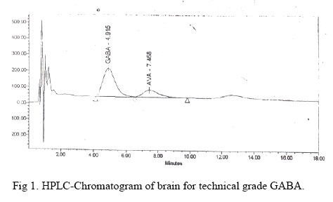
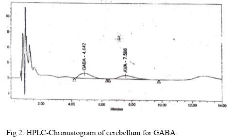
Condition of HPLC System. Waters (USA) 464-pulsed Electrochemical Detector. Column.Novapak (reverse phase) RP-C_18 column (3.9 x 150 mm). Mobile phase.Sodium acetate (pH 5) (0.18M), 55% + acetonitrile (v/v), 45%. Flow rate.1.5 mL/min. Run time.15 min. Applied potential. Glassy carbon electrode (700 mv) vs. Ag/AgCl Reference electrode. Temperature.20ºC. Reagents. i. M (Perchloric acid) HClO4 ii. GABA stock: 1.2, 0.625, 0.313 and 0.156 mM. iii. M Carbonate Buffer. iv. Working reagent: 0.0671 g of OPA dissolved in 50 mL methylalcohol and to which is added 56 μL of TBT (Tertiary butyl thiol). Sample PreparationIn the present experiment, the cerebellum and whole brain without cerebellum portions were separately weighed, homogenized in 0.1 M HClO4 and particulate matter removed by centrifugation and filtration. The clarified supernatant was diluted with 0.1 M HClO4 to give a total dilution factor of 200 for the original brain samples. ProcedureTo 500 μL of diluted brain homogenate were added 20 μL of the AVA (5-amino valeric acid) stock (internal standard) and 40 μL of GABA stock, and mixed thoroughly. Working reagent of 800 μL and the above mixture of 200 μL were added to the sample and mixed thoroughly (kept capped to contain the thiol odour). It was allowed for the reaction to proceed at room temperature for 6 minutes and 50 μL of the derivatized sample was injected, run for 15 minutes and the retention time (RT.) of derivatized sample was found to be at 4.9 min (Fig 1 and Fig 2). The data were recorded and analysis was done by Millennium package (Waters) and GABA concentration was expressed as mg/gm of wet tissue (Table 4). TissuePortion of liver was washed in physiological saline, homogenized and kept for estimation of activities of catalase (CAT) [5], levels of reduced glutathione (GSH) [6] and malondialdehyde (MDA) [7], glycogen [8] and tissue protein [9]. Another portion of liver was collected in ice-cold 1.15% KCl and homogenized within 10 min, centrifuged and the microsomal pellets were separated and was used for estimation of superoxide dismutase (SOD) [10], Cytochrome P450 and b5 [11] contents by double beam-UV-Vis spectrophotometer. Residual Concentration in Brain, Lungs, Liver, Heart, Kidney and TestesThe tissue residual concentration of a-CP was estimated by the modified method of Marie et al. (1982) by Mandal et al. [12]. Tissue sample preparation: Tissue (2 g) was extracted for 4 min with acetonitrile (25 mL) and anhydrous sodium sulfate (0.5 g) using a homogenizer. The extract was filtered through anhydrous sodium sulfate (0.5 g) and the tissue was re-extracted twice with acetonitrile (25 + 12 mL). The extract was clarified by centrifugation and filtered through anhydrous sodium sulfate. The combined acetonitrile extracts were concentrated to 20 mL and partitioned with hexane (2 ´ 10 mL). The hexane phases were discarded and the acetonitrile phase was evaporated to dryness using a rotary vacuum evaporator at 400ºC. The volume was finally made up to 5 mL with acetone for GLC estimation.
Calibration A stock solution of 1 mg/L of a-CP (analytical grade >99%) was prepared as an external standard. The retention times of a-CP was 13.5 min. The data were recorded in a HP 3392A integrator. ApparatusA Hewlett Packard (USA) model 5890A gas chromatograph coupled with a 3392 A (HP) integrator and equipped with a Ni63 electron capture detector was used for analysis of a-CP. Operational parameters were: Injector temperature- 275ºC Oven temperature - 255ºC Detector temperature - 275ºC Flow rate of carrier gas N2 – 70 mL per minute. Column: A 1.8 x 2 mm I.D. glass column packed with 3% OV-101 on chromosorb W.H.P. (80–100 mesh) was used. InjectionStandard and samples (2 μL) were injected into gas liquid chromatograph With 10 μL Capacity Hamilton Syringe. Statistical AnalysisAll the values were expressed as mean ± S. E. M. Statistical analysis was done by using SPSS 7.5. Statistical significance of differences between two means was assessed by unpaired Student’s ‘t’ test. A difference at p<0.05 was considered statistically significant. Result Biochemical and Antioxidants Profile Effect of α-CP on certain biochemical and antioxidants parameters are summarized in Table 1 and Table 2 respectively. α-CP significantly increased the activities of serum AST, ALT, ALP, LDH. (Table 1) In liver it significantly decrease the cytochromeP 450 content and activities of CAT, SOD, and GSH level and increased the MDA level and b5 content. Blood glucose level was significantly increased, where as liver glycogen was significantly decreased. Serum GLB and total protein levels decreased significantly without appreciable alteration of ALB. Brain GABA LevelFig 1 shows typical chromatograms of standard GABA and Fig 2 shows chromatogram of GABA in cerebellum. The concentrations of GABA were decreased significantly (p<0.01) compared to control in cerebellum as well as in whole brain except cerebellum after repeated daily oral administration of a-CP for 60 days at 14.5 mg/kg dose (Table 3). Tissue Residual ConcentrationThe concentration of a-CP in certain organs following consecutive daily oral administration for 60 days have been presented in Table 4. It transpires that the concentration of a-CP in liver, brain, testis, kidney, lungs, and heart were 0.17 ± 0.02, 0.11 ± 0.03, 0.45 ± 0.01, 0.98 ± 0.02, 1.42 ± 0.21 and 0.35 ± 0.02 ppm respectively after 60 days administration. It is also evident from Table 4 that concentration of a-CP was highest in lungs followed by kidney, testis, heart, liver and brain. It is reflected from Table 4 that a-CP like other pyrethroids have a tendency to accumulate in these important organs due to their lipophilic character. DiscussionAnimals showed a sequence of signs of toxicity. Activities of SOD, CAT, GSH and MDA levels in the liver reflect the oxidative status and the serum enzymes like AST, ALT, and ALP represent the functional status of the liver. Chemical-induced cellular alteration varies from simple increase of metabolism to death of cell. The increase or decrease of enzyme activity is related to the intensity of cellular damage. Therefore, increase of transaminase activity along with the decrease of activity of free radical scavengers is the consequence of α-CP induced pathological changes of liver. The severe hyperglycemia may due to the effect of increase in catecholamines level, which causes glycogenolysis, and this may be reason of significant decrease in liver glycogen [2]. The decreased CAT, SOD activities and increased MDA level in liver concomitant with increased serum AST, ALT, ALP activities suggest that α-CP causes hepatic damage and pathogenesis may be through the generation of free radicals. α-CP undergoes metabolism in the liver via esteric and oxidative pathways by the cytochrome P450 microsomal enzyme system [13]. Inhibition of P450 contents by α-CP (Table 2) may cause oxidative stress resulting in decrease the activities of CAT, SOD and glycogen level and increased the level of MDA. And all these may lead to hepatic necrosis. Blood lactate was not measured in this study, but increased activity of LDH may indicate a shift towards anaerobiosis. The present antioxidants status and biochemical changes correlated with histopathological changes of tissues corroborated with the findings of Giray et al. [Error! Reference source not found.] and Manna et al. [2]. The GABA level is more in the cerebellum than the rest of the brain (I.e., without cerebellum) in control rats. α-CP decreased GABA levels significantly both in cerebellum and the rest of the brain in treated rats. The decrease GABA level might be either due to decrease synthesis or increase catabolism of GABA resulting in the inactivation of the Cl-channel leading to excitation, convulsion [1] and even death.
Acknowledgement We acknowledge to Prof. A. Chowdhury and Dr. A. Bhattacharya, Pesticide residual Laboratory, department of Agricultural chemicals, Bidhan Chandra Krishi Viswa Vidyalaya, Nadia, W.B for providing GLC-ECD for tissue residual analysis, and to the Director, Chittaranjan National Cancer Research Institute, Kolkata for instrumental help to estimation of brain GABA, and also acknowledge to the gift of analytical grade a-CP by M/S, Gharda Chemical Ltd., Mumbai, India, to carry out the research work. References
Copyright © 2006 by Razi Institute for Drug Research (RIDR) The following images related to this document are available:Photo images[pt06028f2.jpg] [pt06028f1.jpg] | |||||||||||||||||||||||||||||||||||||||||||||||||||||||||||||||||||||||||||||||||||||||||||
| |||||||||