 |
| About Bioline | All Journals | Testimonials | Membership | News |
|
||||||
|
||||||
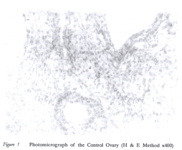
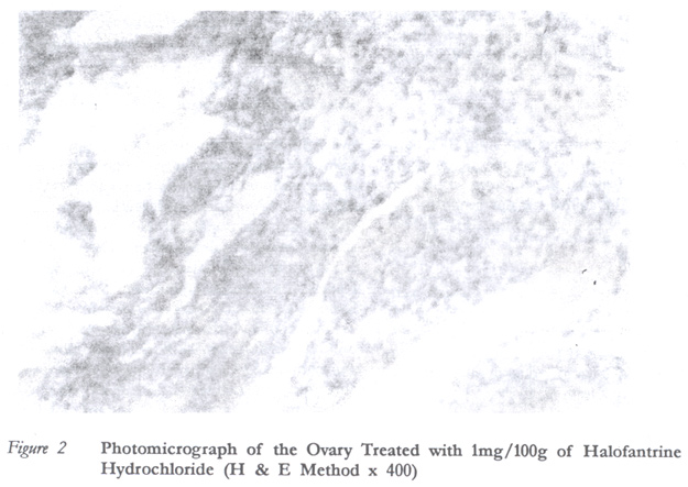
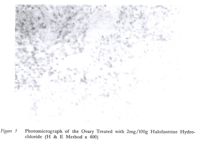
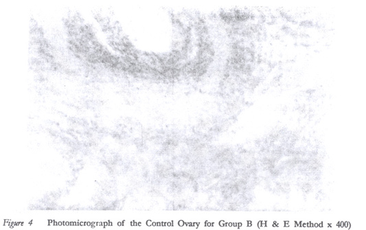
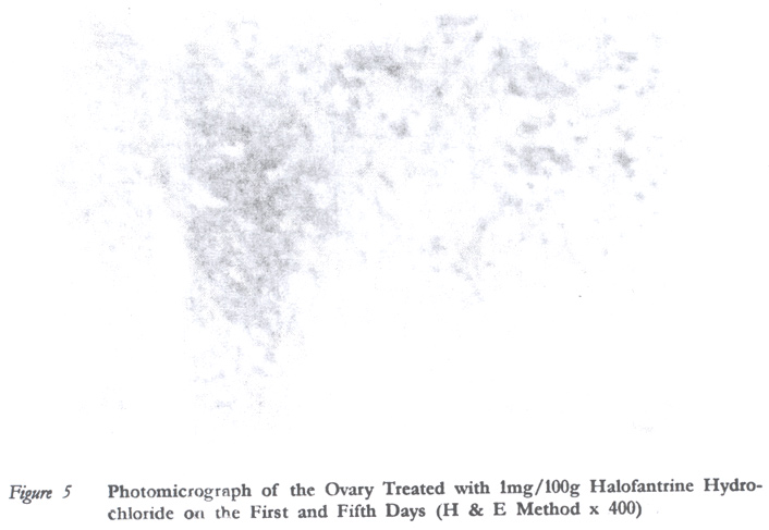
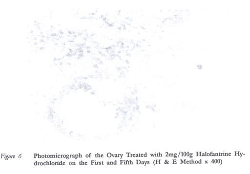
African Journal of Reproductive Health, Vol. 7, No. 1, April, 2003 pp. 113-120 Effects of Halofantrine Hydrochloride (Halfan) on the Histology of the Ovary of Mature Female Wistar Rats JO Adjene1 and FO Agoreyo1 1Department of Anatomy, School of Medicine, University of Benin, Benin City, Nigeria. Email: agoreyo@uniben.edu agoreyofo@yahoo.com Code Number: rh03014 ABSTRACT Halofantrine hydrochloride (halfan) was administered via oral route to matured female wistar rats weighing 180–200g. The Wistar rats were randomly selected and divided into six groups of four rats each, making a total of twenty four rats and coded A1, A2, A3, B1, B2 and B3. Groups A3 and B3 were used as controls. Three consecutive doses of 1mg/100g halfan each were administered to group A1 at six hourly intervals while three doses of 2mg/100g halfan each were administered to group A2 at six hourly intervals. The animals were sacrificed on the 5th day after drug administration by cervical dislocation. Animals in groups B1 and B2 were given doses corresponding to A1 and A2 respectively, but in addition drug administration was repeated the fifth day following the day of first administration. The animals in this group were sacrificed on the tenth day by cervical dislocation, the ovaries were removed, blotted dry, fixed in 10% formal saline for histological processing and studies. Histological changes observed in the ovary include retarded growth of follicles, reduction in size and number of follicles, increase in cytoplasmic vacuolation, constriction of blood vessels, absence of corpora lutae and cases of cellular necrosis. These alterations were more pronounced in those groups sacrificed on the tenth day. (Afr J Reprod Health 2003; 7[1]: 113–120) RÉSUMÉ Effets du chlorhydrate halofantrine (halfan) sur l'histologie de l'ovaire des rats de wistar femelles mûres. Le chlorhydrate halofantrine (halfan) a été administé par voie orale aux rats de wistar femelles mûres qui pesaient 180–200g. Les rats de wistar ont été selectionnés au hasard et divisés en six groupes, quatre rats dans un groupe. Il y avait au total vingt-quatre rats codés A1, A2, A3, B1, B2 et B3. Les groupes A3 et B3 servaient de groupes témoin. On a administré trios doses consécutives de 1mg/100g de halfan au groupe A1 toutes les six heures alors qu'on a administré trios doses de 2mg/100g de halfan au groupe A2 toutes les six heures. Le cinquième jour après l'administration, les animaux ont été sacrifiés par la luxation du rachi cervical. Les animaux des groupes B1 et B2 ont reçu les mêmes doses que ceux des groupes A1 et A2 respectivement, mais en plus, on a répété l'administration des médicaments le cinquième jour après la première administration. Les animaux de ce groupe ont été sacrifiés le dixième jour par une luxation du rachis cervical; on a levé les ovaires, les a séchés et fixés dans 10% de solution salée pour contrôler le procès histologique et pour les étudier. Les modifications histologiques observées dans l'ovaire comprenment le retard de croissance du follicule, la réduction dans la taille et dans le nombre des follicules, l'augmentation de la vacuolisation cytoplasmique, la constriction des vaisseaux sanguins, l'absence de copora lutae et des cas de nécroses cellulaires. Ces modifications étaient plus marquées chez les groupes qui ont été sacrifiés le dixième jour. (Rev Afr Santé Reprod 2003; 7[1]: 113–120) KEY WORDS: Halofantrine hydrochloride, ovarian follicles, Wistar rats INTRODUCTION Malaria is caused by parasites that belong to the genus plasmodium with four different species, namely, Plasmodium falciparum, Plasmodium vivax, Plasmodium malariae and Plasmodium ovale. Malaria infection is common in Central and Southern America, Hispaniola, sub-Saharan Africa, the Indian sub-continent, South East Asia, the Middle East and Oceania. The estimated risk of a traveller acquiring malaria varies markedly from area to area.6 The resistance of Plasmodium falciparum to most antimalarial drugs is very rampant and on the increase. This is due to the remarkable adaptability of parasites to drugs and man's abuse of malaria drugs for prophylaxis and treatment of undiagnosed fever in endemic areas. Resistance to chloroquine has been confirmed in malaria endemic countries except the Dominican Republic, Haiti, Central America, west of the Panama Carnal Zone, Egypt and most countries in Middle East.13 In addition, resistance to both chloroquine and sulfadoxine has been reported in the Amazon Basin area of South America and in sub-Saharan Africa. Resistance to mefloquine has been reported in West Africa.8 Halofantrine is a phenanthrenemethanol antimalarial that is schizonticidal with a high degree of activity against the erythrocytic stage of malaria parasite. It is indicated for the treatment of acute malaria in patents with single or mixed infections of P. falciparum and P. vivax. It is also indicated for the treatment of malaria in patients who can tolerate oral medication and who wish to moderate malaria to less than 100,000 parasites/mm3.15 Halofantrine has not been shown to be mutagenic either in animals or in tests utilising CHO chromosome aberration test and dominant lethal assays in rats. Adequate and well controlled studies in human have been done but it has been found to be embryocidal in rats10 especially when it is above therapeutic dosages. Self-medication is especially common in developing countries and sometimes at dosages above the therapeutic dose. The ovaries are the female organs of reproduction found in a depression called ovarian fossa on the lateral wall of the pelvis. They are greyish-pink, almond shaped and have a smooth surface before regular ovulation begins. The ovary produces the female sex cells, of which one is made available each month throughout the woman's reproductive years except during pregnancy. The ovary also has endocrine action in that it produces progesterone and oestrogen such as estriol, estradiol and estrone. Recent findings show that a drug that is effective in malaria treatment may cause damage to certain organs of the body. It is therefore important for drugs to be avoided and only to be safely administered when necessary. There has been no correlation between teratogenic effect of drug in laboratory animal and man.12 The presence, however, of a negative effect of a drug in experimental animals indicates the possibility of producing similar effects and its embryotoxicity.10 The objective of this experiment is to determine the probable effect of halofantrine on the histology of the ovary of female wistar rats. The findings from the study can be related to human and thus serve as a source of further research on human. MATERIALS AND METHODS Animals Twenty four female wistar rats were purchased at the animal house of the Department of Biochemistry, College of Medical Sciences, University of Calabar, Nigeria. The rats were maintained at a temperature range of 25oC to 30oC and were fed with growers mash obtained from Pfizer Nigeria Limited. Water and feed were provided ad libitum. This continued until the rats weighed between 180 and 200g. They were cared for in compliance with applicable guidelines. Drug Administration The halofantrine used in this research was produced by SmithKline and French Laboratories and obtained from Greso Pharmacy Store in Calabar, Cross River State, Nigeria. The drug suspension was administered to the animals on the basis of their body weight. Usually, 5mls of the drug suspension contains 100mg of halofantrine. The therapeutic dose for the experimental animals was thus 1mg/100g body weight as against the therapeutic dose of humans, which is 10mg/kg six hourly doses. The suspension of halofantrine was administered orally with the aid of an orogastric tube. The rats were divided into two batches and groups as shown below: Group A1 Animals in this group were given 1mg/100g body weight of the drug suspension orally three times at six hourly intervals, making the total dose given 3mg/100g. There were four rats in this group and they were sacrificed on the 5th day. Group A2 Animals in this group were given 2mg/100g body weight of the drug suspension orally three times at six hourly interval, making the total dose given 6mg/100g. There were four rats in this group and they were sacrificed on the 5th day. Group A3 There were four rats in this group and they were given oral doses of physiological (normal) saline (0.9% sodium chloride) solution corresponding to the same amount per body weight of the treated groups and on corresponding drugs. They were sacrificed on the 5th day. Group B1 Animals in this group were given therapeutic doses (1mg/100g per body weight) like those in group A1, but another therapeutic dose of the drug suspension was repeated in them on the 5th day while the animals in group A1 were sacrificed for histological studies. There were four rats in this group and they were sacrificed on the 10th day. Group B2 Animals in this group were given 2mg/100g body weight of the drug suspension like in group A2. Another dosage regimen of the drug suspension was repeated in the animals in this group on the fifth day. They were sacrificed on the 10th day. Group B3 (Control) There were four rats in this group, which were given oral administration of physiological (normal) saline (0.9% sodium chloride) solution corresponding to the same amount per body weight of the treated groups and corresponding number of days. They were sacrificed on the 10th day. Histological Study Animals in group A were sacrificed on the 5th day following the 1st day of administration of the drug while those in group B were sacrificed on the 10th day following the 1st day of the administration of the drug. Their ovaries were dissected, blotted dry and fixed in 10% formal saline for histological studies. The fixed ovaries were dehydrated in ascending grades of alcohol (ethanol), i.e., from 70% to 100% alcohol. The tissues were cleared in xylene, infiltrated in molten paraffin wax at 60oC for one and half hours and were changed twice within the period. The tissues were embedded in metal moulds and orientation was done to allow for coronal section. Serial sections were cut at 5% thick using a rotary microtome. The strips of sections were gently lowered into the surface of a warm water bath. The floated sections were mounted in albumenised slides, and put in an oven maintained at 40oC for 30–40 minutes to dry. The sections were dewaxed in xylene, stained with hematoxylin and eosin and mounted for histological examination. RESULTS In control group A3, the ovary was composed of an outer cortex and an inner medulla (Figure 1). A cross section of the cortex showed follicles at various stages of development, increase in size and number of Graafian follicles and developing corpora lutea. In group A1, the ovary showed growing follicles and cytoplasmic vacuolation. There were cases of cellular necrosis (Figure 2). In group A2, the ovary showed retarded growth of Graafian follicles followed by haemorrhage. There were reductions in size and number of Graafian follicles. Absence of corpora lutea was observed. The medullary stroma was less populated with large intercellular spaces. There were also signs of cellular necrosis. In group B3 (control group) the histological features observed in the ovary were like those found in group A3 (Figure 4). In group B1, the ovary showed retarded growth of Graafian follicles. There was reduction in size and number of Graafian follicles. There was absence of corpora lutea and increased cytoplasmic vacuolation. There were also cases of cellular necrosis (Figure 5). In group B2, the ovary showed early stages of development of Graafian follicles and absence of corpora lutea. Cellular necrosis and cytoplasmic vacuolation were pronounced. This group was also associated with haemorrhage. The medullary stroma was less populated (Figure 6). DISCUSSION In this study, after administering 1mg/100g and 2mg/100g halofantrine, the treated ovaries showed the absence of corpora lutea and reduction in size. The absence of corpora lutea and reduction in size of the remaining follicle following prolonged treatment of rats with oestrogen has been demonstrated.3 It has been observed that emotion could delay or even prevent ovulation in females.9 This study also shows that halofantrine hydrochloride caused reduction in follicle size and cytoplasmic vacuolation. These findings are in agreement with the report of Donham1 on the effect of monosodium glutamate on the ovary, in which the ovaries were characterised by abundant interstitium, arrested follicular development, fibrotic ovaries and permanent sterility. Studies also carried out with some laboratory animals revealed the extent to which various substances such as hormonal defect affect the reproductive system. Macroscopic post-mortem findings in response to oestrogen administration include uterine enlargement, reduction in size of seminal vesicles, testes, prostate glands, atrophy of seminiferous tubules and inhibition of spermatogenesis.4 The occurrence of ovarian cancer has also been reported among women who take diethylsthilbestriol, a synthetic oestrogen drug.11 The presence of compact follicles, increased cytoplasmic vacuolation and cellular necrosis was observed in the treated ovary. These histological alterations were more marked in the group treated with 2mg/100g. These findings are in agreement with the report of Yoshimura14, that PGI2 alters the apical region of the follicular wall through vascular or inflammatory influences that facilitate follicular disruption and immature ovum discharge. Espay2 also found that pre-ovulatory gonadotropic surge may induce mammalian ovulation of initiating inflammatory processes in the wall of mature ovarian follicle. Our study reveals cases of ovarian tissue necrosis in the treated groups, which was dose dependent. Earlier phototoxicity studies by Loizeaux and Grenan7 showed scarring and necrosis of the skin of mice where doses in excess of 80mg/kg halofantrine was administered, while oral administration of 40mg/kg halofantrine hydrochloride produced a slight erythematous response. This study indicates that administration of high doses of halofantrine hydrochloride can induce tissue necrosis and degenerative changes within the ovary of matured wistar rats. This would influence both endocrine balance and reproductive activities, thereby affecting both anatomical and physiological functions of the ovary. Previous work done on the uterine tube of female wistar rats has shown that halofantrine produces cardiac complication,16,17 and uterine epithelial cell distortion was observed in the histological sections of animals treated.18 The contribution of this study to knowledge is that of establishing the cytotoxicity of high doses of halofantrine hydrochloride on the ovary of mature wistar rats. This also gives an insight into possible anomalies that could result when exposed to overdose of this drug. These findings are, however, restricted to the rat. Similar experiments are yet to be carried out in humans, who differ from other species in their biochemical make-up. Further studies to establish this finding will include measurement of blood levels of the drug and its metabolites (main one) as well as its disposition in different tissues and organs of the animal especially the ovary so as to correlate the blood levels of drugs/metabolites on the effects found in the histology of the organ. REFERENCES
Copyright 2003 - Women's Health and Action Research Centre The following images related to this document are available:Photo images[rh03014f4.jpg] [rh03014f6.jpg] [rh03014f2.jpg] [rh03014f5.jpg] [rh03014f1.jpg] [rh03014f3.jpg] |
| |||||||||
