 |
| About Bioline | All Journals | Testimonials | Membership | News |
|
||||||
|
||||||
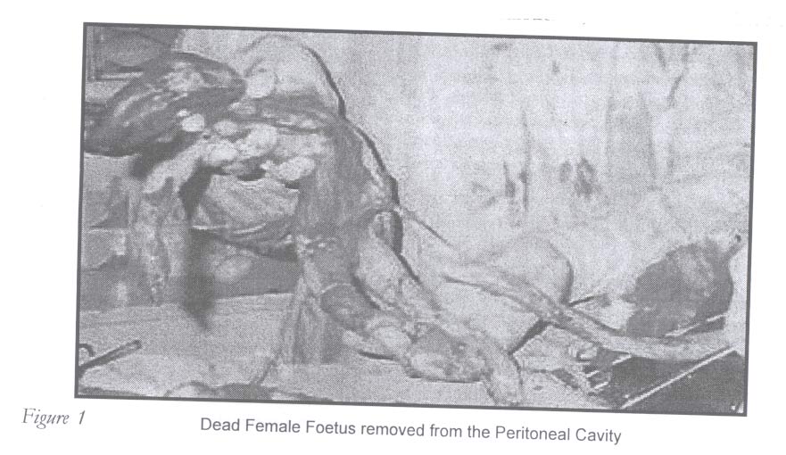
African Journal of Reproductive Health, Vol. 9, No. 1, April, 2005 pp. 162-165 Case Report Heterotopic Pregnancy with Spontaneous Vaginal Delivery at 36 Weeks and Laparotomy at Term - A Case Report Chisara C Umezurike1 and Paul A Feyi-Waboso2 1Department of Obstetrics and Gynaecology, Nigerian Christian
Hospital, Aba, Nigeria. 2Department of Obstetrics and Gynaecology,
Abia State University Teaching Hospital, Aba, Nigeria. Abstract Heterotopic pregnancy, although rare, is occurring more frequently because of an increase in genital infection and the escalating use of new reproductive technologies in infertility patients. The case of a 30-year-old para 2+1 prophetess is presented. She had a spontaneous vaginal delivery at term. Persistent abdominal pain and distension led to suspicion of heterotopic pregnancy. This was confirmed by ultrasonography. Laparotomy revealed a macerated fetus in the peritoneal cavity. The purpose of this report is to sensitise practitioners about the reality and existence of the condition. (Afr J Reprod Health 2005; 9[1]: 162-165) Résumé Grossesse hétérotopique à l'accouchement par voie vaginale spontané à 36 semaines et la laporotomie à terme — a propos des observations. La grossesse hétérotopique, quoique rare, devient plus fréquente à cause de la l'infection génitale et l'emploi croissant de nouvelles technologies par les malades qui souffient de la stérilité. Le cas d'une prophétesse de 30 ans qui était para 2+1 est présenté. Elle a eu un accouchement par voie vaginale spontané à terme. Les douleurs abdominales persistantes et la distension ont fait soupçonner une grossesse hétérotopique. Ceci a été confirmé par l'ultrasonographie. La laparotomie a révélé un foetus macéré dans la cavité péritonial. Ce rapport a pour but de sensibliser les praticiens à la réalité et à l'existence de la condition. (Rev Afr Santé Reprod 2005; 9[1]: 162-165) Key Words:Infertility, infection, laparotomy, pregnancy, heterotopic Introduction Heterotopic pregnancy is the co-existence of intrauterine and extrauterine gestations. Hitherto heterotopic pregnancy was considered a rare condition with occurrence of one in 4000-7000 spontaneous pregnancies.1,2 It is even rarer to carry both the extrauterine and intrauterine pregnancy to as near term as possible.2 In natural conception cycles, heterotopic pregnancy is a rare event occurring in less than 1:30,000 pregnancies.3,4 During the past decade the rising incidence of heterotopic pregnancy following induction of ovulation with clomiphene citrate has been reported.5-7 Also responsible is the increasing use of new assisted reproductive technologies such as in vitro fertilisation (IVF) and embryo transfers (ET) in infertile women.8,9 It has, however, stabilised at 1:100 pregnancies with these procedures.10 The rising incidence of genital tract infection may also be contributory.11 The purpose of this case report is to sensitise practitioners about the reality and existence of heterotopic pregnancy. Case Report Mrs U.N. was a 30-year-old prophetess. She was para 2+1, 2 alive. Her pregnancy in 1996 ended in an emergency caesarean section (CS) at term for fetal distress. She had a spontaneous incomplete miscarriage at eight weeks gestation in 1997, after which she had an evacuation of retained products of conception performed at a mission hospital by a medical practitioner. There were no complications. Her second confinement was in 1998 and she had a spontaneous vaginal delivery at term. She was admitted on 10th July 2001 following referral from a private maternity home where she received her antenatal care. This was uneventful until 36 weeks when she had a spontaneous vaginal delivery of a live female baby of unknown birth weight at the maternity home. The baby developed jaundice during the first day of life and died on the third day. She complained of persistent and increasing abdominal pain, which the midwife had told her was caused by uterine fibroids that would later require surgery. She also observed that her abdomen remained distended and she was, therefore, referred to our centre two weeks after delivery. On admission, her general physical condition was satisfactory. She was not clinically anaemic. Her pulse rate was 84 beats per minute, blood pressure 110/70mmHg and temperature 37°C. Her abdomen was firm and irregular with generalised tenderness. There was an abdomino-pelvic mass, which corresponded to a symphysiofundal height of 36cm. Pelvic examination revealed that the cervix was displaced to the right lateral fornix. The cervical os was closed. The uterus and adnexa could not be delineated. The pouch of Douglas was bulging and firm. Baseline investigations done on admission included full blood count, random blood sugar and serum electrolytes. The haemoglobin was 12.0g/dl. There were no pus or red blood cells on urine microscopy. Endocervical swab culture was negative. Transabdominal ultrasonography revealed a bulky but empty uterus and an extrauterine non-viable fetus in the peritoneal cavity. A diagnosis of heterotopic pregnancy was made. Emergency laparotomy done under general anaesthesia with endotracheal intubation revealed a dead macerated female fetus (birth weight 1.3kg) in the peritoneal cavity. The placenta was plastered to the anterior surface of the uterus and bladder. Having extracted the fetus (Figure 1), the placenta was left intact. Post-operative progress was satisfactory and her post-operative haemoglobin was 10.4g/dl. She was discharged on the 10th post-operative day. However, she was re-admitted 14 days later with fever and anaemia. The haemoglobin was 5g/dl. She was treated for sepsis with intravenous ceftriaxone 2g stat, then 1g daily for seven days and intravenous metronidazole 500mg eight hourly for five days. She was also treated for malaria. After being transfused with three units of blood she was again discharged 10 days later with a haemoglobin of 9.8g/dl. Discussion Heterotopic pregnancy is becoming more frequent because of increased genital infection and especially the wider use of assisted reproductive techniques.11 The patient presented with neither a genital tract infection nor any assisted reproductive technique. As such we infer that she had double ovulation and fertilisation with one embryo implanting in the normal endometrium of the corpus uteri and the second implanting either primarily in the abdomen or secondary to tubal abortion or rupture. Heterotopic pregnancy following ovulation induction and other assisted repro-ductive techniques has been widely reported. Heterotopic pregnancy usually presents a diagnostic difficulty.12,13 This was well illustrated in this case where diagnosis was made only after delivery of the intrauterine fetus when the symphysiofundal height remained 36cm. Persistent abdominal pain should have heightened suspicion of a co-existing abdominal pregnancy during the antenatal period.13 Factors that determine the management of heterotopic pregnancy include the gestational age and clinical presentation of the patient.14 Maternal morbidity and mortality correlate to the management of the placenta following delivery. Authorities dispute whether there is any correlation between the implantation site and the feasibility of placental removal, which depends on the degree of invasion, site of the placenta and other involved organs, and the surgical accessibility of the placental blood supply.15 Interference with it may lead to uncontrollable haemorrhage, and if left in situ, though it is usually the procedure of choice, morbidity from abscess formation is high.16 Case series suggest that safe surgical ligation of the placental blood supply, followed by complete placental removal is feasible in about 60% of cases of abdominal pregnancy, and should be the preferred choice to reduce maternal morbidity and mortality.17,18 If this is not possible then the placenta should be left in situ following ligation of the umbilical cord. Subsequent management is usually expectant, although placental resorption may be aided by use of methotrexate, selective arterial embolization, or second laparotomy.15 Unfortunately, surgical management was difficult in this case because the placenta was plastered to the bladder and the vascular supply was not easily accessible and could not be ligated without compromising the bladder. It was adjudged safer to leave it behind and to allow it to degenerate and become spontaneously absorbed rather than encountering overwhelming haemorrhage in an attempt to remove it.19 The price for this decision, presumably, was her significant post-operative morbidity. In this case expectant management was employed. It is important to maintain a high index of suspicion for heterotopic pregnancy based on clinical signs even in the absence of the epidemiological risk factors. This will improve clinical outcome. References
© Women's Health and Action Research Centre 2005 The following images related to this document are available:Photo images[rh05018f1.jpg] |
| |||||||||
