 |
| About Bioline | All Journals | Testimonials | Membership | News |
|
||||||
|
||||||
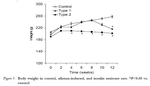
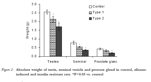
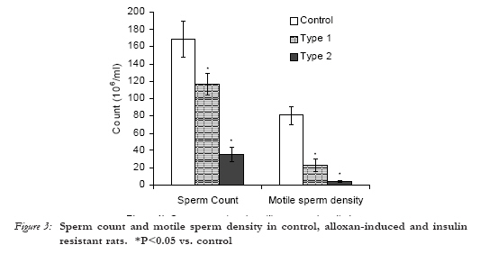
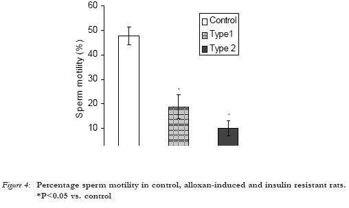
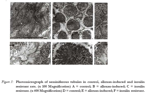
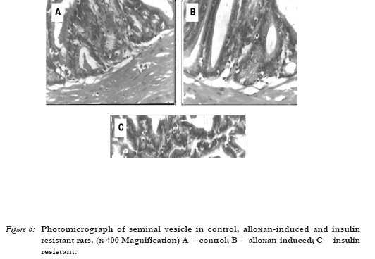
African Journal of Reproductive Health, Vol. 10, No. 3, December, 2006, pp. 106-113 Alloxan-induced and Insulin-resistant Diabetes Mellitus affect Semen Parameters and Impair Spermatogenesis in Male Rats A. P. Arikawe1; A. O. Daramola2; A. O. Odofin3 and L. F. O. Obika4 1Department of Physiology, College of Medicine, University of Lagos, Idi-Araba, Lagos. Nigeria; 2Department of Morbid Anatomy, College of Medicine, University of Lagos, Idi-Araba, Lagos, Nigeria; 3Department of Medicine, College of Medicine, University of Lagos, Idi-Araba, Lagos, Nigeria; 4Department of Physiology, School of Basic Medical Sciences, College of Medical Sciences, University of Benin, Benin City, Edo State. Nigeria. Correspondence: A. P. Arikawe Department of Physiology, College of Medicine, University of Lagos, Idi-Araba, Lagos. PMB 12003, Lagos. Nigeria. Phone number: 0802 360 5282 Email Address: arikawepaul2002@yahoo.co.uk Code Number: rh06043 Abstract The purpose of this study was to determine the effects of diabetes mellitus and insulin resistance on semen parameters, histology of reproductive organs and serum concentrations of testosterone and luteinizing hormone (LH). Male Sprague-Dawley rats weighing 180 - 200g were made diabetic by intravenous injection of alloxan (40 mg/kg) and insulin resistant by chronic fructose feeding (25% fructose) for 12 weeks. Rats were anaesthetized, followed by laparatomy. Blood was obtained by cardiac puncture for measurement of testosterone and LH concentrations by radioimmunoassay. Semen analysis was carried out; reproductive organs were isolated, fixed in Bouin's fluid and processed for histological studies. All semen parameters analyzed were significantly reduced (P < 0.05) in the diabetic and insulin resistant rats compared to control rats. Body weight and weight of reproductive organs were also significantly reduced (P < 0.05). Furthermore, tissue fixation studies revealed changes in the cytoarchitecture of reproductive organs in the diabetic and insulin resistant rats compared to control rats. However, serum concentrations of testosterone and LH were not significantly different in all the groups. We conclude that diabetes mellitus and insulin resistance affect semen parameters and impair distinct phases of spermatogenesis in male rats. Some mechanisms responsible for this impairment are suggested. (Afr J Reprod Health 2006; 10[3]:106-113) Key Words: Diabetes mellitus, insulin resistance, spermatogenesis, testosterone, luteinizing hormone Résumé Diabète sucré provoqué par l'alloxane et le diabète insulimorésistant influent sur les paramètrres du sperme et entravent la spermatogenèse chez les rats mâles. Cette étude avait pour but de déterminer les effets du diabète sucré et la résistance à l'insuline sur les paramètres du sperme, l'histologie des organes de reproduction et les concentrations sériques de la testostérone et l'hormone lutéinisante (HL). Des rats mâles Spraque-Dawley qui pesaient de 180 - 200g étaient rendus diabète à l'aide d'une injection intraveineuse de l'alloxane (40mg/kg) et l'insulinorésistant à travers une alimentation fructose chronique pendant 12 semaines. Les rats ont été anesthésiés et ont subi la laparotomie. Nous avons obtenu le sang à travers une ponction cardiaque afin de mésurer la testostérone et les concentrations LH à l'aide de la méthode radioimmanologique. L'analyse du sperme a été faite; les organes de reproduction ont été isolés, fixés dans le liquide de Bouin avant des les préparer pour les études histologiques. Tous les paramèters du sperme qui ont été analysés ont été réduits de manière significative (P < 0, 05) chez les rats diabetes et insulinorésistants par rapport aux rats témoin. Le poids corporel et le poids des organes de reproduction étaient également réduits de manière significative (P < 0, 05). De plus, les études de la fixation des tissues ont révélé des modifications dans la cytoarchitectonie des organes de reproduction chez les rats diabètes et les rats insulinorésistants par rapport aux rats témoin. Néanmoins, les concentrations du sperme de la testostérone et LH ne variaient pas de manière significative dans tous les groupes. Nous concluons que le diabète sucré et insulinrésistance ont un effet sur les paramètres du sperme et entravent les étapes distinctes de la spermatogènes chez les rats mâles. Nous avons préconisé quelques mécanismes qui sont responsables de cette insuffisance. (Rev Afr Santé Reprod 2006; 10[3]:106-113) Introduction Diabetes mellitus is a constellation of anatomical, biochemical and physiological abnormalities caused by insulin deficiency. Spermatogenesis is an intriguing but complicated biological process1. In mammals, it is composed of three distinct phases of cellular and molecular changes2, 3. These are mitosis of type A spermatogonia into type B spermatogonia and primary spermatocytes4; meiosis of primary spermatocytes into secondary spermatocyctes and haploid spermatids; spermiogenesis i.e. morphogenesis of spermatids into spermatozoa5. In the rat, the entire process of germ cell development, except for the early phase of spermatogenesis from type B spermatogonia up to preleptotene and leptotene spermatocytes is segregated from the systemic circulation because of the blood-testis barrier (BTB) created by tight junctions between Sertoli cells near the basal lamina6. Preleptotene and leptotene spermatocytes must migrate progressively from the basal to the adluminal compartment of the seminiferous epithelium traversing the BTB, while differentiating into haploid spermatids. Without this timely movement of developing germ cells across the seminiferous epithelium, spermatogenesis cannot go to completion and infertility will result. Testosterone in response to LH bathes the seminiferous epithelium and provides a high local concentration of androgens to the Sertoli cells that is necessary for normal spermatogenesis7. It has been reported that diabetes mellitus interferes with reproductive function in laboratory rats8, but the extent and mechanism of this interference is not fully understood. This study was designed to determine the extent to which diabetes mellitus interferes with reproductive function in male rats and some mechanisms of this interference are proposed. We hypothesized that diabetes mellitus would interfere with spermatogenesis and affect semen parameters in male Sprague-Dawley rats. Materials and Methods Animals Male adult Sprague-Dawley rats weighing 180 - 200 g were obtained from the Laboratory Animal Department. Animals were kept under standard conditions of temperature 27ºC - 30ºC, with 12h light/dark cycle and were randomly divided into 3 groups. Group 1 served as control and was fed with normal rat chow. Group 2 served as Type 1 diabetic group and received a single dose intravenous injection of alloxan monohydrate (40 mg/kg bodyweight)9 into the lateral tail vein. Hyperglycaemia was confirmed 72 hours later using Dextrostix Test Strips (Bayer Corporation, U. K.) following the glucose oxidase method10. Four weeks after induction of Type 1 diabetes mellitus, the rats were sacrificed. Group 3 served as Insulin resistant diabetic group and was fed ad libitum on a special diet containing 25% fructose mixed with 75% normal rat chow for 4 weeks11 and continued till the 12th week. Hyperglycaemia was confirmed using Dextrostix Test Strips (Bayer Corporation, U. K.) following the glucose oxidase method10. All animals had free access to drinking water throughout the duration of the study. Rats with blood glucose concentration above 250 mg/dl were used as Type 1 diabetic rats12 while rats with blood glucose concentration above 200 mg/dl were used as Insulin resistant diabetic rats13. Semen Analysis and Isolation of Reproductive Organs At the 12th week, the rats were anaesthetized with ether, followed by laparatomy and blood obtained by cardiac puncture for serum immunoassay of luteinizing hormone and testoterone. Caudal epididymis was carefully isolated and several incisions (1mm) were made on it. It was then suspended in 1 ml normal saline and immediately made up to 10 ml with normal saline. Semen analysis was carried out immediately using the new improved Neubauer counting chamber for determination of the concentration of spermatozoa, sperm motility and sperm density. Testis and seminal vesicle were carefully isolated, weighed, washed in buffered saline and transferred into Bouin's fluid for 72 hours for tissue fixation. The tissues were embedded in wax, mounted and thin sections (6.0mm thick) were cut and stained with haematoxylin and eosin. Hormone Assay Blood was obtained by cardiac puncture for measurement of serum testosterone and LH concentrations by radioimmunoassay in batches with control sera at both physiological and pathological levels using standard quantitative solid phase enzyme-linked immunosorbent assay (ELISA) technique with microwell kits (Syntro Bioresearch Inc., California, U. S. A.). Statistical Analysis Results were expressed as means ± S. E. M. The significance of differences among groups was analyzed statistically by One-way ANOVA (Analysis of Variance), followed by Student's unpaired t - test. Differences were considered statistically significant at P < 0.05 Results Blood glucose Blood glucose concentration was significantly higher (P < 0.05) in alloxan induced and insulin resistant diabetic rats (256.0 ± 22.0; 218.0 ± 24.6 mg/dl) compared with control rats (114.0 ± 4.0 mg/dl). Body weight Body weight increased progressively in all the groups (Figure 1). At the 6th week, it began to decline significantly (P < 0.05) in insulin resistant diabetic rats compared to control rats. Body weight continued to decline significantly between the 8th and 12th week in insulin resistant diabetic rats compared to control rats. On the other hand, significant decrease (P < 0.05) in body weight was observed in alloxan induced diabetic rats from the 10th week following alloxan injection compared to control rats. Absolute weight of reproductive organs Weight of testis was significantly lower (P < 0.05) in alloxan-induced and insulin resistant diabetic rats (2.14 ± 0.16; 1.70 ± 0.20 g) compared to control rats (2.55 ± 0.11 gm). Similarly, the weight of the seminal vesicle was significantly lower (P < 0.05) in alloxan-induced and insulin resistant diabetic rats (0.55 ± 0.04; 0.37 ± 0.04 g) compared to control rats (0.78 ± 0.05 g). However, the weight of the prostate gland was significantly lower (P < 0.05) in insulin resistant diabetic rats (0.20 ± 0.02 g), but not in alloxan-induced diabetic rats (0.32 ± 0.06 g), when compared to control rats (0.43 ± 0.05 g) (Figure 2). Semen Parameters Sperm count was significantly reduced (P < 0.05) in alloxan-induced and insulin resistant diabetic rats (116.0 x 10⁶ ml (± 12.3); 35.4 x 10⁶ ml (± 8.1) compared to control rats (169.0 x 10⁶ ml (± 21.0). Likewise motile sperm density was significantly reduced (P < 0.05) in alloxan-induced and insulin resistant diabetic rats (23.2 x 10⁶ ml (± 7.4); 4.2 x 10⁶ ml (± 1.5) compared to control rats (80.4 x 10⁶ ml (± 10.7); (Figure 3). Percentage sperm motility was also significantly reduced in alloxan-induced and insulin resistant diabetic rats (19 ± 5%; 10 ± 3%) compared to control rats (48 ± 5%); (Figure 4). Testosterone and LH serum concentrations Serum testosterone concentration though lower in alloxan-induced diabetic rats (1.55 ± 0.56 ng/ml) and insulin resistant diabetic rats (1.45 ± 0.45 ng/ml) compared to control rats (2.37 ± 0.45 ng/ml) was not significantly different in all the groups. Likewise, serum LH concentration though not significantly different in all the groups was higher in alloxan-induced diabetic rats (3.42 ± 0.80 ml IU/ml) compared to control rats (1.92 ± 0.02 ml IU/ml) and insulin resistant diabetic rats (1.92 ± 0.04 ml IU/ml). Histology of reproductive organs Histological sections of the testes revealed significant changes in the cytoarchitecture of both alloxan-induced (Type 1) and insulin resistant (Type 2) male diabetic rats (Figures 5 and 6). Alloxan Induced (Type 1) diabetic male rats There was disorganized spermatogenesis in about 15% of seminiferous tubules characterized by a disorderly arrangement of the germ cells. More stricking was the reduction in spermatogenesis (hypospermatogenesis) in more than 50% of seminiferous tubules. Varying degress of severity was noticed with some rats having severe hypospermatogenesis in about 40% of tubules. Leydig cells were also significantly reduced compared to the control group. The epithelium of the seminal vesicles however showed no significant changes. Insulin-Resistant (Type 2) diabetic rats This group of rats showed more severe histological changes compared to the Alloxan induced diabetic rats. There was hyposperma-togenesis in over 90% of tubules with severe hypospermatogenesis in over 60% of tubules. The Leydig cells showed a greater degree of reduction than in alloxan induced diabetic rats and there was laso marked atrophy of the linning epithelium in the seminal vesicles. Likewise, pseudo-stratified columnar epithelium is also depleted in the insulin resistant diabetic rats (Figure 6). Discussion Our results show that alloxan and fructose-induced insulin resistance increase blood glucose concentrations in rats14,15 and cause diabetes mellitus. Body weight did not differ significantly between control rats and insulin resistant diabetic rats fed with chronic fructose diet for 2 weeks and is in agreement with an earlier report13. Significant weight loss was observed in alloxan-induced and insulin resistant diabetic rats. This is also consistent with an earlier report16. Consistent with a previous study17, absolute weight of repro-ductive organs was significantly decreased in alloxan-induced and insulin resistant diabetic rats. Serum testosterone concentrations though lower in alloxan-induced and insulin resistant diabetic rats was not significant when compared to control rats. This is in line with an earlier report in humans18 and in rats12. However, our result is contrary to a previous study19 in which significant decrease in serum testosterone, LH and FSH was reported in streptozotocin induced diabetic rats. The difference could be due to the drug used to induce diabetes The mechanism of impairment of distinct phases of spermatogenesis by diabetes mellitus is presently not well understood. Our results on serum LH and testosterone concentrations eliminate the possibility that the impairment originates from the pituitary-hypothalamic axis and points to the possibility that testosterone is involved in the impairment of spermatogenesis by diabetes mellitus. Moreover, testosterone has been reported to play a vital role in the regulation of fluid dynamics of the testis, commencing with blood flow and vasomotion and ending with the production of seminiferous tubular fluid20. Our results suggest the following mechanisms in the impairment of spermatogenesis by diabetes mellitus: (i) disturbance in the functions of Sertoli cells with or without a disturbance or disruption in the physiology and/or morphology of the blood-testis barrier21,22. (ii) alterations in the microenvironment provided by the Sertoli cells, either directly or resulting from changes in paracrine signals from the seminiferous tubules23. (iii) disruption of ionic channels24. Conclusion In summary, the findings of this study show that diabetes mellitus affect semen parameters and impairs distinct phases of spermatogenesis in male rats. The extent of this impairment is a subject of further research; however some mechanisms have been suggested. Acknowledgement The authors are grateful to Dr. M. O. Ajala, Dr. R. R. Ettarh and Mr. T. Teniola for their technical assistance. References
© Copyright 2006 - Women's Health and Action Research Centre
The following images related to this document are available:Photo images[rh06043f6.jpg] [rh06043f5.jpg] [rh06043f4.jpg] [rh06043f2.jpg] [rh06043f3.jpg] [rh06043f1.jpg] |
| |||||||||
