 |
| About Bioline | All Journals | Testimonials | Membership | News |
|
||||||
|
||||||
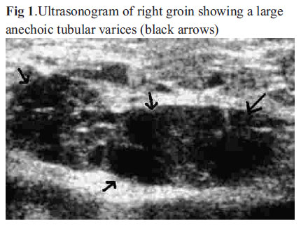
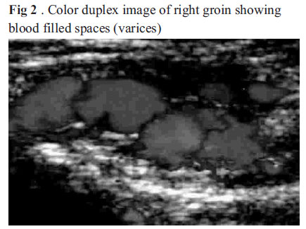
African Journal of Reproductive Health, Vol. 15, No. 2, June, 2011, pp. 163-164 CASE REPORT Round Ligament Varicosities mimicking inguinal herniae in pregnancy - a diagnostic dilemma Sajad Ahmed Salati Dr Sajad Ahmad Salati, Lane 2, Bulbul Bagh, Barzullah, Srinagar, J&K Pin:190005, INDIA E-mail: docsajad@yahoo.co.in Code Number: rh11029 Abstract Round ligament varicosities are rare surgical entities which can occur during pregnancy and mimic inguinal herniae. Correct diagnosis by clinical examination and ultrasonography can avoid an unnecessary surgical operation during pregnancy and its related complications.We present one case of 26 years pregnant lady reporting with bilateral round ligament varicosities. Résumé Le ligament round des varicoses sont des entit és chirurgicales rares qui peuvent se produire pendant la grossesse et peuvent imiter l'hernie inguinale. Un bon diagnostic à travers l'examen clinique et à l'aide de l'ultrasonographie peut éviter une opération chirurgicale pendant la grossesse aussi bien que les complications qui y sont liées. Nous présentons un cas d'une femme enceinte âgée de 26 ans qui s'est présentée avec le ligament round des varicoses bilatérales. Key words: Round ligament, varicosities, inguinal hernia, pregnancy Discussion The round ligaments (ligamentum teres uteri) are two flattened fibro-muscular bands between 10 and 12 cm. in length, situated between the layers of the broad ligament. They are attached to the uterus on either side in front of and below the opening of the fallopian tube. Each ligament leaves the pelvis via the deep inguinal ring, passes through the inguinal canal and continues on to the labia majora where its fibers spread and mix with the tissue of the mons pubis1. The function of the round ligament is to maintain ante-version of the uterus (a position where the fundus of the uterus leans ventrally). When the uterus grows during pregnancy, these ligaments can stretch causing pain in groins, the condition termed as round ligament syndrome2. In this condition, no lumps are formed. However, due to smooth muscle relaxation effect of progesterone, a raised cardiac output causing increased venous return from the limbs, and most importantly progressive obstruction by the gravid uterus, pelvic vein enlargement occurs during pregnancy and sometimes this phenomenon extends to the veins of the round ligament traversing the inguinal canal, resulting in round ligament varicosities3. The exact incidence of round ligament varicosities in pregnancy is unknown as very few cases are reported in literature4. For the authors, this case was the first in their combined surgical practice of about two decades. The presentation of round ligament varicosities is similar to inguinal herniae5, 6. Both the entities present as inguinal lumps (reducible or irreducible) and both may transmit cough impulses. Establishment of correct diagnosis is important and is undertaken with the aid of ultrasonography, followed by color duplex scanning3, 5. Round ligament varicosities appear as large dilated spaces on ultrasonography, and color duplex scanning shows these spaces to be containing blood .In literature, it is the failure of the physician to evaluate groin lumps with imaging which had led to diagnosis of hernia and unnecessary surgical interventions5, 7. Management of round ligament varicosities is conservative because they resolve spontaneously postpartum4,8. Patient needs to be assured and followed up till complete resolution and might require occasional mild analgesic. In some cases, these varicosities might get thrombosed in the postpartum period; this complication produces a firm, tender, irreducible swelling simulating a strangulated inguinal hernia, and a differential diagnosis becomes an urgent necessity, in view of the mandatory surgical treatment in case of hernia9, 10. If however the clinician is aware of thrombosis of round ligament varicosities as a differential diagnosis of painful groin lumps in pregnancy and instead of simply relying on clinical examination, advises Doppler ultrasound imaging, the correct diagnosis can be made confidently. Besides ultrasound, CT scan and MRI have also been mentioned in literature as effective modalities of imaging the thrombosed varicosities11. To conclude, it is stressed that imaging by ultrasound should be undertaken in all pregnant patients presenting with inguinal lumps/complaints to avoid unneccasary operations and complications. References
Copyright 2011 - Women's Health and Action Research Centre, Benin City, Nigeria The following images related to this document are available:Photo images[rh11029f1.jpg] [rh11029f2.jpg] |
| |||||||||
