 |
| About Bioline | All Journals | Testimonials | Membership | News |
|
||||||
|
||||||
Iranian Journal of Reproductive Medicine Vol. 3, No. 2, Spring, 2005, pp. 62-67 Morphologic changes in fresh and vitrified mouse ovaries after retinol palmitate administration Homayoon Babaei,1 Ph.D., Amin Derakhshanfar,2 Ph.D., Seyed Noureddin Nematollahi-Mahani,3 Ph.D., Fathemeh Nabipour,4 M.D., Akram Zeraatpisheh,5D.V.M. 1 Department
of Clinical Science, Faculty of Veterinary Medicine, Shahid Bahonar University,
Kerman, Iran. Code Number: rm05011 Abstract Background: Retinoids have been
suggested to play a role in oogenesis and oocyte survival.
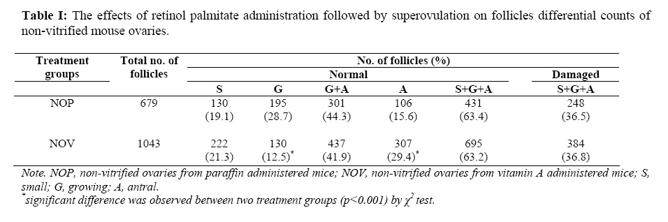
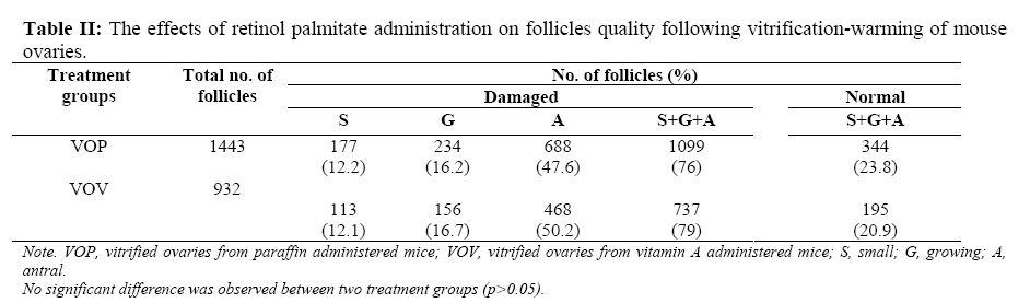
Keywords: Ovary, Superovulation, Retinol Palmitate, Vitrification Introduction Retinol and its cellular metabolites, all-trans retinoic acid and 9-cis retinoic acid are known as retinoids. These compounds are recognized as important regulators of vertebrate development, cellular differentiation and tissue function. In addition, retinoids are essential for normal reproductive processes in both males and females (1-3). Several studies indicate a positive effect of retinol supplementation when diets are inadequate in vitamin A. In litter-bearing species, administration of retinol or β-carotene has been reported to increase embryo survival in mice (4), rabbits (5) and swine (6,7). Retinoids may function as important regulators of oogenesis and oocyte survival in the mouse fetal ovary and positively impact events associated with oocyte maturation in adult farm species leading to improved embryonic survival (8). Thus, embryonal survival may be associated with differences in oocyte development before ovulation (9). Transplantation of cryopreserved ovaries is a strategy for improving the reproductive efficiency of young female patients, suffering from infertility, due to iatrogenic loss of ovarian function, resulting from chemotherapy and/or radiation therapy (10). In an early study, Deansely (1957) reported the successful cryopreservation of ovarian tissue (11) and then, Parrot (1960) reported the birth of live offspring after orthopic transplantation of a slice of cryopreserved mouse ovary (12). Ovarian cryopreservation is mostly carried out by slow freezing (13) but many attempts have been made to improve cryopreservation conditions by using simple and efficient procedures. For this purpose, the vitrification method was applied to the field of biology as a method of cryopreservation and its recent development has greatly simplified the cryopreservation procedures. This approach was first applied for oocyte cryopreservation by Critser et al (14) and recently, Nematallahi-Mahani, et al. (15), Sugimoto et al. (16) and Salehnia et al. (17) showed that vitrification is a useful strategy for the preservation of some mammalian ovaries. Despite many reports on improvement of vitrification procedure, the rate of success is still lower than non-vitrified materials (18). To the best of our knowledge, there are not any reports which evaluate the effect of retinoids on ovaries following vitrification. The current experiment, therefore, was designed to study 1) the effects of retinol palmitate administration on changes in proportion of different follicles in response to superovulation. 2) the effects of retinol palmitate administration on quality of follicles after vitrification-warming of ovaries. Materials and Methods Chemicals were purchased from Sigma Company (St. Louis, MO, USA) unless otherwise indicated. Animals All animals were cared for and used in accordance with the International Guiding Principle for Biomedical Research Involving Animals at Kerman University of Medical Science. The BALB/c Mice were housed under a lighting regimen of 12 hours light to 12 hours dark and temperature-controlled conditions (22 ± 2°C). Food and water were freely available at all times. Ten, 4 week old female mice were used in this study. Experimental Design To determine whether retinol palmitate is capable of increasing follicles quality and lowering follicular damage following vitrification of ovaries, the mice (n=10) were randomly assigned to either paraffin (n=5) or vitamin A (n=5) administered groups. Vitamin A administered animals were injected 250 IU of retinol palmitate (Osvah, Iran), dissolved in 0.1 ml of paraffin oil intraperitoneally on days one and ten. Paraffin administered mice only received 0.1 ml of the vehicle (paraffin oil). The animals were superovulated with 10 IU PMSG (Folligon®, Intervet) simultaneously with the second injection of paraffin or retinol palmitate (19). Forty eight hours after PMSG injection, the mice were killed by cervical dislocation and ovaries were collected from both groups. The collected left ovaries from both paraffin and vitamin A administered mice were considered as non-vitrified (NOP, non-vitrified ovaries from paraffin administered mice; NOV, non-vitrified ovaries from vitamin A administered mice) and the effects of retinol palmitate administration on changes in follicles differential counts were evaluated. In addition, the collected right ovaries from both paraffin and vitamin A administered mice underwent vitrification (VOP, vitrified ovaries from paraffin administered mice; VOV, vitrified ovaries from vitamin A administered mice) and the effects of retinol palmitate administration on quality of follicles after vitrification-warming were evaluated. Cryopreservation of Ovaries by Vitrification We adopted the method of Vajta et al. (1998) with some modification (20). Briefly, the vitrification solution consisted of VS1 {10% ethylene glycol (EG) and 10% DMSO in holding medium (TCM-199 + 20% FBS: HM)} and VS2 {20% EG and 20% DMSO in HM}. The ovaries were equilibrated in VS1 for 15 minutes at room temperature, and then in VS2 for 2 minutes. Each ovary was loaded into plastic straw (outer diameter: 5mm) with the least volume of the vitrification solution at room temperature, followed by plunge directly into liquid nitrogen. Warming and Cryoprotectant dilution The vitrified samples were thawed rapidly by immersing the end of the tubes in a thawing solution composed of 1.0 M sucrose in HM for 10 minutes. The temperature of the media used for warming was held at 37°C. Histopathological Evaluation The fresh (non-vitrified) and the recovered vitrified ovaries were fixed in 10% buffered formalin, embedded in paraffin wax, serially sectioned at 6μm, stained with hematoxylin & eosin and analyzed under light microscope. Each ovary yielded approximately 600 sections, and differential follicle counts were obtained from every 10th section to provide a 10% sample selection (approximately 60 sections per ovary). Three ovarian follicle classes (small, growing, antral) were identified in the tissue sections according to Thomas et al. (1997), (21). Briefly, 'small' follicles consisted of an oocyte surrounded by one unbroken monolayer of granulosa cells. 'Growing' follicles had an oocyte surrounded by a multilayered, solid mantle of granulosa cells. 'Antral' follicles were characterized by a central oocyte encircled by a fluid filled space and bordered by several layers of granulosa cells. The number of normal and damaged follicles at each follicular class was also recorded. In addition, structural normality (follicle and stromal cell morphology, even distribution of granulosa cells, intact theca and appearance of oocytes) was evaluated. Statistical Analysis The SPSS ® software for windows (standard version, 10.0.1) was used for data evaluation (SPSS Inc, Chicago, IL, USA). Data were analysed by Pearson's Chi-square test. Results The number of morphologically normal follicles as well as changes in different follicular classes was calculated for the non-vitrified ovaries after retinol palmitate versus paraffin administration followed by superovulation (Table I). No statistical difference due to retinol palmitate injection was observed for the percentage of small follicles between the two non-vitrified groups (19.1% vs. 21.3%). The rate of growing and antral follicles was nearly identical in the two groups (44.3% vs. 41.9%). While the proportion of the antral follicles in the NOV group was statistically higher in comparison to the NOP group (29.4% vs. 15.6%; p<0.001). There were not any statistical differences in the rate of damaged follicles between the two non-vitrified groups. When the retinol palmitate group was compared with the control after vitrification-warming, no positive effect was found due to retinol palmitate administration. As shown in Table II, the proportion of the damaged follicles did not show any significant difference between two vitrified groups (VOP: 76% vs. VOV: 79%). Most histopathological features of both non-vitrified groups were normal. The cytoplasm of the oocytes was clear and normal and all the granulosa layers and theca interna and externa were intact and firmly attached to the related basement membranes. The stromal cells were normal with distinct boundaries, prominent nucleus and nucleolus and pinkish cytoplasm. In contrast, in both vitrified groups the stromal cells had distinct margins with foamy cytoplasm. The detachment of the granulosa layers from the basement membrane and the deformity of the oocytes was also seen (Figure 1). Discussion The results of the present study show clearly that retinol palmitate administered mouse produce more antral follicles in response to superovulation. In addition, we did not observe any positive effect of retinol palmitate on follicle quality after vitrification of ovaries. Cryopreservation of oocytes and embryos by vitrification has been widely conducted because of its simplicity and high survival rate of oocytes and embryos as well as cryopreservation of ovarian tissues (17). Sugimoto et al. (2000) observed that rat ovarian follicles survived after vitrification and transplantation with a decrease in the number of healthy antral follicles (approximately 30%) (16), and according to another report by Baird et al. (1999), about 28% of primordial follicles survived after transplantation of frozen/thawed ovarian tissue(22). In contrast, in the present investigation, the percentage of normal follicles including 'small', 'growing' and 'antral' in both vitrified groups (Table II) was lower than the above mentioned reports. Higher rate of damaged follicles in both vitrified groups shows that vitrification may induce some short-term deleterious effects on ovarian tissues. Transplantation of ovaries following vitrification may decrease such harmful effects of cryopreservation through change in the milieu of follicles (23), and autorepair of follicles under the control of various tropic factors (24). This finding is in agreement with the studies of Sugimoto et al. (16), and Schmidt et al. (25) which reported that continuous follicle maturation occurred in cryopreserved ovarian autografts following transplantation. Antioxidant agents such as alpha-tocopherol as well as vitamin A improve the survival rate of follicles (8, 26, 27). However, the effect of retinoids on follicles quality following cryopreservation stress has not been yet investigated. Schweigert & Zucker (1988) suggested that concentrations of vitamin A in follicular fluid might serve as an indicator of follicular quality in cattle (28). Vitamin A concentrations were observed to be highest in large non-atretic follicles and lowest in small atretic follicles. Some reports from in vivo and in vitro studies in domestic livestock species lend supported that retinoids may target the oocyte and influence the subsequent development of embryos (7, 29-31). Improvement of follicular quality that is gained through retinol palmitate administration may in turn lead to better tolerance against deleterious effects of vitrification following transplantation. However, we did not observe any useful effect of retinol palmitate on follicle quality after vitrification of ovaries. There are some evidences that vitamin A may influence oocyte and embryo development. The results obtained by Lima et al. (2004) demonstrated that the addition of retinol to the embryo culture media had a significant positive effect on bovine early embryonic development (32). Rabbits that had higher blood levels of vitamin A produced more oocytes and embryos in response to superovulation than those that had lower levels (33), and mouse treated with vitamin A before superovulation had higher ovulation rate and produced more zygotes (19). As our results show, the percentage of antral follicles in retinol palmitate administered mice was statistically higher than the control group following superovulation. This may be the probable mechanism of the positive effect of retinol palmitate on folliculogenesis after superovulation, which describes the results of our study and the reports mentioned above. In contrast, Shaw et al. (29) and Brown et al. (34) did not show any effect of vitamin A on the response of ovaries to superovulation in cows. Transport and metabolism of retinoids are mediated and regulated by specific binding proteins (35, 36). Retinol is transported systemically and intercellularly by retinol-binding protein (RBP). Within target cells, retinol binds cellular retinol-binding protein (CRBP) which functions in retinol accumulation and presentation to dehydrogenases for conversion to retinal and retinoic acid (37). Brown et al. (2003) reported that within the ovarian follicles, strongest CRBP was observed in granulosa cells of preantral follicles (38). This evidence demonstrates that exogenous retinoids may have better effect on growing and antral than small follicles. Furthermore, retinoids have been shown to stimulate steroidogenesis by granulosa cells in vitro and synergistically enhance the ability of FSH to induce LH receptors (39). Antrum formation and final growth are entirely FSH/LH dependent (40). Together, these reports suggest that retinoids play a role in moving normal preantral toward antral follicles and may explain increasing the proportion of antral follicles in response to superovulation that is shown in the present study as well. Conclusion In conclusion, the results of the present study demonstrate that when retinol palmitate is administered to the mice, growing follicles developed to antral follicles in response to superovulation. However, short term harmful effects of vitrification on ovaries may not be overcome by retinol palmitate administration. Further in vitro investigations are needed to evaluate long term effects of vitrification in retinoids treated animals. Acknowledgements Special thanks to Mr. Ali Haddad Narafshan for revising the English of the manuscript. References
© Copyright 2005 - Iranian Journal of Reproductive Medicine The following images related to this document are available:Photo images[rm05011t1.jpg] [rm05011f1.jpg] [rm05011t2.jpg] |
| |||||||||
