 |
| About Bioline | All Journals | Testimonials | Membership | News |
|
||||||
|
||||||
Iranian Journal of Reproductive Medicine Vol. 3, No. 2, Spring, 2005, pp. 74-78 The effects of cumulus cells on in vitro maturation of mouse germinal vesicle stage oocytes Reza Mahmoudi,1Ph.D. student, Aligholi Subhani,1Ph.D., Parichehr Pasbakhsh,1Ph.D., Farid Abolhasani,1Ph.D., Iraj Amiri,2 Ph.D., Mozhdeh Salehnia,3 Ph.D., Farideh Etesam1 Ph.D. 1 Department of Anatomy, Faculty of Medicine, Tehran University of Medical Sciences, Tehran, Iran.
Code Number: rm05013 Abstract Background: In
vitro maturation (IVM) of oocytes is a promising technique to reduce the costs
and avert the side-effects of gonadotropin stimulation for in vitro
fertilization (IVF). The pregnancy rates from oocytes matured in vitroare
much lower than those of in vivo stimulation cycles, indicating that
optimization of IVM remains a challenge.
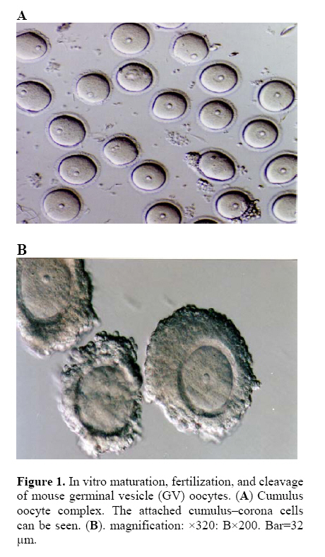
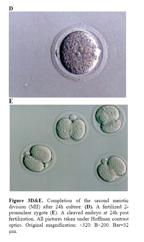
Key Words: Immature oocyte, In vitro maturation, Mouse, Cumulus cells Introduction In vitro maturation (IVM) of mammalian oocytes has become an efficient method to produce mature oocytes in order to use in assisted reproductive techniques such as In Vitro Fertilization (IVF), Intra cytoplasmic sperm injection (ICSI), and cloning. Induction of ovulation to obtain mature oocytes for IVF has become a routine procedure in many infertility clinics. Some women, however, rather fail to respond to the hormonal stimulation or are at risk of ovarian hyperstimulation (1). In vitro maturation of oocytes offers an alternative to obtain mature oocytes in these cases (2, 3). Immature human oocytes exhibit acceptable meiotic competence to metaphase II (MII), but their subsequent developmental competence remains disappointingly low. Only 40–80% of fertilized IVM oocytes progress through early cleavage, and of those that do cleave and are transferred, 15% implant to form a viable fetus (3-6). Oocyte maturation is often conceptually dividedinto nuclear and cytoplasmic processes. Nuclear maturation isa term that refers to the resumption of meiosis from the germinal vesicle (GV) stage and progressionto MII. Cytoplasmic maturation is a more general termthat refers to other maturational events (not directly relatedto meiotic progression) that prepares the oocyte for fertilizationand preimplantation development (7, 8). Within the follicle, the regulation of oocyte development and cellular function is dependent on gap junctional communication, paracrine communication and interaction with elements of extracellular matrix (9-11). Cumulus cells play an important role on oocyte maturation, since they provide and transfer several known and unknown factors that are essential for normal nuclear and cytoplasmic maturation of oocytes and subsequent embryonic development after fertilization (12-14). Moreover, cumulus cells also are known to play an important role in regulation of meiotic progression of oocytes (15). During the growth and accomplishment of meiotic competence of oocytes (before initiation of meiosis), cumulus cells are responsible for maintenance of nuclear arrest at GV stage via elevating intercellular cAMP level in the oocytes by transferring an inhibitor signal through gap junctions (16, 17). Initiation of meiosis is also related to cumulus-function, as there are evidences that cumulus cells secrete a meiosis-inducing factor (18). According to the mentioned data, this study was proposed to investigate the effects of cumulus cells on in vitro maturation, fertilization and developmental rates of mouse GV stage oocytes. Materials and Methods Collection of GV oocytes Oocytes were obtained from 6-8 weeks old Blab C female mice. The animal were kept under controlled condition (12hr light, 12hr dark) and fed with water and pellets ad libitum. The mice were stimulated by an i.p. injection of 10 IU pregnant mare serum gonadotropin (PMSG). The animals were killed 44hr later by cervical dislocation and the ovaries were removed into TCM-199 (Sigma)supplemented with 10% fetal bovine serum (FBS). The GV-stage oocytes of ovarian follicle were released by puncturing with a 28G sterile needle under a stereomicroscope. A total of 654 oocytes were obtained from 30 ovaries and they were used for in vitro maturation. The average number of collected oocytes was 21.8 per ovary. In Vitro Maturation (IVM) The collected GV-stage oocytes were divided into two groups: Group I: denuded oocytes (DO) (Figure1A) and Group II cumulus oocyte complexes (COC) (Figure1B). Each group were placed in 25µl micro drops TCM-199 supplemented with 10% FBS, 0.23 mM sodium pyrouvate, 50mg/l streptomycin, 60 mg/l penicillin and 1µg/l epidermal growth factor (EGF), over laid with embryo-tested light mineral oil (Sigma) for 24hr in a humidified atmosphere of 5% CO2 at 37ºC. At various intervals from the onset of incubation, oocytes were observed by inverted microscopy and morphological changes in the nucleus or the extrusion of first polar body (MII) were used as the criterion for nuclear maturation of GV-stage oocytes. After IVM, oocytes in group II were exposed to 3 mg/ml hyaluronidase in TCM199 and cumulus cell were removed by repeated pipettingand the matured oocytes were collected for in vitro fertilization. In Vitro Fertilization (IVF) Sperms were collected from epididymes of blab C male mice aged 12 weeks. The sperm suspension (1 ×10⁶ motile spermatozoa/ml) was capacitated for 2hr in one ml T6 media supplemented with 16 mg/ml BSA. The in vitro matured MII stage oocytes from each treatment group were placed into 0.9 ml T6 and 0.1 ml capacitated spermatozoa was added to it. After 5hr incubation, the oocytes were washed through three droplets of T6 medium. Then, the oocytes were cultured in a droplet of T6 (25µl) under mineral oil. They were assessed for cleavage to the 2-cells stage after 24hr. Statistical analysis Collected data were analyzed by chi-square test. The differences in the values of maturation, fertilization and developmental rates, were considered significant when p<0.05. Results In vitro maturation of mouse oocytes Table Ishows the number of oocytes arrived to MII stage (Figure 2C) after 24hr culture. The maturation rate of COC was significantly higher than that of DO (p<0.05). In vitro fertilization and development of mouse oocytes As shown in Table II, the number of fertilized oocytes (Figure 3D) were significantly higher in COC than those of DO (p<0.05).But the difference between the number of fertilized oocytes which developed to 2-cells stage (Figure 3E) was not significant (p>0.05) in two groups. Table I: Maturation rate of mouse oocytes after 24hr culture. DO = Denuded oocyte, COC = Cumulus oocyte complex, GV= Germinal vesicle oocyte. GVBD= Germinal vesicle breakdown, MII=metaphase II. Significant differences between DO and COC are indicated by an asterisk (p<0.05).
Table II: Fertilization and developmental rates of mouse oocytes. Significant differences between DO and COC are indicated by an asterisk (P<0.05).
Discussion Cumulus cells have been considered to play an important role in oocyte maturation by keeping the oocyte under meiotic arrest, inducing meiotic resumption and by supporting cytoplasmic maturation. These functions have been attributed to their gap junctions and their specific metabolizing capabilities (12-15). Physical contact between oocyte and cumulus cells has been considered necessary for the transfer of nutrients and factors essential for oocyte development (16-19). In addition, dissociated cumulus cells have been reported to produce paracrine factors, which resume meiosis in denuded oocytes (20). The beneficial effects of cumulus cells on oocyte maturation and early development were reported in different species (12, 14, 19-23). Our study confirmed these mentioned results and demonstrated that maturation and fertilization rates were significant by higher in cumulus-oocyte complex compared with denuded oocytes (p<0.05). But there was no significant difference in cleavage rates. However, the obtained data in this study are different whit some other published results. For example Nishi et al (2003) investigated on the mice GV oocytes and reported that maturation, fertilization and developmental rates of DO were 64.4, 51.7 and 43.2%, and cumulus-oocyte complex (COC) were 76.4, 70.5 and 50.5%, respectively. The maturation rate (MII) and fertilization rate (2PN) of COC was significantly higher than that of DO (p<0.05) (25). On the other hand, Aono et al (2003) investigated on murine GV oocyte and reported that the maturation (MII) and cleavage rate (2-cell) of COC were 90.2%, 94.1% respectively (25). In our study, these amounts are higher than Nishi et al investigation in both COC and DO, and lower than that of Aono et al. This difference maybe is due to the animal, oocyte collection method and medium that used. Conclusion In conclusion, our study indicates that in vitro maturation and fertilization and cleavage rates of mouse cumulus-oocyte complex (COC) are more successful than denuded oocytes. Further studies should explore the factors in cumulus cells that are responsible for these results. We think by exploring and extracting these factors and using them in culture media, oocytes maturation and embryonic preimplantation development invitro can be improved. References
© Copyright 2005 - Iranian Journal of Reproductive Medicine The following images related to this document are available:Photo images[rm05013f3d-e.jpg] [rm05013f1.jpg] [rm05013f2c.jpg] | ||||||||||||||||||||||||||||||||||||||||
| |||||||||
