 |
| About Bioline | All Journals | Testimonials | Membership | News |
|
||||||
|
||||||
Iranian Journal of Reproductive Medicine Vol. 5, No. 4, Autumn, 2007, pp. 159-163 In vitro maturation media, cysteamine concentration and glutathione level affect blstocysts development in mouse Amaneh Mohammadi Roushandeh1Ph.D., Parichehr Pasbakhsh2Ph.D., Zohreh Alizadeh3Ph.D., Mehryar Habibi Roudkenar4 Ph.D. 1 Department of Anatomy, Medicine faculty, Tabriz
University of Medical Sciences, Tabriz, Iran. Received: 29 August 2007; accepted: 27 December 2007 Code Number: rm07031 Abstract Background: Preparation
of oocytes is one of the critical factors that determine the developmental
competence of embryos produced by in vitro fertilization (IVF). Objective: In
this study, the effect of cysteamine, type of media and glutathione (GSH) level
on blastocysts development after in vitro maturation of mouse oocytes were
investigated. Key words: Cysteamine, IVM, Blastocysts, GSH, Oocyte, Mouse. Introduction Preparation of oocytes is one of the critical factors that determine the developmental competence of embryos produced by in vitro fertilization (1). In vitro-matured (IVM) oocytes are known to have a lower developmental competence after IVF to in vivo-derived oocytes (2). IVM oocytes have been used in some laboratories because their use makes it feasible to obtain a large number of oocytes from ovaries at relatively low cost. Many factors influence the karyoplasmic and cytoplasmic maturation of oocytes in vitro, including co-culturing with follicular cells such as cumulus cells or granulosa cells, duration of maturation, and type of media (3-8).However, the developmental ability of oocytes matured in defined media still tends to be lower than that of oocytes matured in media supplemented with chemicals such as amino acids or antioxidants (9, 10). TCM199 and MEME are two commercially available media used for human oocyte culture (11). For a great understanding of the effects of the different media and its supplements on IVM, subsequent embryo and fetal development, more studies are necessary. Cysteamine is a thiol compound that is known to be a scavenger of hydroxyl radical, and may contribute to maintaining the redox status in oocytes (12, 13). Addition of Cysteamine to a maturation medium increased GSH synthesis in bovine oocytes (14) and enhanced in vitro development of porcine embryos derived from intracytoplasmic sperm injection. It was shown that basic culture medium alone could influence rates of oocyte maturation (15) but addition of thiols compounds, like cysteamine to culture medium could have different effects depending on the concentration, the species and the type of oocyte (16). In this study, the effects of two culture media with different concentration of cysteamine on germinal vesicle breakdown (GVBD) and MII development, 2 cell embryo and blastocysts formation were investigated. Synthesis of GSH after administration of cysteamine to culture medium was measured and effects of its level on IVM, IVF and IVC rate was assesssed. Materials and methods Superovulation Oocytes were obtained from immature female ICR mice with age 4 weeks that were kept under controlled light and temperature conditions with free access to water and food. They had 12 hour light and 12 hour dark condition.Animal experiments were approved by the ethnical committee of Tehran Medical University and performed with accordance with the university guidelines. Prepubertal ICR mice were primed with 5 IU of PMSG. The ovaries were removed 45 hr later and placed in TCM 199 or MEME according to subsequent culture. IVM Cumulus oocytes complexes (COCs) were retrieved directly from the follicles under a stereomicroscope by two 27 gauge needles. IVM medium consisted of MEME and TCM199 supplemented with 10% fetal Bovine serum, 75mIU FSH, 10ng/ml EGF (Epidermal growth factor, sigma) and different concentration of Cysteamine (Sigma, USA) 0, 50, 100, 200 and 500µM. the in total 15-20 oocytes were transferred to 50µl droplet covered with Mineral oil and cultured in 37ºC and 5 % CO2. For IVM in each group 150 oocytes were cultured. In vivo MII oocyte In vivo ovulated (IVO) (17) oocytes were obtained from mice previously injected with 5 IU of PMSG to stimulate follicular development, followed 46 h later by 5IU HCG. MII oocytes were obtained from ovarian tube 16 h after HCG. IVF Epididymal sperm suspensions were prepared from adult mice and preincubated for 2 hr in IVF medium to ensure capacitation. IVF medium consisted of HTF medium was supplemented with 3% BSA fraction V. After IVM, MII oocytes were washed in IVF medium, and placed in 100 µl micro-droplets under mineral oil, then 2×10⁶ spermatozoa/ ml were added. Sperm and oocytes were incubated for 4 hours after which oocytes were removed, washed, and placed in 50 µl micro-droplets of in vitro fertilization medium( HTF with 4mg/ml BSA). After 24 hours, numbers of two cells embryos were counted. In vitro culture Two cells embryos were washed in culture medium and cultured for subsequent development. In total 10-15 embryos were cultured in 50 µl KSOM medium. GSH assay For GSH assay, Oocytes (germinal vesicle and MII stage) were washed three times in the stock buffer (0.2 M sodium phosphate buffer containing 10 mM EDTA, PH=7.2), and groups of 10-15 oocytes in 5μl of stock buffer were transferred to 1.5-ml microfuge tubes. Samples were stored at -80°C until assay. The intracellular concentration of GSH in oocytes was determined using 5, 5'-dithio – bis (2 - nitrobenzoic acid) - glutathione disulfide (DTNB-GSSG) reductase recycling assay (9). Briefly, 700 μl of 0.33 mg/ml NADPH and 100 μl of 6 mM DTNB in the stock buffer, and 190 μl of distilled water were mixed in a microfuge tube. Then 10 microliters of 250 units/ml glutathione reductase (Sigma, USA) was added to initiate the reaction and the absorbance was monitored continuously at 412 nm using a spectrophotometer (Ziess, Japan) for 2 min and the quantity was determined from a standard curve which was constructed. The experiments were repeated 3 times with 10-15 oocytes for each group. Statistical analysis Statistical analyses of data from five replicate trials werecarried out by INSTAT view. Comparisons between groups of oocytes were performed using Chi-square and ANOVA with Tukey-Kramer Multiple Comparison Test as a post test. All valuesare expressed as Mean ± SD. A probability of p<0.05 was considered to be statistically significant. Results IVM rate Rates of GVBD and MII development are shown in Table 1. In all Cysteamine groups exept 500 µM, rate of GVBD improved in two media. Rate of GVBD and MII in TCM199 and 100µM cysteamine was higher than control group significantly (p<0.05) (Table I). In MEME the development of GVBD and MII was higher in 200 µM and rate of MII was significantly higher than control group (p<0.05). IVF and IVC rate Two cells embryos and blastocysts development are shown in Table1. Rate of two cell embryos was increased in the presence of cysteamine in a dose dependent manner. In the presence of 500 µM of cysteamine, rate of IVF was decreased more probably because of cytotoxicity effect. Rate of IVF was increased in TCM199 with 100 µM and in MEME with 200 µM cysteamine significantly (P<0.05). Blastocysts development was increased in the presence of cysteamine in TCM199 significantly (P<0.05). Rate of blastocysts in TCM199 with 100 µM was 45% and in MEME with 200 µM was 39%. Results showed that IVC rate in TCM199 was higher than MEME (Table I). Table I.Percentage (%) of IVM, IVF and IVC in different groups in two media of TCM199 and MEME.
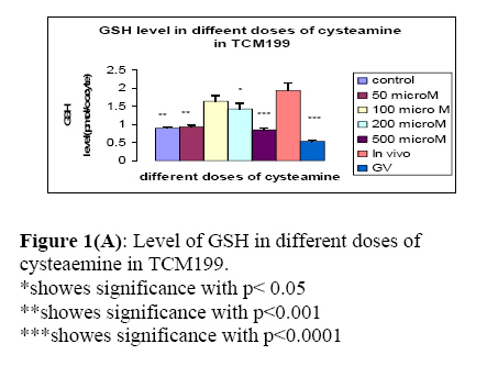
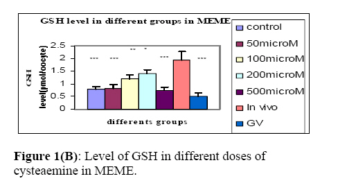
*- Cys: cysteamine GSH analysis GSH level was compared between oocytes cultured in the presence of cysteamine in two media and those matured in vivo. GSH level has been shown in different groups in two media in Figure 1a and Figure 1b. Intracytoplasmic GSH level in samples of oocytes obtained from mouse at oocyte collection time (0 h) and after IVM (24 hours cultured in TCM199) were analyzed. Also the level of glutathione in in-vivo MII oocytes was measured. Level of glutathione in In vivo oocytes was significantly higher than other groups (Figure 1a). The results indicate that the level of glutathione in MII oocyte was significantly higher in GV stage oocytes (p<0.001). Also the level of Glutathione in 100 and 200μm cysteamine were significantly higher than the control group in TCM199 and MEME respectively (p<0.05). In this experiment, for comparison of GSH synthesis in different IVM media, its level at oocyte collection time (0 h) and after IVM (24 hours cultured in MEME) were analyzed. Level of glutathione in in-vivo oocytes was significantly higher than other groups. Discussion The results of the present study revealed that cysteamine improved the rate of IVM, in vitro fertilization and blastocysts development in dose dependant manner. In this study, 100µm cysteamine in TCM199 and 200 µm in MEME had the highest rate of IVM and IVF and blastocysts development. Gasparinnini et al (2006) showed that 50 µm cysteamine increased rate of IVM and blastocysts in buffalo oocytes. In the study, administration of cysteamine increased the GSH synthesis (18). Difference species could be the possible reason between this result and our result. De Matos et al (2003) showed that 50, 100, 200 µm of cysteamine had no effects on IVM development but the rate of 2 cell embryo and blastocysts were increased (16). In our study, addition of cysteamine had dose dependant effects on IVM and IVC. It is noted, that they used different medium in their study (16). Our results showed that GSH synthesis increased in the presence of cysteamine during IVM of mouse oocytes. Moreover, our study demonstrate that culture conditions stimulate a significant increase in oocyte GSH content during meiotic progression, from GV stage through MII in oocytes. The augmentation has beneficial effect on the rate of GVBD and MII developmental competencies of mouse oocytes. In the present study, the addition of cysteamine not only improved the GSH content in MII oocytes but also had positive effect on the maturation rate of mouse Germinal vesicle oocytes. GSH is the major non-protein sulfydryl compound in mammalian cells; it serves as a reservoir for cysteine and plays an important role in protecting the cell from oxidative damage. GSH content increases during development and oocyte maturation in the ovary, as the oocyte approaches the time of ovulation, and protects it in later stages of Fertilization (19). After fertilization, GSH participates in sperm decondensation in parallel to oocyte activation, and in the transformation of the fertilizing sperm head into the male pronucleus (20). Mercaptoethanol and cysteamine reduce cystine to cysteine and promote the uptake of cysteine enhancing glutathione (GSH) synthesis. Supplementation of mercaptoethanol, cysteine and cystine to IVM medium increased the intracellular GSH content of bovine oocytes and improved embryo development and quality, producing more embryos reaching the blastocyst stage on day 6 than embryos matured in unsupplemented medium (21). In vivo level of glutathione in MII oocytes was significantly higher than that of in vitro condition in our study. in consistence with our finding, Zheng et al (1988) using hamster oocytes showed that the significant increase in GSH occurred during the transition from a nuclear or fibrillar GV to the time that chromosomes was condensed and spindle began to form (13). Luciano et al (2006) showed that in the presence of 100µm cysteamine the level of GSH was not increased in equine compared to control. Difference between our results and luciano might be because of species, different medium and contents of medium (22). TCM199 improved GVBD and MII development better than MEME. Presence of 100µm cysteamine in TCM199 and 200µm in MEME had positive effects on IVM and IVF rate. In general terms, MEME is a less complex medium than TCM199. Both contain a similar range of inorganic salt, but MEME has a higher glucose concentration, fewer non-essential amino acids and a relatively small number of vitamins. TCM199 has a lower glucose concentration, both essential and non- essential amino-acids, a large range of vitamins and several other components such as cholesterol and ribose. Amino acids are known to be beneficial for embryo development, and oocyte maturation (11). Ruth et al (et al) showed that MEME had better effects on human MII development compared to TCM199. This result was not in consistent with the present study. Difference in type of oocyte (human oocyte) and absence of cysteamine in culture medium changed the results (11). Conclusion Overall, our result indicated that cysteamin plays a positive role in improvement of oocyte and embryo development. However, further studies are required to clarify its mechanisms. References
© Copyright 2007 - Iranian Journal of Reproductive Medicine The following images related to this document are available:Photo images[rm07031f1b.jpg] [rm07031f1a.jpg] | ||||||||||||||||||||||||||||||||||||||||||||||||||||||||||||||||
| |||||||||
