 |
| About Bioline | All Journals | Testimonials | Membership | News |
|
||||||
|
||||||
Iranian Journal of Reproductive Medicine Vol. 5, No. 4, Autumn, 2007, pp. 177-181 Effects of melatonin on histopathological changes after experimental ovarian torsion-detorsion in cat Fatemeh Mostajeran1 M.D., Maryam Naderi1 M.D., Shahriar Adibi2 D.V.M. 1 Department of Obstetric and Gynecology, Isfahan University of Medical Sciences,
Isfahan, Iran. Received: 11 May 2007; accepted: 27 December 2007 Code Number: rm07034 Abstract Background: During the detorsion of
a torsioned ovary, oxidant agents are released and melatonin as an antioxidant
can reduce ischemia. We studied the histopathological changes after using
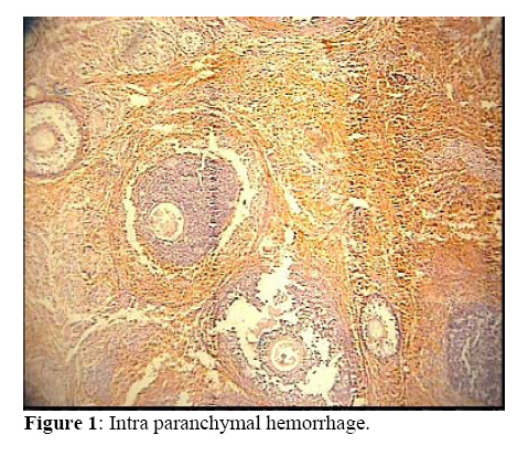
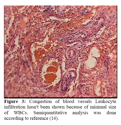
melatonin on experimental torsioned ovary in cat. Key words: Melatonin, Saline, Ischemia, Reperfusion, Cat. Introduction Ovarian torsion is a serious gynecologic problem and is most common in premenarchal girls and women of childbearing age (1-3). Because most of these women desire future fertility, the removal of an ovary could later adversely effect their reproductive life. On the other hand, when future fertility desired, rapid diagnosis is important and conservative management includes detorsion of the Involved segment (3 ,5). During the detorsion process an excess amount of molecular oxygen is supplied to the tissues and abundant amount of ROS (reactive oxygen species) are produced. Accumulation of activated neutrophils and release of Ros such as O20-, O0H, H2O2 which interact with proteins, lipids and nucleic acids resulting in loss of cell membrane integrity and structural or functional changes in proteins (6-10). Melatonin is a biologically relevant indole compound and a free radical scavenger antioxidant which protect cells against the damage induced by several oxidative agents (11-14). The protective effects of Melatonin on ovarian ischemia-reperfusion has not yet been investigated until Turkoz et al in Malataya (2004) demonstrated that Melatonin administration reduced the morphological changes by inducing Ischemia/ Reperfusion(I/R). In particular,Polymorphonuclear Neutrophilic (PMN) infiltration, edema, hemorrhage and vascular dilatation were much lower but hadn't been totally prevented (15). To determine whether treatment with Melatonin modifies ischemic indices,we examined its effects on an invivo model of adnexial Ischemia/ Reperfusion injury in cats . The aim of the present study was to investigate the effects of Melatonin on histopathological change in I/R injury in cat ovaries. Materials and methods To determine whether ischemia followed by subsequent reperfusion induces ovarian damage and the effects of Melatonin on the injured ovaries, we created a model of adnexal I/R using cats. The permission for the animal tests and experiments was given by the center of animal researches of professor Torabynegad Institution of Isfahan University. All surgical procedures were performed while the cats were under general anesthesia. In total twenty cats with almost similar weights were subjected to right unilateral 360° clockwise adnexial torsion by a ring forceps which induced ischemia by occlusion of the tuboovarian vesseles for 3 hr (15-17). Melatonin (10mg/kg) that is a white lipophil powder was dissolved in 1% ethanol (total volume =10 cc) just before use and injected intra peritoneally 30 min before reperfusion in the Melatonin group while saline was administered (10cc) in the saline group. Reperfusion was achieved by releasing the occlusion and restoring the circulation for 3 hr. After 3 hr, cats in both groups were reanesthesized, laparatomy was performed and right ovaries were surgically removed. The ovaries were preserved in formalin for histopathological examination. Histopathological examination At the end of each experiment, the ovaries were removed and fixed in 10% neutral buffered formalin solution and then were embedded in paraffin as usual. Serial sections were cut using the microtome at a thickness of 4 μm and were stained with hematoxylin and eosin.The histologic sections were examined for the presence of interstitial edema, vascular congestion, hemorrhage and PMN infiltration, using a microscope. The slides were coded and semiquantitative analysis of the sections was performed without knowledge of the treatment protocol. The changes seen were graded as follows (16): Grade I: Mild edema / Mild
vascular congestion/No hemorrhage /No PMN. Statistical analysis The Statistical package for social sciences (SPSS) version 10 was used for statistical analysis. Individual group parameters were assessed with Manwhitney test. The spearman's test determined the correlation between ischemic indices. Results Macroscopically, torsioned ovaries had a cherry–red color. Microscopic examination of ovaries in both groups showed: interstitial edema, vascular congestion, hemorrhage and acute infiltration by PMNs. Table I. The distribution of histoligical grades of the ovaries in 2 groups of cats.
(There was no grade 0 or normal) The Manwhitney test demonstrated edema and vascular congestion in saline group were more severe than Melatonin group (p–value=0.009), also hermorrhage and leukocyte infiltration were more obvious in saline group (p–value=0.018). The histological changes, graded as described in the methods, are summarized in Table I. Thehistological grades of the ovaries in melatonin. Group were lower than those of the saline group (p-value=0.015). Further more, there was a correlation between edema and vascular congestion (r =1, p-value =0.001), hemorrhage and PMN infiltration (r=1, p-value=0.001) and the other ischemia indices, for example vascular congestion and hemorrhage (r =0.857, p-value< 0.001). Discussion Ovarian injury resulting from torsion and detorsion resembles the phenomenon of Ischemia – Reperfusion injuries in other organs (1-3). It has been demonstrated that oxygen free radical generation is a critical mechanism causing injury in post ischemic cells and tissues , therefore oxygen , despite its vital properties, can be the most toxic elements known to human (6,7,18,19). Detorsion of the ovary, a conservative management that save it for future fertility ,is one of the most important factors in future injury (1,3) Lipid peroxidation by active oxygen radicals can alter both membrane structure and function thus reperfusion injury may increase vascular permeability. Neutrophil infiltration might be regarded as another source of free radicals in the ischemic tissue, because they stimulate inflammatory mediators such as Tumor Necrozing Factor (TNF) and Interluekin-1(IL1)which are involved in the pathogenesis of I/R injury. Edema and increased permeability occurred in vivo after ischemia reperfusion, due to disruption of endothelial cell junction integrity (20, 21). In the present study, Melatonin significantly prevented hemorrhage and greatly reduced the number of PMN and degree of vascular congestion and edema. These effects of Melatonin may occur not only because of its anti oxidant and Ros scavenger properties, but also its stimulatory effect on endogenous anti oxidant enzyme such as: catalase and SOD (super oxide dismutase) which bind to cell membrane receptors and decrease Ca and CAMP concentrations. Thus it is superior to other scavengers (22-26). The results of the histopathological parameters in our study indicate that administration of Melatonin has beneficial effects in the prevention of reperfusion injury of ovary. Hara et al (1996) demonstrated that melatonin given in advance of severe exercise prevented the reduction in muscle GSH (an antioxidant enzyme in normal tissues) (27). Another study reported that melatonin preventes the paraquat induced reduction in GSH in both lung and liver (28).Although melatonin scavenging actions have been demonstrated in a number of tissues (29-33), only Turkoz et al demonstrated the effects of melatonin on reducing ischemic indices such as xanthine oxidase (p-value <0.001), MDA (Malondialdehyde) (p-value <0.001) , acute PMN infiltration, edema and Vascular dilatation in ovaries . In this study, Melatonin administration reduced the morphological changes by induced I/R; in particular, PMN infiltration, edema and vascular dilatation were much lower but had not been totally prevented by i.p Melatonin administration (15). In addition melatonin has an ameliorating effect on oxidative stress-induced renal tubular damage in diabetic nephropathy via its antioxidant properties (34). High dose of melatonin (50mg/kg), physiologically, biochemically and morphologically can be useful to normalize contractility injured by oxidative stress in intestinal ischemia/reperfusion (35). Conclusion We demonstrated that Melatonin reduced tissue damage induced by I/R in cat ovaries. This means that administration of Melatonin could be helpful in protection of ovaries from torsion – detorsion induced damage in humans. References
© Copyright 2007 - Iranian Journal of Reproductive Medicine The following images related to this document are available:Photo images[rm07034f2.jpg] [rm07034f3.jpg] [rm07034f1.jpg] | |||||||||||||||||||||||||||||
| |||||||||
