 |
| About Bioline | All Journals | Testimonials | Membership | News |
|
||||||
|
||||||
Iranian Journal of Reproductive Medicine Vol. 6, No. 2, Spring, 2008, pp. 77-82 Pre and post natal exposure of 50 Hz electromagnetic fields on prostate glands of rats: an electron microscopy study Amir Afshin Khaki1 Ph.D., Arash Khaki2 D.V.M., Ph.D., Shahram Garachurlou2 Ph.D., Fereshteh Khorshidi3M.D. , Nazila Tajadini1M.Sc., Navid Madinei3M.D. 1Department of Anatomical Sciences, Tabriz University
of Medical Sciences and National Public Health Management Center (NPMC),
Tabriz, Iran. Received: 8 January 2008; accepted: 20 June 2008 Code Number: rm08013 Abstract Background: Men are unavoidably exposed to ambient electromagnetic
fields (EMF) generated from various electrical gadgets and from power
transmission lines. Prostate gland plays an important role in secretion of
semen as largest accessory gland of male reproductive system. It seems that
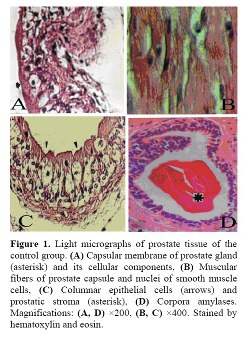
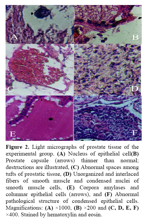
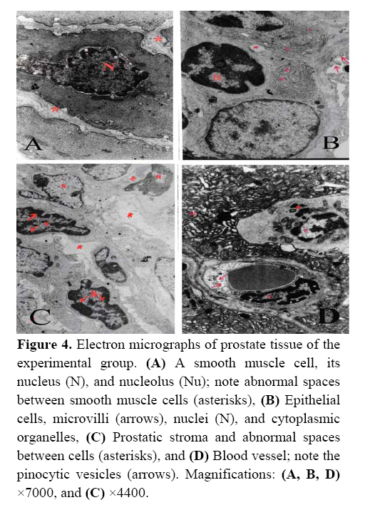
protection of this gland against EMF is important in spermatogenesis process. Key words: Electromagnetic fields, Prostate, Rats. Introduction With the increased use of power transmission lines and modern electrical gadgets, concern about public health due to chronic exposure to electromagnetic fields (EMF) has come into a sharper focus (1). Many people are always exposed to EMF as results of working with various electrical gadgets. EMF are classified according to their frequency and long wave. EMF Frequency differs due to the sources. Among these sources we can name: printers, vacuum cleaners, cellular phones, television sets, hair dryers,electric razors, microwaves and etc (1). As the use of computer technology grows all over the world, we must anticipate more reports on disorders of normal life due to exposure to EMF (2). Numerous studies have been done about relationship of EMF with occurrence of somatic disorders like: sterility, spontaneous abortion, prematurity, mental retirement, congenital malformation, and genetic diseases. Many studies have been done about effect of EMF on abortion in onset of pregnancy (3). Previous studies showed that there was a penetrable barrier around seminifer tubules in testis of mammals (4). Studies with electron microscope revealed that there was a tight connection between sertoli cells which makes a tight barrier against penetration of outside materials to these tubules (4). Biochemistries and physiological studies confirmed importance of the role of sertoli cells as regulators in spermatogenesis (4). Some of these investigations have revealed adverse effects of EMF in fertility in men (5). An investigation showed premature-aging in genital organ of rats, as a result of devastating effect of EMF on spermatogenic cells and decrease of testosterone secretary cells (6). Flow cytometry method studied the effect of 50 Hz EMF on spermatogenesis process and showed devastating and adverse effects of these fields in growth differentiation of spermatogenic cells (7). Our EM and LM studies previously confirmed adverse effects of EMF on sertoli cells in testis of rats (destruction of nucleus and cells organelle and cells connection were clearly observed) (8). Given the importance of prostate in male reproductive process, in this study we examined the probable effects of EMF on prostate in rats. Based on our review of literature, there was no investigation about effects of non–ionizing radiation on prostate gland by electron microscopy (TEM). The aim of this study was to demonstrate the exact effects of nonionizing radiation on prostate gland as the largest accessory gland of male reproductive system which plays an important role in secretion of semen. Previous studies showed importance of semen secretion in maintenance, nutrition and support of spermatozoids. Materials and methods Animals A total of 50 male and 50 female Wister rats (about 15 week-old) procured from animal house were used for the study. Rats of the same sex were housed together (five per cage) and kept in quarantine for a week to rule out any disease. Rats were fed on compact food in the form of granules and water. This food was consisted of all essential ingredients including vitamins and minerals. Environmental conditions (temperature and humidity) in all animal holding areas were continuously monitored. Temperature was maintained in the range of 20oC–30oC and relative humidity was monitored at 35-60%. Fluorescent light was provided on a 12 hr light/dark cycle and put on from 8 a.m. till 8 p.m. Lights were located at distance of three meters from the cages so that these did not interfere with the EMF of the experimental design. EMF exposure The equipment was based on Helmholtz coil which works following Fleming's right hand rule. It produced an alternate current of 50 Hz frequency creating an EMF of 80 G. The intensity of EMF could be controlled by a transformer. The equipment had two main parts. In the first, there were two copper coils placed one above the other separated by a distance of 50 cm. Between the coils (the exposure area) there was a cylindrical wooden vessel, the interior of which had a chamber for keeping the cages of experimental animals. The second part was the transformer which checked the input and output voltage with a voltmeter and current with an ampere meter. The equipment was calibrated by gausmeter. To prevent increase of temperature inside the chamber, a fan was fitted at the top and the temperature was checked. At a time two cages could be put within the chamber with 7-8 rats per cage. The rats were selected at random into breeding pairs. Each of the 50 breeding pairs habituated in separate cages in order to allow monogamous mating. Females were observed for the sign of pregnancy i.e., vaginal plugging on the next day. Of the 50 breeding female rats, 30 rats at random were selected for exposure to EMF as experimental group and 20 as control group (unexposed). The male rats used for breeding were subsequently returned to the animal house. One hundred and seventy-two pups which received EMF exposure in utero (i.u.) were delivered by the experimental female rats (gestation period approximately 3 weeks). In the control group, 83 pups were born from 10 control female rats. From both experimental and control groups 30 and 20 pups each were randomly chosen. The experimental pups were exposed to EMF till five weeks of postnatal (p.n.) age. They were exposed 8 hours per day for 5 weeks (means: 56 days postnatal period). All rats were free in the cage during exposure time. The temperature was the same for both control and exposed rates. At the end of this period, 15 pups from each group (termed experimental group and control group) were sacrificed. At the termination of the stipulated exposure period as per experimental design, the rats were anaesthetized with chloroform and 10% formalin was injected through the inferior vena cava. The prostate were removed and fixed in formalin for light microscopy. Haematoxylin and eosin were used to stain the histological sections (6 mm thick). EM Study For transmission electron microscopy, the tissue samples were cut into pieces (2×2 mm) and fixed in 2.5% glutaraldehyde and 1% paraformaldehyde in 0.1 M phosphate buffer (pH 7.4) for 6-8 hr at 4oC. They were washed and postfixed in 2% OsO4 for 1 hr at 4oC. The tissue was dehydrated through ascending grades of ethanol and embedded in araldite CY212. Semithin sections (1 µm) were cut and stained with toluidine blue. Ultrathin sections (60-70 nm) were cut, mounted onto copper grids and stained with uranyl acetate and alkaline lead citrate. They were observed under a Philips CM10 transmission electron microscope. We used t-test method for statistical analysis in this study. Results Light Microscope (LM) Control group: Prostate gland has made up of 30 to 50 tubular alveolar glands, with C-shaped capsule around the gland (Figure 1-A), made of fibro-elastic connective tissue, with fibers of smooth muscle (Figure 1-B). Prostate gland epithelial cells seemed cubical (inactive) or pseudo stratified (active) (Figure 1-C), with euchromatic nucleus. Blood vessels have cubical endothelial cells with clear nuclei (Figure 1-A). Increase of corpus amylace that was a measure of prostate oldness, was called prostatic salts (Figure 1-D). Experimental group: Prostate capsule in experimental groups was thinner than control, in some parts, it had been torn apart (Figure 2-B). Empty spaces were seen between tubuloalveolar glands (Figure 2-B, C). Smooth muscle fibers were irregularly speared out in different directions (Figure 2-D). Nucleus of these cells was dense and heterochromatic (Figure 2-D). Epithelial cells were cubical with heterochromatic nuclei (Figure 2-A, B). Some of epithelial cells with dense nuclei had clustered together and made vivid pathologic structures in prostate tissue (Figure 2-F). Endothelial cells of blood vessels showed dense nuclei and abundant RBC demonstrated inside the vessel (Figure 2-C). Corpus amylace were observed in prostate of experimental group more than control group (Figure 2-E). Electron Microscope (EM) Control group: There was a fibro muscular wall around secretary cells of prostate gland, with oval shaped smooth muscle cells (Figure 3-A). Their nuclei seemed euchromatic and located centrally (N). There were many ribosomes, mitochondria (M) and myofilament in cytoplasm of smooth muscle cells membrane (Arrow) (Figure 3-A). Columnar secretary epithelial cells with round nuclei were seen (Figure B). REr, mitochondria (M), ribosomes and Golgi apparatus were demonstrated in their cytoplasm. Round secretary granules were abundant and occupied in the apical part of the cells. Many microvilli (M) were demonstrated in apex of cells. Basal cells of prostate gland with columnar nucleus occupied a large part of cytoplasm. Nucleus membrane seemed indented. Abundant round mitochondria in cytoplasm were seen (Figure 3-C). Endothelial cells of blood vessels had oval shaped nuclei with clusters of ribosomes in cytoplasm. Mitochondria and Golgi apparatus were seen clearly in the cytoplasm with many pinocytotic vesicles (Figure 3-D). Experimental group: Nuclei of smooth muscle cells seemed heterochromatic, and nucleus membrane were indented. Many empty spaces were demonstrated between the smooth muscle cells. Vacuolization were detected in the cytoplasm of these cells and between them too. Myofilaments were separated from each other. Mitochondria were dilated and nonenergised (Figure 4-A). Secretary epithelial cells were cuboids and inactive (Figure 4-B). Number of microvillis was decreased significantly (Arrow). Nuclei (N) of these cells seemed heterochromatic, and mitochondria (M) were dilated. Secretary granules in apical part of cells were decreased clearly. They were in charge of secretary activities of epithelial cells (Figure 4-B). Empty spaces (*) were abundant between the stromal cells and nuclei (N) were heterochromatic. Nuclei membrane showed a lot of breaking (Figure 4-C). Oval nuclei of endothelial cells of blood vessels (N) were heterochromatic and vacuolization occurred clearly in cytoplasm of these cells. Mitochondria were dilated and pinocytotic vesicles were more than control group (*). Empty spaces were abundant between them (Arrow) (Figure 4-D). Discussion The potential of EMF adversely affecting the health of the human population is an issue which continues to receive a great deal of attention in both public and scientific forums. Earlier, the harmful effect of ionizing radiations like X-rays, gamma rays etc. have been demonstrated on gonadal tissue. We revealed that the fibro muscular capsule of prostate has become thinner than control and this was possibly caused by tear of collagen and reticular fibers of prostate capsule. This observation was similar to one that examined EMF effects on seminifer tubules of testis of rats that had caused thinness and tear of some parts (8, 9). Fibro muscular Capsule was thinner than control group and was probably responsible for not expelling the lymph and edema in prostate that leads to hyperemia and increase in the weight of prostate in experimental group rats. These results confirmed conclusion of other researchers that had announced; exposure to EMF cause increase of prostate weight and other glands of male genital organ (10-12). EM findings were corresponding to the LM results too. The number of pinocytotic vesicles increased which were in charge of transport of substances between the inside and outside of endothelial cells of blood vessels and it may be responsible for edema in prostate gland. Evaluation and comparing the blood vessels in control and experimental group rat showed that in experimental group squamous endothelial cells demonstrated dense nuclei. Hyperemia was seen in the vessels, too. Vivid spaces among tissues and epithelial cells of prostate were observed. Spaces were big with blister-like appearance. These results were demonstrated previously under exposure of ionizing radiation (13). This study demonstrated that nonionizing radiation had same effects. Blister like appearance showed abundant due to many empty spaces between epithelial cells of this gland. These results were similar to condition of seminifer tubules in testis of aged rats reported by other researchers (14, 15). Therefore it can conclude that non-ionizing radiations were able to cause premature-aging in prostate gland too. Increases in number of corpus amylace were seen more in experimental group rats. It was another important signs of aging in prostate of experimental group rats. These results corresponded with another study about effect of EMF on sertoli cells in rat (6). Destructive effects of EMF on smooth muscle cells of prostate gland were seen clearly. Nuclei of smooth muscle cells in experimental group were dense and heterochromatic and had not consequence like control group. Muscle fibers were irregularly spread out in different directions. Considering the important role of these fibers in discharging the ejaculation of prostate to prostatic urethra, the experimental group rats can not efficiently do this. Probably it cause decrease of semen fluid and it can have adverse effect on spermatogenesis process and nutrition and spermatozoid storage. EM findings were confirmed the LM results too. It showed that myofilaments were irregular and separated and diverse from each other in experimental smooth muscle cells. In experimental group rats, smooth muscle could not do their role perfectly and probably secretion of semen decreased. It showed harmful effect on spermatogenesis, and affected its process, probably. Epithelial cells in experimental group seemed cuboidal and inactive, also nucleus of these cells were heterochromatic; probably it showed cellular damage and cell death. Similar to these effects previously have been observed in the study of the non-ionizing EMF on sertoli cells in testes of rats (6). The clusters of epithelial cells with dense nucleus, abnormally gathered together, made pathological structures in prostate tissue that was probably due to cell debris of epithelial cells. It can be a measure for cell death that leads to decrease in activity of the biggest gland of male genital system and may lead to sterility in male rats. EM studies confirmed LM findings, about epithelial cells too. Secretory epithelial cells were cubic and inactive with heterochromatic nuclei and dilated mitochondria. Number of microvilli and secretory granules decreased clearly. Probably it showed that these cells were going toward cell damage and cell death. Prostate couldn’t play its secretory role properly and it could make a defect in spermatogenesis process. This defect may lead to infertility in male rats. This study confirmed the idea of other studies about unfavorable effects of EMF on sertoli cells in seminifer tubules (6, 16). EM studies showed vacuolization in this gland which was an important sign of cell damage. Stromal cells damages were important too, because of their role in changing the epithelial cells. Mitochondria were dilated and could not do cell oxidation. It can conclude that non-ionizing radiations made cell damage in prostate of rat, which may lead to sub fertility. EMF produces biological stress and free radicals which can make the susceptible animal population prone to congenital malformations, tissue and cell damage or death (17). Long term exposure to EMF may be linked to even higher levels of oxidative stress with corresponding changes mentioned above (18, 19). This study concluded similar destructive effects of ionizing and non-ionizing radiations on morphology of prostate gland of rate. Given the direct and undeniable role of prostate and vesicle seminal in spermatogenesis process, and effect of non-ionizing EMF on this gland in rat, it can be suggested that EMF are able of interrupt the normal process of spermatogenesis and probably cause sterility in men. Acknowledgment This research project was done under financial support and supervision of Tabriz University of Medical Sciences, Tabriz, Iran. Reference
© Copyright 2008 - Iranian Journal of Reproductive Medicine The following images related to this document are available:Photo images[rm08013f4.jpg] [rm08013f2.jpg] [rm08013f1.jpg] [rm08013f3.jpg] |
| |||||||||
