 |
| About Bioline | All Journals | Testimonials | Membership | News |
|
||||||
|
||||||
Iranian Journal of Reproductive Medicine Vol. 6, No. 3, Spring, Summer, pp. 149-155 Effect of Cnidoscolous aconitifolius (Miller) I.M. Johnston leaf extract on reproductive hormones of female rats Musa Toyin Yakubu1 Ph.D., Musbau Adewumi Akanji1 Ph.D., Adenike Temidayo Oladiji1 Ph.D., AbdulWaheed Olajide Olatinwo2 M.B.B.S., Abdulfatai Ayoade Adesokan1 Ph.D., Monsurat Oyenike Yakubu3 RN, RM, Bamidele Victor Owoyele4 Ph.D., Taofik Olatunde Sunmonu1 Ph.D., Moyosore Salihu Ajao5 M.Sc. 1Department of Biochemistry, Faculty of Sciences, University of Ilorin, Ilorin, Nigeria. Received: 16 March 2008; accepted: 4 September 2008 Code Number: rm08024 Abstract Background:
The increase in the rate of infertility in women has prompted the need to
search for plants with antifertility potentials. Key words: Cnidoscolous aconitifolius, Prolactin, Follicle stimulating hormone, Luteinizing hormone, Estradiol, Progesterone, Fertility, Conception, Contraceptive. Introduction Plant-derived chemicals that influence endocrine activities in both humans and animals have received a great deal ofattention due to their possible beneficial as well as adverse effects (1). Some of these plants are known to possess anti-fertility effect through their action on hypothlamo-pituitary-gonadal axis or direct hormonal effects on reproductive organs resulting in inhibition of ovarian steroidogenesis (2). Examples include Momordica cymbalaria, Goniothalamus spp which are used traditionally to control fertility as well as abortifacient during early months of pregnancy (3). Cnidoscolous aconitifolius(Miller) I.M. Johnston,(Family – Euphorbiacae) known as tree spinach (English), efo iyana ipaja, or efo Jerusalem (Yoruba) is commonly found growing in western part of Nigeria. It is an ornamental, evergreen, drought deciduous shrub of 3 to 5 m tall (4). The large (32 cm long and 30 cm wide) palmate lobed leaves are alternately arranged. The leaves are commonly eaten as vegetable (5). The shoots and leaves of C. aconitifolius are used as a laxative, diuretic, circulation and lactation stimulants. It has also been recommended for diabetes, obesity, acne, kidney stones and eye problems (6). Most of the studies on the plant have been on the nutritive values of the leaf meal in broiler chicken and antimicrobial activity of the essential oil against Escherichia coli and Salmonella typhi (7, 8). According to our ethnomedicinal survey, the plant is used in managing myriad of ailments and the leaves are widely consumed within many localities in Nigeria. This is however, without information on its effect on reproductive hormones of the females. This study was therefore designed to provide information on the effect of aqueous extract of C. aconitifolius leaves on female reproductive hormones. Several studies (9-11) have shown that chemical compounds including plant extracts could alter the concentrations and functions of female reproductive hormones. Materials and methods Plant material and authentication The plant samples were collected from a single population within the premises of the main campus of University of Ilorin, Ilorin, Nigeria, and were authenticated at the Forestry Research Institute of Nigeria (FRIN), Ibadan, Nigeria. A voucher specimen (FHI 107768) was deposited at the FRIN herbarium. Experimental animals Female albino rats (Rattus norvegicus) of Wistar strain weighing 152.58±3.52 g were obtained from the Animal Holding Unit of the Department of Biochemistry, Faculty of Science, University of Ilorin, Ilorin, Nigeria. Assay kits The assay kits for prolactin, progesterone, estradiol, follicle stimulating and luteinizing hormones were supplied by Diagnostic Automation Inc., Calabasa, CA, USA. All other reagents used were of analytical grade and were prepared in volumetric flask using glass-distilled water. Phytochemical screening Chemical tests were carried out on the C. aconitifolius leaf extract as described for alkaloids, tannins, phlobatannins and anthraquinones (12), phenolics, flavonoids, cardenolides and dienolides (13), glycosides, saponins, steroids and triterpenes (14). Quantitative analyses of the detected phytochemicals were carried out for phenolics (15), flavonoids (16), alkaloids (17), tannins (18), saponins (19), anthraquinones, triterpenes and phlobatannins (20). Preparation of extract The procedure described by Yakubu et al (21) was employed. Briefly, 194.10 g of the wet leaves of C. aconitifolius were oven dried at 40oC for 48 h until a constant weight of 37.58 g was obtained. The dried leaves were pulverized using Blender/Miller III, (model MS - 223, China) and the resulting powder was stocked in a plastic container. Twenty grams of the powder was extracted in 1 L of cold distilled water for 48 h at room temperature with constant shaking on a shaker (Stuart Scientific Orbital Shaker, UK). The extract was filtered with Whatman No. 1 filter paper and the resulting filtrate was concentrated on steam bath to give 4.88 g of the residue (brownish-black slurry) which is equivalent to a yield of 24.40 ±1.88%. Calculated amount of the residue (2.42, 4.84 and 9.68 g) was separately reconstituted in 100 ml of distilled water to give the required doses of 250, 500 and 1000 mg kg-1 body weight respectively. Animal grouping and extract administration A total of 60 female rats of proven maturity were housed in clean metabolic cages of dimensions 33.0×20.5×19.0 cm contained in well-ventilated standard housing conditions (temperature: 28–31oC; photoperiod: 12 h natural light and 12 h dark; humidity: 50–55%). The cleaning of the cages was done daily. The animals were allowed free access to rat pellets (Bendel Feeds and Flour Mills Ltd., Ewu, Nigeria) and tap water. They were acclimatized for two weeks before the commencement of the experiment. The rats were completely randomized into four groups of 15 each as follows: Group A: Control, received 6.5 ml kg-1
body weight of distilled water (vehicle). The various groups were orally administered with 1 ml each of distilled water and the extract (250, 500 and 1000 mg kg-1 body weight) once daily (09:00–09:45 h) using plastic syringes attached to metal oropharyngeal cannula. Five rats from each group were sacrificed 24 h after 1, 3 and 7 days of their respective daily doses. This study was carried out following approval from the Departmental Ethical Committee on the Care and Use of Experimental Animals for Research. Preparation of serum The procedure described by Yakubu et al (21) was employed. Briefly, under ether anaesthesia, the veins after being slightly displaced (to prevent blood contamination by interstitial fluid) were cut with a sterile scapel blade and 5 ml of the blood was collected into clean and dry centrifuge tubes. The blood was then left for 10 min to clot at room temperature. The tubes were thereafter centrifuged at 33.5 g x 15 min using Uniscope Laboratory Centrifuge (Model SM800B, Surgifriend Medicals and Essex, England). The sera were later aspirated with Pasteur pipettes into clean, dry, sample bottles and were then used within 12 h of preparation for the hormonal assay. Hormonal assay The procedure described in the hormone assay kits was used according to the principle highlighted by Tietz (22) for prolactin, estradiol and progesterone while that of Uotila et al (23) was used for luteinizing and follicle stimulating hormones. Statistical analysis Results were expressed as the mean of five replicates±SD except for the phytochemical screening. Means were analyzed using a one-way ANOVA and values at p<0.05 were considered statistically significant (24). In all the Figures, bars carrying letters different from the control for each day are significantly different (p<0.05). Results Phytochemical screening of the extract revealed the presence of alkaloids, saponins, phenolics, tannins, flavonoids, anthraquinones, phlobatannins and triterpenes with alkaloids constituting the highest while triterpenes was the lowest. Others such as glycosides, steroids, cardenolides and dienolides were not detected in the extract (Table I). Table I. Phytochemical constituents of aqueous extract of Cnidoscolous aconitifolusleaves.
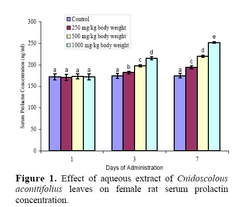
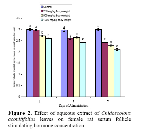
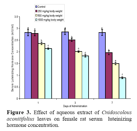
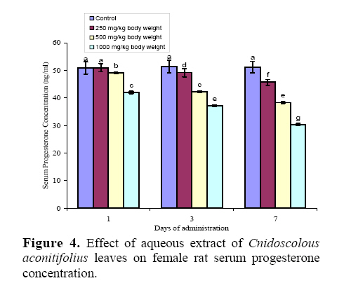
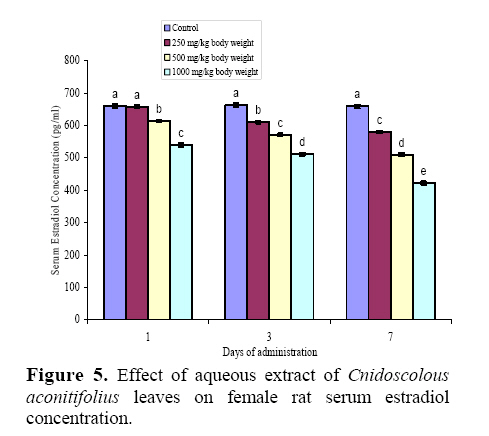
n = 3 ± SD The effects of administration of aqueous extract of C. aconitifolius leaves at 250, 500 and 1000 mg kg-1 body weight for 7 days on the concentration of serum reproductive hormones in the female rats are depicted in figure 1, 2, 3, 4, and 5. Compared with the control, administration of the extract produced increase (p<0.05) in serum prolactin concentration only after the third dose. The single dose administration did not produce any significant effect on the serum hormone (figure 1). The concentrations of follicle stimulating hormone (FSH), luteinizing hormone (LH), progesterone and estradiol in the serum were reduced by the extract (figure 2, 3, 4, and 5). While the concentrations of FSH, LH and progesterone were reduced on all the days, the least dose (250 mg kg-1 body weight) on the first day did not produce any significant change (p>0.05) in the concentration of the hormones (figure 2, 3, and 4). However, by the end of the treatment period, the concentrations of FSH, LH and progesterone had reduced by 23, 64 and 41% respectively of the control value in the highest dose group. Except for the 250 mg kg-1 body weight of the extract on the first day, all the other doses produced reduction in the serum estradiol concentration. By the end of the experimental period, the extract at 250, 500 and 1000 mg kg-1 body weight had reduced the serum hormone by 12, 23 and 36% respectively of the control value (figure 5). Discussion Maturation of pre-ovulatory follicles and ovulation are under the combined and balanced influences of ovarian and extra ovarian hormones. Imbalances or alterations in these hormones lead to irregularity in the ovarian functions and duration of estrous cycle (25). These hormonal imbalances may be caused by numerous chemical agents contained in plant extracts. Phytochemical screening has revealed many bioactive as well as toxic agents of plant extracts that can affect the regulation of oestrous cycle, conception and reproduction (11, 21). Alkaloids and flavonoids have been shown to reduce plasma concentrations of LH, estradiol and FSH (26-28). Therefore, the presence of these phytochemicals may account for the alterations in the levels of the circulating hormone observed in this study. Prolactin helps to initiate breast development by inducing lobuloalveolar growth of the mammary gland. It also stimulates lactogenesis. Dopamine serves as the major-inhibiting factor or break on prolactin secretion (29). The enhanced level of prolactin observed in this study may be attributed to the effect of the extract probably acting as a dopamine antagonist. High prolactin levels tend to suppress the ovulatory cycle by inhibiting the secretion of both follicle-stimulating and gonadotropic-releasing hormones (GnRH) (29), which are necessary for ovulation. Such increase in prolactin may inhibit ovulation and promote the loss of menstrual periods which will hinder conception. The elevated level of prolactin in this study justifies the folkloric use of the plant in stimulating lactation. Follicle stimulating hormone is the central hormone of mammalian reproduction, essential for gonadal development and maturation at puberty as well as gamete production during the fertile phase of life (30). It stimulates the growth and maturation of ovarian follicles by acting directly on the receptors located on the granulosa cells. The reduction in the levels of FSH by the extract may hamper folliculogenesis and delay maturation of the follicle in the pre-ovulatory phase (31). It is possible that the extract might have exerted its effect on the anterior pituitary or the hypothalamus since the secretion of FSH is regulated by the gonadotropic releasing hormone secreted by the hypothalamus. The reduction in the levels of the hormone may adversely affect conception in the female animals. This study agreed with the work of Benie et al (11) where administration of Afrormosia laxiflora, Pterocarpus erinaceus and Cola nitida stem bark decreased the release of the gonadotropins (LH and FSH). Luteinizing hormone stimulates secretion of sex steroids from the gonads. In females, ovulation of mature follicles in the ovary is induced by a large surge of LH secretion during the pre-ovulatory periods. Several authors have demonstrated that LH release surges at the proestrous stage are responsible for ovulation (32, 33). Any substance capable of inhibiting this release could provoke disruption of ovulation by decreasing the number of mature follicles or induce an oestrous cycle disruption at rest (11). Therefore, the reduction in the serum LH levels may be explained by an inhibitory effect of the extract on the release of LH which may trigger disruption of ovulation. This may result in impairment of oestrous cycle; hamper conception and normal reproduction in the females. Several studies have demonstrated that flowers of Malvaviscus conzatii as well as Cynomorium coccineum and Withania somnifera can hinder gonadotropin release and induce similar effect on the oestrous cycle (34, 35). Our findings agreed with that of Jarry et al (36) where triterpenenoid glycoside in methanolic and lipophilic extracts of Cimicifua racemosa (Black cohosch – English) was responsible for the reduction in LH concentration. In addition, Benie et al (37) also reported that the extract of Combretodendron macrocarpum blocked the rat oestrous cycle in luteal stage and also decreased plasma levels of LH and FSH. Therefore, it is possible that C. aconitifolius contains anti-gonadotropic substance(s) which may affect the oestrous cycle and hamper reproduction in females. Progesterone which is produced in the ovaries, placenta, and adrenal glands, helps to regulate the monthly menstrual cycle, prepare the body for conception and pregnancy (38) as well as stimulate sexual desire. The hormone also encourages the growth of milk-producing glands in the breast during pregnancy. High progesterone levels are believed to be partly responsible for symptoms of premenstrual syndrome (PMS), such as breast tenderness, feelings of bloat and mood swings. The feedback inhibition of GnRH secretion by estrogens and progesterone provides the basis for the most widely-used form of contraception. Such feedback inhibition of GnRH prevents the mid-cycle surge of LH and ovulation. The reduction in the levels of serum progesterone by C. aconitifolius leaf extract may have consequential effect on conception in females; impede ovulation which may result in annovulation and sequelae. Studies have shown that water based extract of combined Lepidagathis longifolia and Phyllagathus rotundifolia reduced progesterone and estradiol concentration in pseudopregnant and non-pregnant rats (39, 40). Alkaloids have equally been reported to inhibit the synthesis of cellular progesterone (41). Therefore, the reduced level of progesterone by C. aconitifolius may not be unconnected with the alkaloidal component of the extract. Estradiol stimulates the growth of the uterine lining, causing it to thicken during the pre-ovulatory phase of the cycle. It is well established that estradiol is directly responsible for the growth and development of reproductive organs. In synergy with FSH, estradiol stimulates granulosa cell proliferation during follicular development (9). Plants with estrogenic property can directly influence pituitary action by peripheral modulation of LH and FSH, decreasing secretion of these hormones and blocking ovulation (42). Thus, the reduction in the serum concentration of estradiol observed in this study may be attributed to a decreased aromatase activity or substratesupplementation during estrogen synthesis (43). Consequently, such decrease in estradiol levels may hamper ovulation, preparation of the reproductive tract for zygote implantation, and the subsequent maintenance of the pregnancy state (44). Our findings contrast that of Ota et al (45) which demonstrated that herbal Shakuyaku (Paeoniae radix), Keihi (Cinnamomi cortex) and Botanpi (Moutan cortex) stimulated the aromatase activity in human granulose cells and increased estradiol secretion in vitro. Kadohama et al (46) reported that several plant alkaloids inhibit aromatase activity, thus altering the potential for steroid production and reproductive performance. Therefore, the alkaloid in the extract may be responsible for the reduced level of estradiol probably by inhibiting aromatase activity. Thus, it is possible that the aqueous extract of Cnidoscolous aconitifoliuscontain biologically active phytochemicals which may be endocrine-disrupting. Such substances in the plant extract may induce hormonal imbalance or disorders such as infertility and contraception in hormone-dependent organs like the ovary and mammary glands. Our findings in this study have important implications for female contraceptive development. Plant products as contraceptive will be more acceptable for economic reasons and side effects that are less than chemical agents. References
© Copyright 2008 - Iranian Journal of Reproductive Medicine The following images related to this document are available:Photo images[rm08024f5.jpg] [rm08024f4.jpg] [rm08024f3.jpg] [rm08024f2.jpg] [rm08024f1.jpg] |
| |||||||||
