 |
| About Bioline | All Journals | Testimonials | Membership | News |
|
||||||
|
||||||
Iranian Journal of Reproductive Medicine Vol. 7, No. 2, Spring, 2009, pp. 73-77 Can Zeta sperm selection method, recover sperm with higher DNA integrity compare to density gradient centrifugation? Noush Afarin Khajavi1M.Sc., Shahnaz Razavi1Ph.D., Mohammad Mardani1Ph.D., Marziyeh Tavalaee2M.Sc., Mohammad Reza Deemeh3M.Sc., Mohammad Hossein Nasr-Esfahani2, 3Ph.D. 1Department of Anatomy,
Isfahan University of Medical Sciences, Isfahan, Iran. Received: 23 September 2008; accepted: 12 May 2009 Code Number: rm09013 Abstract Background: Sperm selection for ICSI based on morphology and
motility might not be relevant to chromatin integrity. Thus sperm selection
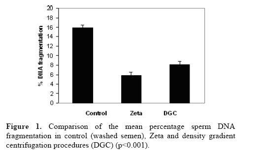
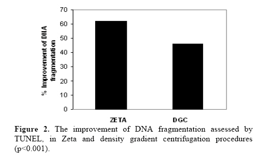
based on sperm characteristics has been suggested. Key words: Zeta method, Density Gradient Centrifugation method, DNA fragmentation. Introduction Male factors account for approximately 50% of infertility. Intra cytoplasmic sperm insemination (ICSI) has become the first choice for treatment of these patients. Although ICSI is routinely carried out for these patients, nevertheless, there is still much concern regarding insemination of spermatozoon with chromatin anomalies (1, 2). Several studies have shown that, the quality of sperm chromatin plays an important role in fertilization, embryo development, post genomic activation and pregnancy rate. Thus, selection of mature sperm with intact DNA is an important step for ICSI procedure. An ideal sperm preparation technique should: 1) recover high number of sperms with normal morphology, 2) recover sperms with intact chromatin, 3) minimize the risk of reactive oxygen species (ROS) generation (excessive ROS could adversely affect DNA integrity and sperm function) and 4) be efficient in removal of seminal plasma which contains factors that prevent sperm capacitation and thereby reduce fertilization potentials (3, 4). The most common procedure used for sperm preparation includes swim-up method (4), discontinuous density gradient centrifugation (5), and glass wool filtration method (6). Sperm preparation with the use of density gradient centrifugation has become the standard technique in most ART settings. In this procedure, coated silica particles with different concentration are placed in a conical tube, to produce a gradient and then semen sample is loaded on the top of the upper layer and then through centrifugation. Cells are separated according to the weight to volume ratio. Therefore, this procedure will select live motile sperm with mature condense chromatin. Since, there is correlation between sperm condensation and morphology, thus, this procedure also selects sperm with normal morphology (5, 7). Spermatozoa from infertile men often have multiple structural and functional defects (8). The standard semen analysis, which includes sperm concentration, motility and morphology, can not be considered as a ‘sensitive’ biological marker and do not address the integrity of the genetic material of the male gamete and sperm functional defects (9). Therefore, researchers have proposed to select sperm on the bases of different functional aspects for ICSI procedure (9). Thus, several sperm selection procedure has been introduced. These include: 1) hyaluronic acid (HA) binding method based on presence of HA receptor (10), 2) electrophoresis (11) or Zeta method (12) based on sperm surface charges, and 3) magnetic cell sorters based on sperm apoptotic markers (Annexin V) (13). Recently, Zeta method has been introduced for sperm selection based on sperm surface charges. Literature study reveal that sperm with high negative surface electrical charge of -16 to -20 mV, named Zeta potential are mature and contain intact chromatin and low DNA damage. The Zeta potential decreases with capacitation or exposure to uterine neuraminidase and follicular fluid (12). Chan et al have shown that it is possible to select sperm according to this electrical charge. The efficiency of Zeta method to recover normal, motile and absence of excessive histones has been previously reported (12). Therefore, the aim of this study was to compare the efficiency of Zeta and density gradient centrifugation sperm selection methods to recover sperm with low DNA fragmentation. Materials and methods Sperm analysis and sperm processing The study received the approval of the Institutional Review Board of Isfahan Fertility and Infertility Center and Royan Institute. Semen samples were obtained from 63 patients referred to Isfahan Fertility and Infertility Center. Routine semen analysis was carried out by light microscopy according to World Health Organization (WHO) criteria (14). Each semen sample was divided into three equal portions. One portion was washed with Ham's F10+10% albumin and was used as control group, the second portion was used for Zeta method and the third portion was used for density gradients centrifugation (DGC). Zeta method Zeta method was carried out according to Chan et al (2006). Briefly, sperm samples were diluted 5 million in 1 ml. Semen were centrifuged and the supernatant were discarded, making sure minimum amount of medium containing serum remained in the tube. Then the pellet was mixed with 1ml of serum free medium and exposed to positive surface charge. To induce a positive charge, the tube was placed inside a latex glove up to the cap and grasping the cap, the tube was rotated two or three turns and rapidly pulled out. Each tube was kept at room temperature for 1 minute to allow adherence of the charged sperm to the wall of the centrifuge tube. Tubes were hold by the cap to avoid grounding of the tube. After 1 minute the tubes were centrifuged at 200g for 5 minutes. Then, the medium and pellet were discarded in order to discard non adhering sperm and other cells. The surface of tube was washed by 0.2ml of Ham's F10 + FCS 10% in order to neutralize the charge on the wall of the tube and detach the adhering sperm. The collected medium at the bottom of each tube was repipetted and used to rinse the wall of the same tube several times to increase the number of recovered sperm (12). Aliquots of the detached sperm were analyzed by TUNEL assay for DNA fragmentation. Sperm preparation by density gradients PureSperm gradients 40 % and 80 % were used for the experiment. All procedures were conducted under sterile conditions. Using a sterile pipette, 2.0 mL of the "lower layer" (80% PureSperm gradient) was transferred into a conical centrifuge tube. Using a new sterile pipette, 2.0 mL of the "upper layer" (40% PureSperm gradient) was gently dispensed on top of the lower layer. A liquefied semen sample was then placed on top of the upper layer and the tube was centrifuged for 20 minutes at 300g. The upper and lower layers were carefully aspirated without disturbing the pellet. Using a transfer pipette, 2-3 mL of Ham's F10 +10% HSA was added to the pellet and the resuspended pellet was centrifuged for 7 minutes at 300g. The supernatant was then removed and the pellet was suspended in a volume of 0.5 mL of Ham's F10 + FCS 10%. Aliquots of the detached sperm were analyzed by TUNEL assay for DNA fragmentation (15). TUNEL assay For the TUNEL assay, a detection kit (Apoptosis Detection System Fluorescein; Promega, Mannheim, Germany) was used. Sperm suspensions were centrifuged for 10 minutes at 300g and 4°C. The supernatant was discarded, and the remaining pellet was washed in phosphate-buffered saline (PBS), pH 7.4. A droplet of this sperm suspension was smeared onto slides, air-dried, and fixed by immersion in freshly prepared 4% methanol-free formaldehyde in PBS for 25 minutes at 4°C. Then, the slides were washed in fresh PBS for 5 minutes at room temperature, treated with 0.2% Triton X-100 in PBS for 5 minutes, and rinsed twice in PBS for another 5 minutes at room temperature. Excess liquid was removed by tapping the slides. The procedure was carried out according to the manufacturer’s instructions. The samples were covered with cover slips, and 300 randomly selected spermatozoa were analyzed with an Olympous fluorescent microscope (BX51, Tokyo, Japan) with the appropriate filters (460–470 nm) at 1000 magnification. The percentage of green fluorescing sperm (TUNEL positive) was determined. Negative controls without TdT enzyme were prepared for each batch of analyzed slides (16). Statistical analysis A Kolmogorov-Smirnov Z test was used to assess the normal distribution of data. Student t- tests were carried out using the Statistical Package for the Social Studies (SPSS 11.5, Chicago, IL) software to compare results between different groups. The efficiency of each method to recover sperm with intact DNA was calculated by subtracting the mean percentage of DNA fragmentation in the processed group from the mean percentage of DNA fragmentation in the control group divide by the mean percentage of DNA fragmentation in the control, multiply by 100 (8). Results The study involved 63 patients who referred to Isfahan Fertility and Infertility Center. In this study, DNA fragmentation was assessed by TUNEL. Figure 1 shows the descriptive analysis of DNA fragmentation in control, Zeta and DGC groups. The mean percentages of DNA fragmentation in these groups were 15.83±8.14, 5.89±3.92 and 8.17±4.23 respectively. The percentage of sperm with DNA fragmentation has reduced significantly by Zeta method compared to the control (p< 0.001). Furthermore, the percentage of DNA fragmented sperm has reduced significantly in the DGC procedure compared to the control (p< 0.001). We also compared the percentage of DNA fragmented sperm between DGC procedure and Zeta method groups. The mean percentage of DNA fragmented sperm between two procedures were significantly different (p< 0.001). In addition, we evaluated the efficiency of each method with respect to control group. The efficiency of Zeta method was 62% while for the DGC procedure was 46% (Figure 2). Discussion An ideal sperm preparation procedure should select morphologically normal and motile sperm from the ejaculate. It should also results in minimum DNA damage during selection process. In fact, no procedure may embrace the latter criteria (17, 18). Human spermatozoa are characterized by remarkable morphological heterogeneity and also by the presence of various degrees of nuclear maturation. Furthermore, it has been suggested that a low DNA fragmentation in spermatozoa of the subfertile men may be responsible for inducing alteration in sperm shape (19, 18). Nevertheless, sperm morphology assessed according to the World Health Organization (14) or strict criteria (20) provides the parameters of conventional semen analysis which best correlate with fertilization in vitro. On the other hand, many studies have demonstrated that infertility patients with male factor infertility possess hidden anomalies in the composition of their sperm nuclei, displaying a higher level of loosely packaged chromatin and damaged DNA (19, 21). Several methods have been used to select normal spermatozoa prior to assisted reproductive technology , e.g. the swim up technique (4), discontinuous percoll density gradient centrifugation (5), glass wool filtration (6) and sperm selection through albumin gradients(7).The most common methods used, are swim up and gradients separation techniques. Recently, other methods have been introduced for sperm selected based on different sperm characteristics (4). Therefore, the aim of this study was to compare the efficiency of Zeta method with density gradients centrifugation to recovered sperm with low DNA fragmentation. In the present study, in DNA density gradient centrifugation method, this value reduced to 46%. Therefore, the results of this study indicate that the Zeta procedure may be considered as an effective procedure for selection of sperm exhibiting minimal DNA damage. This difference between the Zeta and DGC can be contributed to the fact that the former procedure selects sperm based on a functional aspect of sperm, the surface negative charge. These results were in accordance with previous reports by Ainsworth et al 2005. They also selected sperm based on sperm surface charge using an instrument called Sperm Sorter. Therefore, procedures which select sperm according to the electronegative charge may have more criteria for an ideal sperm selection procedure (11). Conclusion Zeta procedure is simple, inexpensive and does not require complicated equipment and may be considered as an ideal method for selection of sperm with intact DNA for ICSI procedure. In addition combination of these procedures may have cumulative beneficial effect for ICSI which remains to be evaluated. Acknowledgment The authors express their gratitude to the Royan Institute for its financial support, as well as the staff of Isfahan Fertility and Infertility Center and Isfahan School of Medical Sciences for their kind collaboration. References
© Copyright 2009 - Iranian Journal of Reproductive Medicine The following images related to this document are available:Photo images[rm09013f2.jpg] [rm09013f1.jpg] |
| |||||||||
