 |
| About Bioline | All Journals | Testimonials | Membership | News |
|
||||||
|
||||||
Iranian Journal of Reproductive Medicine Vol. 7, No. 2, Spring, 2009, pp. 85-89 Effects of extremely low-frequency magnetic field on mouse epididymis and deferens ducts Farzad Rajaei1,2 Ph.D., Mehdi Farokhi2 M.Sc., Nazem Ghasemi2 M.Sc., Ali Asghar Pahlevan3 Ph.D. 1Infertility Research Centre, Faculty of Medicine,
Qazvin University of Medical Sciences, Qazvin, Iran. Received: 20 December 2008; accepted: 16 May 2009 Code Number: rm09015 Abstract Background: Considerable attention is focused on effects
of electromagnetic field (EMF) and its increasing use in everyday life.
Appliances and various equipments are sources of electromagnetic fields with a
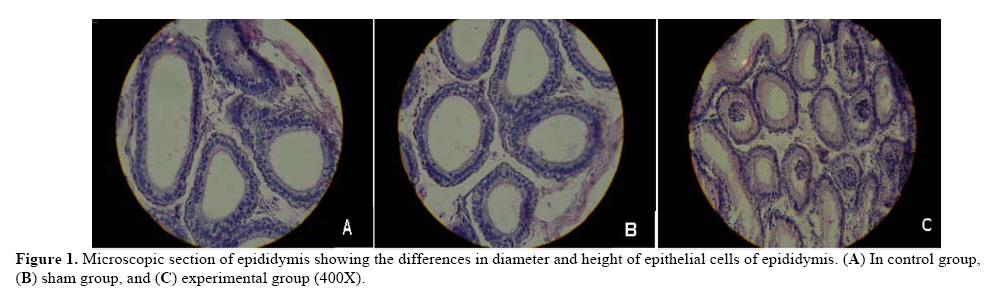
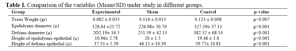
wide-range of technical characteristics. Key words: Electromagnetic field, Mouse, Epididymis, Deferens duct. Introduction The wide-spread application of electromagnetic field (EMF) in everyday life has produced many concerns on health effects of daily environmental exposure to electromagnetic radiation. There are many types of equipment emitting such radiation among them, some could have therapeutic application in humans (1). EMF can effect on cells via some mechanisms. For instance, electromagnetic radiations effect on cytoplasmic membrane could cause a change in functional potential due to biochemical change followed by a change in concentration of ions trafficking within the membrane (2). In addition, the physical reaction between EMF and chemical bonds among atoms could lead to formation of free radicals in the body of living creatures (3, 4). There are many data about EMF effects on reproductive system; however there are no morphometric studies about the effects of EMF on epididymis and deferen duct. For example, a study by Al-Akhras demonstrated that exposure to these radiations could affect the sex hormones and also other fertility parameters (5). There are many reports indicating of detrimental effects of exposure to EMF on reproductive system and development of fetus (6, 7). The deferens duct is important reproductive organ so that the ejaculation disorder due to its disorder classify as one of the major causes of infertility among men (8). It is worth mentioning that the role of this duct in boosting fertility and promoting the functionality and survival of sperms should not be neglected (9). Furthermore, the epididymis duct is among the environment which causes the most changes in proteins of sperm membrane, as the sperms spend a long time in epididymis following discharge from testes and before ejaculation. The majority of essential changes in sperms to achieve the fertility potential and motility are acquired while passing through the epididymis duct (10). In particular, it is the primary part of epididymis duct in which the suitable environment is provided for sperm maturity and this internal surrounding is associated with the function of epithelial cells (11). Regarding the role of deferens and epididymis ducts in fertility and the increased application of EMF-generating equipment, the present study was aimed at targeting the long-term effects of such radiations on these organs. Materials and methods This was an experimental study carried out at Qazvin University of Medical Sciences (Iran). Thirty BALB/C mice weight between 24-28 grams purchased from Razi Vaccine and Serum Manufacturing Institute (Tehran). The animals were kept in a humid atmosphere (65-70%) at 25°C at comparative biology department while a time interval of 12 hours of access to light and darkness during each day for a total of one week was provided for acclimatization. The EMF was produced using a system providing an intensity of 0.5 mT and a frequency of 50 Hz (The La fight signal model device was produced by SharifUniversity of Technology, Iran). Prior to utilization of the EMF-generating apparatus, the power system, frequency producer, the animal chamber, and all connections were fully checked by Teslameter (Leybold, Germany) for accurate performance. Also, the intensity of EMF-generating apparatus was examined before and during the study using the apparatus-associated Tesla meter. Initially, the animals were randomly divided into 3 groups marked as experimental, sham, and control groups. The experimental group was exposed to EMF (0.5 mT, 50 Hz) for 4 hours a day and a total period of 2 months. The sham group was left in similar environmental conditions inside the EMF-generating apparatus with no exposure to radiation. The control group was also kept in a similar setting but outside the EMF-generating apparatus. At the end of the study period, the animals were sacrificed through cervical dislocation followed by removal and weighing of the right testicles in the first place with further sampling from epididymis and deferen duct of the right side. Samples were fixed in 10% formalin (Merck, Germany) for 48 hrs and processed in tissue processing device (Shandon-citadel 1000). The samples were embedded in melt paraffin (Merck, Germany) and the slices with diameter of 5-µm sections were produced by rotator microtome (Shandon-AS 325). Out of many sections from each sample, the sections no. 4, 8, 12, and 16 were chosen and stained with H and E technique for microscopic examinations (Zeiss, Germany). Finally, 40 microscopic fields from a similar number of slides in each group were randomly selected and photographed using a digital camera. The diameter and the length of epithelium height in epididymis and also the deferens duct were measured using computer morphometric software “Image tools (3.0 SDK)” followed by analysis of data with ANOVA and Tukey post hoc follow-up tests. Results Our data showed that the weight of testicles in experimental group was significantly reduced compared to sham and control groups (p<0.007). While the epididymis diameter in experimental group was considerably decreased compared to control group (p<0.001), no difference between the experimental and sham groups was observed (Figure1). Comparing the mean diameter of deferens duct in experimental group with sham and control groups was indicative of a significant decrease in experimental group (p<0.001). Our data also showed that the mean height of epithelial cells of epididymis in experimental group was accompanied with a significant decline compared to sham and control groups (p<0.001). Moreover, the mean height of epithelial cells of deferens duct in experimental group compared to other two groups, as shown in table 1, was significantly decreased (p<0.001) (Figure2). Discussion The preponderance of evidence suggests that the low-power, low-frequency, electromagnetic radiation associated with household current does not constitute a short or long term health hazard, and whilst some biophysical mechanisms for the promotion of cancer have been proposed (such as the electric fields around power lines attracting aerosol pollutants) (12, 13). Nevertheless, some research has implicated exposure in a number of adverse health effects, leukemia (14), neurodegenerative (15, 16) and miscarriage (17). The results of present study on epididymis and deferens duct showed that the diameter and height of epithelial cells in experimental group were significantly decreased compared to sham and control groups. There are no morphometric studies about the EMF effects on epididymis and deferens duct in literature and all studies have been focused on semen fluid parameters and sexual hormones. For instance, the study by Tablado in 1998 demonstrated that when rats were exposed to EMF (0.7 T), the sperms collected from the epididymis region of experimental group were showing morphological changes such as tail twisting, head disorder, and disfiguration of middle part (18). In another study by Tablado et al it was shown that when the pregnant mice were exposed to EMF (0.5-0.7 T) since the 7th day of pregnancy to birth time, no apparent histopathological change in testes and epididymis of fetus was demonstrated (19). Our data were indicative of considerable changes in epithelial cells and diameters of deferens and epididymis ducts which could be associated with irregularities in development process and the fertility potential of sperms (9, 10). The recent studies have reported that the EMF can change the intracellular processing like enzyme activity, cytoskeleton and cell nucleus by altering the cell membrane and glycoproteins. Hence, the decreasing in cell height and duct diameter in the present study could be due to synthesize disorder of proteins which involved in cell structure (20). In addition, Tablado et al investigated the effect of EMF (0.7 T) following a time period between 10-35 days. Later, the examination of sperms obtained from mouse epididymis in experimental group showed that there was a significant increase in speed and the number of viable sperms in epididymis duct compared to control group (21). Wilson et al showed that exposure to EMF (50 Hz, 0.1 T) could results in reduction of testes weight (22). Study by Kim et al also confirms the reduction of testes weight following the exposure to EMF (23). These findings are in accordance with those found in our study and perhaps the reason for reduction of testes weight could be attributed to an increased cellular death following the exposure to electromagnetic radiation. Al-Akhras et al in 2006 investigated the effects of exposure to EMF (50 Hz, 25 mT) on fertility parameters and sex hormones in rats and showed that there was no significant reduction in testes weight, nevertheless; the weight of seminal vesicles and the number of sperms were decreased. Also, the levels of testosterone, FSH, and LH were increased (5). Furthermore, the study by Sang Leeet al in 2004, in which BALB/C mice were exposed to EMF (0.1 and 0.5 mT, 60 Hz) showed that no change in testes mass was found in experimental group; nonetheless, the germinal cells in their study were showing apoptosis with morphological changes in seminiferous tubules (24). Finally, it could be deduced that perhaps the damaging effects of EMF radiation is associated with an increase in body temperature (25) and free radicals formation (3, 4); both of which could be considered as detrimental agents to body tissues especially the reproductive system. Hence, in reducing the side-effects of these radiations on reproductive system, avoidance to unnecessary application of such EMF-generating appliances is recommended. In conclusion, the present study showed that the EMF radiation could negatively affect the male reproductive system in mice through decreasing the height of epididymis and deferens duct. However, regarding the effects of such radiation on human reproductive system, further studies are needed. Acknowledgement We are grateful to deputy for research at Qazvin University of Medical Sciences for funding of present study and also Dr Safari for providing us with EMF-generating equipments. References
© Copyright 2009 - Iranian Journal of Reproductive Medicine The following images related to this document are available:Photo images[rm09015f1.jpg] [rm09015f2.jpg] [rm09015t1.jpg] |
| |||||||||
