 |
| About Bioline | All Journals | Testimonials | Membership | News |
|
||||||
|
||||||
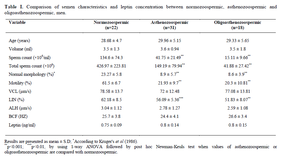
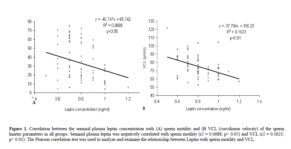
Iranian Journal of Reproductive Medicine Vol. 8, No. 3, Summer, 2010, pp. 95-100 The Leptin concentrations in seminal plasma of men and its relationship to semen parameters Seyed Gholam Ali Jorsaraei1 Ph.D., Hiroaki Shibahara2 M.D., Ph.D., Ayustawati2 M.D., Ph.D., Yuki Hirano2 M.D., Ph.D., Tatsuya Suzuki2 M.D., Eisa Tahmasbpour Marzony3 M.Sc., Mahtab Zainalzadeh1 M.D., Mitsuaki Suzuki2 M.D., Ph.D.
Corresponding Author: Seyed Gholam Ali Jorsaraei, Fertility and Infertility Research Center (Fatemeh Zahra), Babol University of Medical Sciences, Babol, Iran. Email: alijorsara@yahoo.com Received: 16 September 2009; accepted: 25 April 2010 Code Number: rm10018 Abstract Background: Leptin is a polypeptide hormone secreted by white adipose tissue in proportion to body energy. Although the participation of leptin in female reproduction is well established, any role in male reproductive function is at best tenuous. Key words: Leptin, Sperm quality, Male infertility, Seminal plasma, Fertilization rate. Introduction Leptin is a 167 - amino acid polypeptide hormone, identified in 1994 by positional cloning in the mouse and human (1). This hormone is secreted by the white adipose tissue in proportion to body energy (fat) stored. Leptin functions as a satiety factor in the regulation of body weight (2-5). Importantly, in addition to its well-known role in energy balance, leptin was soon identified as a permissive regulator of human reproductive maturity and serve as mediator in a wide range of neuroendocrine systems, including the reproductive axis (2, 5-9). So far, many studies have pointed to a direct role of leptin in the control of male reproductive function (10-11). However, in contrast to its well proven effects in female fertility (14, 15), the actual role of the hormone in the regulatory network controlling male reproductive function has been a matter of debate (11). Some studies support the role of serum leptin in the regulation of gonadal functions in men indirectly via the central neuroendocrine system (16) and directly via peripheral tissue membrane receptors (17). New reports suggest that leptin plays an important role in relaying energetic status to reproduction (11). It may be hypothesized that leptin in uncapacitated sperm is involved in the accumulation of energy substrates, which would be spent during capacitation (11, 18). Leptin is expressed in the seminiferous tubules and in seminal plasma and also directly acts on testis (4, 19), but its cellular origin in these contexts is not exactly defined. The most likely source has been shown either seminal vesicle or prostate tissue (9). On the contrary, the leptin receptors have been identified in the Leydig cells (8, 20). Besides, recent study found that human ejaculated spermatozoa secrete leptin that can affect some events tightly related to this process (11). Leptin secretion by sperm suggested that the sperm has ability to modulate its metabolism, according to its energy needs, independently by systemic leptin expression. This may represent a protective mechanism in male reproduction to guarantee the accumulation of energy substrates to maintain the gamete fertilizing capability (11, 12). Therefore, it was suggested that testes may contribute to leptin secretion and low levels of seminal leptin may be a risk factor for idiopathic male infertility. Nevertheless, the role of leptin in male reproductive function and sperm quality or capacitation has not been completely assessed and it was considered that more studies are required to clarify this issue. This study focused primarily on leptin concentration in the seminal plasma of normozoospermic, asthenozoospermic and oligoasthenozoospermic men. The associations of seminal plasma leptin concentration with sperm quality were evaluated. Materials and methods Semen analysis Semen specimens were provided by 71 men (age between 22-40 years old) that referred to Jichi Medical University Hospital for in vitro fertilization-embryo transfer (IVF-ET) treatment. All semen samples were collected by masturbation after 2-3 days of sexual abstinence in a sterile plastic jar. After liquefaction, semen specimens were evaluated for sperm concentration, motility and motility characteristics: VCL (curvilinear velocity), ALH (amplitude of lateral head displacement), BFC (beat cross frequency) and LIN (linearity), according to the guidelines of the World Health Organization (20). Morphology smears were scored using the Kruger’s strict criteria (21). All sperm specimens were evaluated with computer-assisted semen analysis (CASA) system (Hamilton Thorne Research, Beverly, USA), as described previously (23). After analysis of sperm parameters, patients were divided into three groups: normozoospermic (n=22), asthenozoospermic (n=31) and oligoasthenozoo-spermic (n=18) men. Asthenozoospermia was indicated by a sperm concentration of >20×106/ml, normal morphology of >4% and motility of <50% and oligoasthenozoospermia was indicated by a sperm concentration of <20×106/ml, normal morphology of >4% and motility of <50%. Normozoospermia was indicated by a sperm concentration of >20×106/ml, normal morphology of >4% and motility of >50%. Measurement of seminal plasma leptin After semen analysis, the seminal fluids were separated by centrifugation (8000 rpm, for 15 min) and were stored at -200 C until use. Leptin concentration in seminal plasma was measured by ELISA (active human leptin; Diagnostic system laboratories, Texas, USA) by using an automatic immunodiagnostic analyzer (Personal lab analyzer, Azwell, Tokyo, Japan) that previously described by Ayustawati et al (24). Statistical analysis An independent t-test was considered to compare the scores of each of the measures and some of the parameters data between groups. The ANOVA model was utilized for statistical analyses of leptin concentration between all groups. The Pearson correlation test and linear regression was used to analysis and examines the relation between the seminal leptin with semen parameters. p<0.05 was considered statistically significant. Results The results of the classic semen analysis are shown in table I. A trend was observed for a lower leptin concentration in seminal plasma of normozoospermic compared with asthenozoo-spermic and oligoasthenozoospermic men. The mean concentrations of leptin in seminal plasma of 22 normozoospermic men were 0.75+/-0.09 ng/ml, while in 31 asthenozoospermic and 18 oligoasthenozoospermic men were 0.8+/-0.14 ng/ml and 0.8+/-0.15 ng/ml, respectively (Table I). A significant correlation was observed between seminal plasma leptin concentrations with sperm motility (Figure 1A; p<0.05) and the VCL of sperm kinetic parameter (Figure 1B; p<0.01). Discussion In the last few years, several reports have shown that leptin is present in unexpected organs, such as the stomach, muscle and placenta or in fluids such as milk (25-27), and in the field of reproduction (20). Leptin acts at the hypothalamic level and the entrance to puberty (26, 29). Normal leptin secretion is necessary for reproductive function to proceed and may be a signal allowing for the point of initiation of the end progression toward puberty (30-32). Recently, it was hypothesized that the net effect of leptin upon male reproductive function may depend on the circulating level of the molecule (33). Besides, the available evidence is suggestive of a tightly regulated, complex mode of leptin action in different levels in the male gonadal axis that involves not only stimulatory but also inhibitory effects (34-37). Predominant stimulatory effects are observed at leptin levels above a minimal threshold; in contrast, direct inhibitory actions may take place in the presence of a significantly elevated leptin concentration (11). The presence of leptin in the genital tract, including the seminiferous tubules or seminal plasma, may influence the mechanisms involved in the sperm maturation (37), capacitation (11, 39) or mobility development of spermatozoa (4). However, in this study, there was a negative signification between seminal leptin and sperm motility. So far, most studies indicated both positive and negative effects of leptin in gonadal functions (38). Some authors indicated that the concentration of leptin in seminal plasma of men with normal semen sample is significantly lower than pathological groups (4) and has a negative correlation with the motility of human spermatozoa (3). Steiman et al (16) measured leptin in the serum of normozoospermic, oligoasthenoterato-zoospermic (OAT) and azoospermic men. The mean concentrations of serum leptin in these groups were 5.0+/-0.5 µg/l, 7.1+/-4.6 µg/l and 7.6+/-0.8 µg/l, respectively. This data suggested that azoospermic men have significantly higher levels of leptin in their serum than fertile men, but the mean of serum leptin in OAT group were not significantly different in comparison with both fertile and azoospermic groups. In our study, a trend was observed for a lower leptin concentration in seminal plasma of normozoospermic compared with asthenozoospermic and oligoasthenozoo-spermic men. Therefore, it is suggested, that asthenozoospermic or oligoasthenozoospermic men have enough leptin concentration in their seminal plasma for sperm capacitation or any other processes of fertilization. These results suggested that spermatozoa can not be the only main source for seminal leptin. Accomplished to recent studies, the source of leptin in seminal plasma is likely either seminal vesicle or prostate tissue in addition to spermatozoa (9, 40). On the other hand, it is possible that testes tissue contributes to seminal leptin production or secretion (9). In this study the presence of leptin in human seminal plasma was observed, of course we couldn’t find which organs is able to secret leptin, but it is obvious that leptin plays roles in spermatogenesis or sperm capacitation, and facilitate ovarian cycle (27, 28). Therefore it can be a marker of the quality of the follicle and viability of the embryo (39). Several reports indicated that leptin concentration in seminal plasma did not show any relationship with the spermiogram parameters, such as concentration, motility, vitality, morphology, head alteration and volume (9). But other studies reported a negative correlation with the motility of human spermatozoa (4) which is in agreement with our study. In the present study, there was a significant negative correlation between seminal leptin and sperm motility. Therefore, it appears that, the role of seminal leptin in male reproductive function is less well known and the relationship between seminal leptin and gonadal function is still unresolved. In conclusion, our data showed that seminal plasma leptin concentration was negatively correlated with sperm motility and VCL of the sperm kinetic parameter. References
© Copyright 2010 - Iranian Journal of Reproductive Medicine The following images related to this document are available:Photo images[rm10018t1.jpg] [rm10018f1a-b.jpg] |
| |||||||||
