 |
| About Bioline | All Journals | Testimonials | Membership | News |
|
||||||
|
||||||
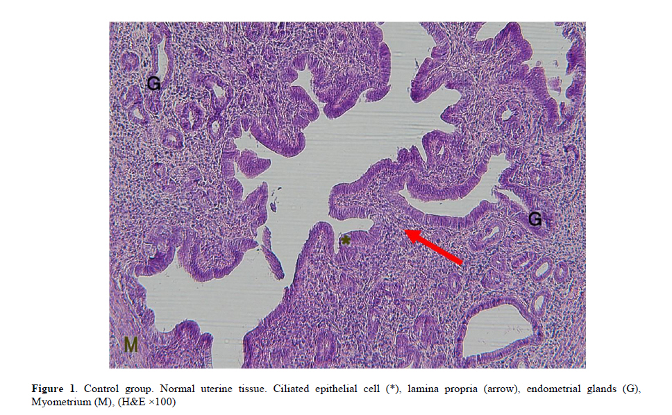
Iranian Journal of Reproductive Medicine Vol. 8, No. 3, Summer, 2010, pp. 111-118 The effect of morphine administration on structure and ultrastructure of uterus in pregnant mice Maryam Dehghan1,2 M.Sc., Mokhtar Jafarpour1 Ph.D., Alireza Mahmoudian1 Ph.D.
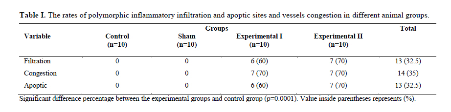
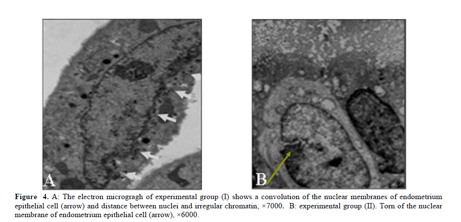
Corresponding Author: Maryam Dehghan, Department of Anatomy, School of Medicine, Shahid Sadoughi University of Medical Sciences, Yazd, Iran. Email: sun_beluga@yahoo.com Received: 8 October 2009; accepted: 26 April 2010 Code Number: rm10020 Abstract Background: Maternally administered opiates such as morphine represent a serious human health problem. Opioid abuse may have unfavorable effects on reproductive organs. Key words: Mice, Morphine, Endometrium, Uterus, Structure, Ultrastructure. Introduction Morphine is the most important alkaloid of opium family which is found as much as 10% in opium. Opioid abuse affects the body systems, and the prevalence of opioids abuse is high worldwide. Several studies have shown that addiction to narcotics analgesics product menstrual dysfunction in women (1). The researchers found that systemic morphine inhibited the visceromotor response to uterine cervical distention in the central nervous system (2). The study of opioid substitution abuse in pregnancy in a Northern Ireland Maternity Unit showed that the number of heroin addicts is increasing and had viable outcomes of high risk pregnancies (3). Investigators explored the effect of different opioids on the myometrium of pregnant rat and showed that opioids may decrease contractions in uterine muscles (4, 5). Other studies showed that some local anesthetics and opioids caused significant alteration by suppressing oxytocin on contractility of the pregnant rat myometrium (6, 7). Several studies have shown that morphine has effects on embryonic abortion (8, 9). A study about the effects of morphine during pregnancy and lactation in mice has shown that morphine induced hypothermia and hyperactivity in Rockland-Swiss mice (10). Morphine disrupts ovarian cycles and may reduce fertility. Also, chronic morphine influences noradrenergic mechanisms in the hypothalamus; which may be responsible for reduced reproductive activity (11). It is also found that the opioid has effects on glucose and eicosanoid metabolism in isolated uterus of ovariectomized and non-ovariectomized rats with restricted diet (12). Previous studies have shown that morphine induced inhibition of ovulation in normal cycling rat (13). In addition, it inhibited spontaneous uterine contractions and synthesis and output of prostaglandin from the uterus (14). Oxytocin is important in parturition, and the use of morphine causes the interruption of parturition with inhibition of oxytocin secretion (15). Other studies examined relevance of an opioid, noscapine, in reducing cystogeneses in rat experimental model of polycystic ovary syndrome (PCOS). The results showed that PCOS induced rat with ovulation blockade, persistent estrus, but in 3-4 days post noscapine administration, folliculogenrsis was followed by ovulation and reduced cystic manifestation and restored ovary and uterus weight (16). Previous studies have shown that morphine exposure caused a reduction of fetal weight and length, and reduced placental blood flow during the second week of pregnancy. It also reduced both cortical thickness and the numbers of neurons in fetal frontal cortex (17). Chronic morphine induced a supersensitivity state in the uterus from both progesterone and estradiol treated mice, also induced a further increase of the contractile effect of adrenaline (18). Previous studies have shown the morphine effects on the preovulatory discharge of pituitary gonadotropins in the cycling rats (19). Effects of morphine abuse on uterine histological and cytological structures are not clear. If morphine administration induces structural changes in uterus, it may lead to uterine factor infertility, embryonic abortion and implantation failure. Therefore, the present study was conducted to focus on the effects of morphine on histological and cytological structures of uterus in pregnant mice. Materials and methods Animals This was an experimental study carried out at Mashhad University of Medical Sciences, Iran. Forty female BALB/c mice (30-35 g) purchased from Razi Vaccine and Serum Manufacturing Institute in Tehran. The animals were housed 10 per cage with 12/12h light-cycle with free access to water and food. The temperature was 24℃, with a relative humidity of 45-55%. All experiments were approved by the University Ethical Committee Experimental procedure Three female mice were crossed with one male and a vaginal plug was designated as day 0 of pregnancy (E0). The pregnant females were divided into four groups: two experimental groups of I and II, one sham, and one control group. 5 mg/kg and 10 mg/kg morphine (10mg/1ml Sigma, USA) were injected via intra-peritoneal (IP), daily for 15 days into each experimental group, respectively (20). The same volume of saline was used for IP injection for sham group, and the control group did not receive any injection. On the 15th day of gestation (E15), the pregnant females were sacrificed and both uterine horns were removed from each animal. Tissue preparation for light microscopy (LM) Uterine tissues (5mm×5mm) were fixed in 10% formalin for 48 h. The tissue was briefly washed with saline. The tissue passed through a series of ascending concentrations of ethanol and rinsing with xylene. The samples were embedded in melt paraffin and the slices with diameter of 5−𝜇𝑚 sections were produced by microtome (Shandon-AS 325). The sections from each paraffin blocks were stained with Hematoxylin and Eosin (H&E). The sections then studied and photographed with light microscopy (BX51, Tokyo, Japan). All chemicals were purchased from Merck Co., Germany. Tissue preparation for transmission electron microscopy (TEM) The uterine tissue was washed in 0.1 M phosphate buffer (Berkshire, UK) to remove mucus and blood from the surface. The tissue was cut into pieces of approximately 1mm³. The tissue was then fixed for TEM in 3% glutarardehyde (Berkshire, UK) in 0.1 M phosphate buffer at pH 7.2 for 2 h, following post–fixation in 1% osmium tetroxide (Berkshire, UK) for 60 minutes. After dehydration in an ascending series of ethanol (Merck Co, Germany), specimens were placed in propylene oxide (Berkshire, UK) and embedded in Epon/Araldite resin (Berkshire, UK). Ultrathin sections (60-80 nm) were contrasted with uranyl acetate and lead citrate and examined by electron microscopy (Zeiss, Gottingen, Germany). Statistical analysis The data were analyzed based on Chi square and fisher tests. P-value of <0.05 was considered as significant. Results In light microscopy, the data showed that uterus of control group which received no treatment did not show any significant structural changes (Figure 1). But, in experimental groups with different dosages of morphine (5mg/kg and 10mg/kg), polymorphic inflammatory infiltration with some apoptic sites and congestion of vessels (Figure 2A, B) were observed in both endometrium and myometrium. The rate of polymorphic inflammatory infiltration and apoptic sites were 60% in the experimental group I, 70% in the experimental group II and 0% in both sham and control groups (Tables I). The rate of congestion of vessels in the experimental groups was 70%, while this was 0% in the sham and control groups (Table I). No significant difference was demonstrated in polymorphic inflammatory infiltration and apoptic sites and vessels congestion between experimental groups. Also, no significant difference was demonstrated in histological changes of uterus between sham and control groups. But, there was a significant difference in polymorphic inflammatory infiltration and apoptic cells and vessels congestion between the experimental group I and control group (Table I). In addition, there was a significant difference in polymorphic inflammatory infiltration and apoptosis and vessels congestion between experimental group II and control (p=0.0001; Table I). The results of TEM showed that endometrial epithelial cells of control and sham groups were normal (Figure 3). However, torn and convolution of the nuclear membrane of endometrial epithelial cells and a distance between nuclei and irregular chromatin were observed in experimental groups (Figure 4A, B). Discussion Morphine abuse during pregnancy is one of the most important and preventable risk factors for an array of adverse pregnancy outcomes, including premature delivery and abortion. Despite widely known risks, nearly 90% of the drug-abusing women are of childbearing age (23). Several studies indicated that morphine injection during pregnancy led to weight loss, growth retardation, and embryonic abortion (8, 9, 24). Recent study has indicated that epidural morphine analgesia induced abortion in second trimester of pregnancy (25). Morphine and methadone reduce the structural and functional integrity of the secondary sex organs by producing a pronounced reduction in serum testosterone levels (26). Several studies have shown that maternal plasma oxytocin concentration was higher and the number of uterine contractions was more frequent in animals with intermittent doses of morphine (27). Morphine stimulates nitric oxide release in human endometrial glandular epithelial cells (28). In the present study, electron microscopy was utilized to study the ultrastructural variations of uterine tissue after administration of morphine. Our results are consistent with previous studies (27, 28) that showed no significant structural changes in control or sham groups. However, morphine administration caused histological lesions, such as polymorphic inflammatory infiltration and apoptic sites and vessels congestion. Also, cytological lesions, such as torn and convolution of the nuclear membrane of endometrial epithelial cells and distance between nuclei and irregular chromatin were noted. Our results are consistent with previous studies (29) that have shown opioids drugs and peptides inhibited estradiol-induced uterine DNA synthesis. Vessels congestion or abnormal accumulation of blood in vessels results from impaired venous outflow from a tissue. Congestion of capillary beds is closely related to the development of venous edema. So, congestion and edema usually run together, therefore inflammatory infiltration and edema induced unfavorable bed of endometrium for implantation and embryo implantation may fail. Previous studies showed a reduction in the ability of embryo to implant in endometriosis patients (30-32). In a study about the effects of fentanyl on placental passage and uterine, there was not any significant deleterious changes in maternal or fetal cardiovascular system, and uterine blood flow and uterine tone were also unaffected (33). Morphine depresses both active amino-acid uptake by human placental villi and transplacental amino-acid transport by reason of the drugs' influence on placental cholinergic and opiate systems. Morphine stimulates opiate kappa receptors and depresses acetylcholine release. Acetylcholine causes dilation of blood vessels and maintains placental blood flow by the activation of endothelial muscarinic receptors which may cause the fetal intrauterine growth retardation (34). Other study has shown that lymphocytic infiltrations may be one of the causes of spontaneous abortion and the effects of disruption at the chorio-decidual interface, observed in elective abortion, provide a clue to initial signs of loss of pregnancy (35). In addition, cytological lesion, such as torn and convolution of endometrial nuclear membrane lead to the unfavorable bed for implantation. These results are in agreement with the findings of Hapangama et al (36) that showed the specific alteration in the regulation of endometrial cells are associated with reproductive failure. In long standing congestion, the poorly oxygenated blood causes chronic hypoxic injury death and apoptic sites. Our findings are consistent with previous report that women addicted to narcotics analgesics product faced menstrual dysfunction (1). Histological and morphological effects of valproic acid and oxcarbazepine indicated apoptic and degenerative effects on rat uterine and ovarian cells. Valproic acid also prevents implantation of embryo to the uterus and causes abortion via endometrial eosinophil infiltration (37). Morphological and biochemical effects of misoprostol on the uterine cervix by electron microscopy have shown that Misoprostol induced splitting and disorganization of the collagen fibers and the granular endoplasmatic reticulum appeared enriched and dilated and the nuclear chromatin was clearly dispersed (38). Gestational morphogenesis of the uterine epithelium by light and transmission electron microscopy indicated prominent changes including a reduction in the number of cell layers, reduction in cell size, reduction in the connective tissue intervening between epithelium and blood vessel endothelium, and increase in blood vessel number (39). Effect of nicotine administration on histology of some vital visceral organs, indicated that nicotine administration induced follicular and endometrial degeneration in ovary and uterus (40). Ultrastructural alterations in human decidua miscarriages indicated that decidual cells showed a moderate degree of degeneration and mononuclear cell infiltration was found to be significant (41).Naeye et al (42) studied the effect of heroin during pregnancy. Their results showed that nearly 60% of mothers and their new born infants had evidence of acute infection, and infants were small for gestational age. In addition, Vucinovic et al (23) have shown that children born to pregnant addicts had a high rate of perinatal morbidity and preterm delivery. Opioids caused perinatal complications such as placental hemorrhage. The findings, in the present study are inconsistent with other researchers that have pointed to interstitial edema, mononuclear cell infiltration and atrophy in patients with drug addiction (43). Others reported that morphine abuse caused placental insufficiencies for amino-acid transport, which may be responsible for fetal intrauterine growth retardation (44). In conclusion, our data indicated that morphine administration caused histological and cytological lesions in endometrial epithelial cells in mice. These abnormalities may lead to uterine factor infertility, leading to implantation failure. Acknowledgment We thank Dr Mohammad Ali Khalili for his critical reading and editing of the manuscript. Also, the authors thank Mr. Hassan Alavi and M. Hassan Mofidpour and M. Iraj Jafari Anarkooli for their technical assistance. References
© Copyright 2010 - Iranian Journal of Reproductive Medicine The following images related to this document are available:Photo images[rm10020f4a-b.jpg] [rm10020f2a-b.jpg] [rm10020f3.jpg] [rm10020t1.jpg] [rm10020f1.jpg] |
| |||||||||
