 |
| About Bioline | All Journals | Testimonials | Membership | News |
|
||||||
|
||||||
Iranian Journal of Environmental Health Science & Engineering,Vol. 2, No. 1, 2005, pp. 6-12 Isolation of Biosurfactant Producing Bacteria from Oil Reservoirs*A Tabatabaee 1, M Mazaheri Assadi 2, A A Noohi 1,V A Sajadian 3 1Faculty of Science, Research and Technology, Islamic Azad University, Iran 2 Biotechnology Center of Iranian Research Organization for Science and Technology, Iran 3Research Institute of Petroleum Industry, Tehran, Iran *Corresponding author: Tel: +98 21 77425001, E-mail: akram_tabatabaee@yahoo.com Code Number: se05002 ABSTRACTBiosurfactants or surface-active compounds are produced by microoaganisms. These molecules reduce surface tension both aqueous solutions and hydrocarbon mixtures. In this study, isolation and identification of biosurfactant producing bacteria were assessed. The potential application of these bacteria in petroleum industry was investigated. Samples (crude oil) were collected from oil wells and 45 strains were isolated. To confirm the ability of isolates in biosurfactant production, haemolysis test, emulsification test and measurement of surface tension were conducted. We also evaluated the effect of different pH, salinity concentrations, and temperatures on biosurfactant production. Among importance features of the isolated strains, one of the strains (NO.4: Bacillus.sp) showed high salt tolerance and their successful production of biosurfactant in a vast pH and temperature domain and reduced surface tension to value below 40 mN/m. This strain is potential candidate for microbial enhanced oil recovery. The strain4 biosurfactant component was mainly glycolipid in nature. Keywords: Biosurfactant producing bacteria, Biosurfactant, Emulsification, Microbial enhanced oil recovery (MEOR), Glycolipid INTRODUCTIONBiosurfactant or surface-active compounds are a heterogeneous group of surface active molecules produced by microoaganisms, which either adhere to cell surface or are excreted extracellulary in the growth medium (Fietcher 1992; Zajic and Stiffens, 1994; Makker and Cameotra, 1998). These molecules reduce surface tension and Critical Micelle Dilution (CMD) in both aqueous solutions and hydrocarbon mixtures. These properties create microemulsion in which micelle formations occur where hydrocarbons can solubilize in water or water in hydrocarbons (Banat, 1995). Several types of biosurfactant have been isolated and characterized, including glycolipids, phospholipids, lipopeptides, natural lipids, fatty acids, lipopolysacharides and other fully characterized. The majority of known biosurfactants are synthesized by microorganisms grown on water immiscible hydrocarbons, but some have been produced on such water-soluble substrates as glucose, glycerol and ethanol (ABU-Ruwaida et al., 1991). Chemically-synthesized surfactants have been used in the oil industry to aid clean up of oil spills, as well as to enhance oil recovery from oil reservoirs. These compounds are not biodegradable and can be toxic to environment. Biosurfactant have special ad- vantage over their commercially manufactured counterparts because of their lower toxicity, biodegradable nature, and effectiveness at extreme temperature, pH, salinity and ease of synthesis. They are potential candidate for much commercial application in the pharmaceutical and food processing and oil recovery industries (Banat, 1995; Desai et al., 1997; Makker and Cameotra, 1998). This paper describes the isolation of a potent biosurfactant producing bacteria for use in Enhance Oil Recovery. MATERIALS AND METHODSMicrobial Culture Isolation of biosurfactant producing bacteria was performed according to Francy et al., (1991) but with there modifies: microorganisms from oil reservoirs [(P (1, 2, 3, 4,5, 6) in west ofIran and E(E 0, 1, 2, 3,4,E1P2, E3 P4) and B (B1, B3)] were isolated by adding 1ml of oil samples to 99ml of SNB medium in 250ml flasks. The SNB medium used in this study was containing of 500ml of Nutrient Broth and 500 ml mineral salt solution. Mineral salts solutions used in this study were a modification of a mineral salts solution developed by Knetting and Zajic. It contained per liter distilled water, KH2PO4, 20g; K2HPO4, 5.0g, (NH4) 2SO4, 30g; NaCl, 0.1g; FeSo4. 7H2O, 0.01; MgSO4. 7H2O, 0.2; CaCl2.2H2O, 0.01; and MnSO4. 7H2O, 0.2 g; CaCl2. 2H2O, 0.01; and MnSO4.H2O, 0.002 g, glucose, 0.03%, yeast extract, 0.03% pH was adjusted to 7.2 (Francy et al., 1991). The mixture was placed on a reciprocal shaker at 200rpm for 2h at 30°C to produce a well-dispersed suspension and to isolation Biosurfactant Producing Bacteria. The mixture (suspension) was diluted serially in SNB and plated in triplicate on one-half SNB with 2% Bacto Agar (SNA) and GYA to isolate heterotrophs and ENDO, EMB, Mackankey Agar to characterization the Gram negative from Gram positive bacteria. The plates were incubated at 30°C. After numbers of colonies were stabilized, viable cell counts of all of strains were determined. Streak culture was performed on isolates to obtain pure isolates. Forty five bacteria were isolated and isolates were maintained on Nutrient Agar slants (Francy et al., 1991). Analitical Methods A) Hemolysis test The first screening test for identification and isolation of BPB is hemolysis test. The bacteria strains for hemolytic activity by plating cell on to Blood Agar and incubated at 30°C for 72 h (Bicca et al., 1999). B) Emulsification test (E24) Several colonies of a pure culture were suspended in test tubes containing 2ml of MSS after 48 h incubation, 2ml hydrocarbon (oil) was added to each tube and mixture vortexed at high speed for 1min, they were leaved to stand for 24 h. The E24 index is given as percentage of height of emulsified layer (cm) divided by total height of the liquid column (cm) (Francy et al., 1991; Bodour et al., 2004). C) Measurement of surface tension Preculture of bacteria strains were prepared in NB in OD600=1.1ml of Inocula were added to 100ml MSS and 1% filtered oil as hydrocarbon source. The mixtures with control samples were incubated at 30°C on shaker at 150 rpm for 3 d. The surface tension was meseared using a KRUSS F6 tensiometer (ABU-Ruwaida et al., 1991). D) Investigation of salt different concentra-tions effect on surface tension The effect of salinity on surface tension was determined by adding different concentrations (1-15%) of NaCl to the minimal cooper’s medium. The mixtures were incubated at 30°C on shaker at 150 rpm for 3 d (Prommachan et al., 2001). E) Investigation of pH effect on surface ten-sion The effect of pH on surface tension were carried out by changing pH (4.2-9.2) of Minimal Cooper’s medium (pH=7.2). The cultures were incubated at 30°C on shaker at 150 rpm for 3 d (Yakimov et al., 1995). F) Investigation of temperature effect on sur-face tension Different temperatures (25-50ºC) was investigated on surface tension. The cultures with control samples were placed on shaker at 150 rpm for 3 d in selected temperature (Yakimov et al., 1995). G) Kinetics of surfactant production The kinetic studies were carried out in batch culture (strain 4) for production of biosurfactant from pure substrate. These studies were carried out in 500 ml flasks with 100ml mineral salt solution and 1% oil. In these experiments, timely variations in biomass surface tension, emulsification, Critical Micelle Dilution (CMD-1, CMD-2) were recorded over a period of 72 h. Biomass was measured by dry weigh method. Surface tension and CMD were measured by Ring method by using du Nouy tensiometer. Emulsification was also estimated by Cooper and Golden berg method (1986). J) Biosurfactant Extraction Culture broth of strain 4 grown on 2% glucose was centrifuged for15min at 10,000×g to obtain cell-free supernatant. The biosurfactant produced by strain4 were extracted and characterized with chloroform-methanol (3). The chloroform-methanol was removed. The remained biosurfactant was analyzed by TLC. The TLC tank was filled with solvent mixture (chloroform-methanol aceticacid-water, 25:15:4:2). The lipid components were detected as brown spots on the plate after spraying with chromosulfuric acid and carbohydrate compounds were detected as red spots on the plate after spraying with α-naphtol and sulfuric acid (David, 1987; ABU-Ruwaida et al., 1991). RESULTSMicrobial Isolates The screening program resulted in 45 aerobic cocci and bacilli (Gram positive and negative) bacterial isolates. Depicted results of cell counts (Table 1) showed that the maximum bacterial cell is present in oil well No. P5, P6, E1, E1P2 with 106cells/ml of crude oil. Table 1: Colony count of oil samples, cells/ml
Haemolytic activity All of the isolated strains were tested for hemolytic activity. Among the isolates, 35 strains showed hemolytic activity. Selected strains were used for further screening (Bicca et al., 1999). Emulsifying ability (whole broth) Screening of microbial isolates performed in order to check the abilities of crude oil emulsifying bacteria. Among 35 strains, 23 had 70% emulsification ability. These strains were selected for further studies. Identification of the best biosurfactant pro-ducers Out of the isolated strains, 8 showed the ability to reduce culture-broth surface tension to values below 40 mN/m (Table 2). The isolated cultures were designated as strain 4 (Bacillus. sp), Strain5 (Bacillus. sp) and strain 30 (Gram negative bacilli, rod shape). Table 2: The best biosurfactant producers
Table 3: Effect of different concentrations of salt on surface tension
Effect of pH on surface tension The surface appreciable effect on surface tension. But maxitension of whole broth of selected strains main-mum of surface tension reduction was at pH tained nearly constant at all tasted pH (4.2-9.2) range from 6.2-7.2 (Table 4). at 30º C, indicating that pH variation has no appreciable effect on surface tension. But maximum of surface tension reduction was at pH range from 6.2-7.2 (Table 4). Table 4: Effect of different pH on biosurfactant production
Effect of temperature on surface tension All of the strains reduced surface tension in tasted temperatures but the best temperature for selected strains was between 30-40 (Table 5). Table 5: Effect of different temperatures on biosurfactant production
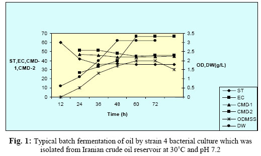
Depicted results in Fig. 1, shows, the production of the biosurfactant and biomass during growth of strain 4 on mineral salt solution containing 1% oil at different time intervals and surface tension and Critical Micelle Dilution (CMD-1and CMD-2). Maximum biomass production (about 3.1g/lit dry weight) was achieved after 60 h of growth. The surface tension of culture broth dropped rapidly after inoculation and reaching its lowest value (36 mN/ m) during the exponential phase after about 3648 h of growth. The CMD plot, a measure of biosurfactant concentration, showed that insufficient surfactant was initially present to form micelles. At 14-36 h of growth, the surfactant concentration started to increase, reaching its maximum after about 36 h. EC values increased with increasing biomass formation, reaching their optimum at about 36h. Biosurfactant nature Preliminary analysis of the biosurfactant produced by the strain 4 indicated the presence of glycolipid and or neutral lipids on the basis of Rf-value (0.6). An alphanaphtol/sulfuric acid was also detected indicating that the lipid extract contains a carbohydrate substance. DISCUSSIONAll strains were tested for haemolytic activity, which is regarded by some authors as indicative of biosurfactant production and used as a rapid method for bacterial screening (Bernheimer and Avijad, 1970; Banat, 1995a; Lin, 1996). After haemolysis test, stabilization of an oil and water emulsion is commonly used as a surface activity indicator. Several studies focused on high emulsifying abilities (Francy et al., 1991; Bicca et al., 1999; Bodour et al., 2004). Identification of biosurfactant producing bacteria can be further confirmed by measurement of surface tension. Reduction of surface tension measurements by isolated bacteria from Iranian crude oil reservoirs indicates the production of surface-active compounds. Similar results obtained by Banat et al (1991). They isolated several bacteria which showed the ability to reduce culture-broth surface tension to values below 40 mN/m. Salt concentration also affected biosurfactant production depending on its effect of cellular activity which is very near to the results obtained by Yakimov et al (1995). He isolated Bacillus licheniformis BAS50 which grew and produced a lipopeptide surfactant when cultured on a variety of substrate at salinities of 13% NaCl. Depicted results showed that the biosurfactant production was optimal at 5% NaCl. Environmental factors and growth conditions such as pH, temperature, agitation and oxygen availability also effect biosurfactant production through their effects of cellular growth or activity. Comparable results were obtained by Kim et al. (1997). They found the surface tension reducing activity of Bacillus subtillis C9 was stable to pH over the range of pH of 5.0-9.5. Also Abu-Ruwida et a1. (1991b). observed biosurfactant production of Rhodoco-ccus was at 6.5-7.2 that determined by surface tension. In this study, strains reduced surface tension in tasted temperatures but the best temperature for selected strains was between 3040°C. Also Abu-Ruwaida et al., 1991 found the optimum biosurfactant production of Rhodoco-ccus.sp at 37ºC. According to investigation of kinetic of biosurfactant production results indicate that the biosurfactant biosynthesis from oil occurred predominantly during the exponential growth phase, suggesting that the biosurfactant is produced as a primary metabolite accompanying cellular biomass formation. Similar result obtained by Abu-Ruwaida et al. in 1991 for Rhodococcus, strain ST-5. According to surface tension reducing and emulsification characteristics of strain 4 and its stability over a wide range of pH and temperatures and high salt concentrations suggest that strain 4 is suitable for use in oil fields such as MEOR and removal of oil pollutions. ACKNOWLEDGEMENTSAuthor thank Dr Moazzami at the Biotechnology Center of Iranian Research Organization for Science and Technology and Mr Hamzeloo at Tehran University for assistance in this project. REFERENCES
© Tehran University of Medical Sciences Publications 2005 |
