 |
| About Bioline | All Journals | Testimonials | Membership | News |
|
||||||
|
||||||
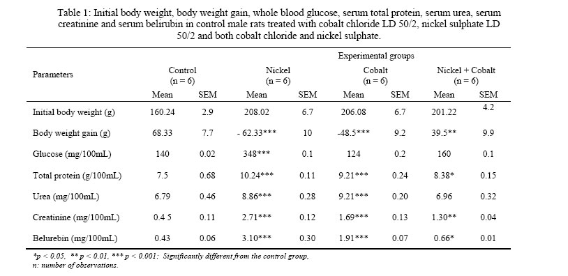
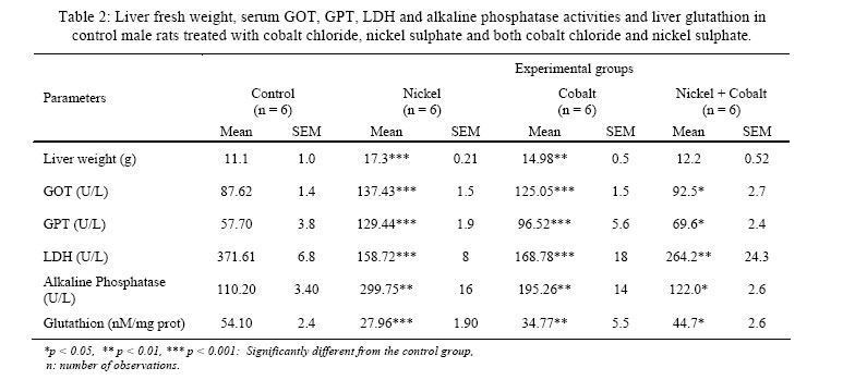
Iranian Journal of Environmental Health Science & Engineering,Vol. 3, No. 1, 2006, pp. 65-69 COMBINED EFFECT OF WATER CONTAMINATION WITH COBALT AND NICKEL ON METABOLISM OF ALBINO (WISTAR) RATS *Z. Kechrid, F. Dahdouh, R. M. Djabar, N. Bouzerna Laboratory of Biochemistry and Microbiology Application, Department of Biochemistry, Faculty of Sciences, University of Annaba, Algeria Received 2 August 2005 Code Number: se06010 ABSTRACT The contamination of water by metal compounds is a worldwide environmental problem. Concentration of metals is widely related to biochemical values, which are used in diseases diagnosis due to environmental toxicity. The sub-chronic combined effects of nickel and cobalt on body weight gain and biochemical parameters were determined and compared with those of Ni (2+) or Co (2+) alone in 6 weeks male albino (Wistar) rats. Animals were given drinking solutions of NiSO4 6H2O [Ni (II) cation, LD50/2] or CoSO4 6H2O [Co (II) cation, LD50/2]. For the combined treatment (Ni + Co), the rats received both Ni (II) cation (LD 50/2) and CO (II) cation (LD50/2). Nickel and cobalt treatment decreased body weight gain. The nickel sulphate increased also the glucose level. The two heavy elements produced hepatic and renal damage, characterized by increased activity of alanine and aspartate transaminases (GPT, GOT) and alkaline phosphatase. However lactate dehydrogenase activity (LDH) was decreased. In addition, serum urea, serum creatinine, serum total protein and serum bilurebin concentrations were significantly elevated. In general the combined effect of Ni-Co was slightly less toxic than nickel or cobalt alone, suggesting antagonism between these toxicants. Key words: Nickel, cobalt, GOT, GPT, LDH, toxicity INTRODUCTION Cobalt and nickel are essential trace metals in the human diet. They are also major components of the alloys employed in the plate and screw used for connecting bones in orthopedic surgery and in the manufacture of artificial organs (Kocijan et al., 2004). Cobalt is used also as coloring agents for pottery, ceramics, and glass. However, excessive amounts of these transitional metal ions are toxic. For example, cobalt and nickel salts have been reported to induce convulsions (Papp et al., 1987), and to cause DNA strand breaks (Christie and Tummolo, 1989), and to be organ toxic (Xie et al., 1995). Cobalt salts are thought to promote the oxidation of reduced glutathion (Iscan et al., 1994) to produce reduction in the number of hepatic hem proteins such as cytochrom P 450 and to interfere with heme metabolism by accelerating its breakdown and inhibiting its synthesis (Nakamura et al., 1975). In addition numerous authors havestudied the impact of nickel on health. It can cause dermatitis to certain persons (Accominoti et al., 1998). Particle of nickel may cause some morphological transformations in numerous cellular systems and chromosomal aberrations (Coen et al., 2001). The salts of nickel as particles of nickel can be allergens and carcinogens in man while forming the oxygenated radicals (Lansdown, 1995). This cytotoxicity was investigated in numerous micro organisms (Wu et al., 1994). Nickel was also found to be responsible on many sexual disorders (Chakroun et al., 2002). Cobalt was found also obviously harmful on the prenatal development of mice, rats and rabbits (Szakmary et al., 2001).The prevention of cadmium toxicity by pretreatment with zinc has been explained on the basis of induction of cadmium thionein (Webb, 1972). Nickel was utilized as preventive effect of cadmium (Tandon et al., 1984) and cadmium as preventive effect against nickel toxicity (Khandelwal and Tandon, 1984). It was also found that cadmium chloride is responsible for the protection against acute toxicity of mercuric chloride (Webb and Magos, 1976). In addition the influence of ascorbic acid on nickel induced hepatic lipid peroxidation in rats was investigated (kusal et al., 2001). Thus, the goal of the present study was to determine the toxicity effect of nickel or cobalt alone or in combination. Therefore, body weight was recorded and the activity of serum enzymes (GOT, GPT, alkaline phosphatase, LDH), total protein, urea, creatinine and bilerubin, were used for the evaluation and measurement of damage and protection of liver and kidney. MATERIALS AND METHODS Animals treatment Male wealing normal albino (Wistar) rats with a body weight of 150 – 200 g were supplied by Algiers Pasteur Institute. Rats were housed in plastic cages and located in air-conditioned room (humidity was around 70 %, temperature was 22 ± 2 C and 12 h light/dark cycle), and fed with laboratory stock diet (ONAB factory, EL-Harrouch, Algeria), and water ad-libitum for 1 week. The acclimated animals were divided into four groups of six animals each. Group I served as an untreated control. Group II rats were administrated with hydrated nickel sulphate (Sigma), NiSO4 6H2O[Ni (II) cation LD 50/2] in distilled water at a dose 132 mg/ kg body weight by gavage on alternate days until the end of the experiment. Group III rats treated also orally with hydrated cobalt chloride (Sigma), CoCl2 6H2O [Ni (II) cation, LD 50/2] at a dose 383 mg/kg body weight (National Institute for Occupational Safety and Health, 2003), and group IV rats were given both nickel sulphate and cobalt chloride until the end of last dose after six weeks. The control subjects and the treated animals were weighed every morning. The slaughter of animals was carried out in the morning by decapitation to avoid the effect of stress. The blood was transferred into ice cold centrifuged tubes and a portion taken for whole-blood glucose analysis, which was performed promptly after decapitation. The remaining blood was centrifuged for 10 minutes at 3000 rpm and the serum was utilized for GOT, GPT, alkaline phosphatase, LDH, total protein, bilurebin, urea and creatinine assays. Livers were rapidly excised, washed and stored at -20 C° for glutathion analysis. Analytical methods The whole blood glucose was measured in 10 µl samples of fresh whole blood by the glucose oxidase (EC 1.1. 3. 4) method, using an YSI Model 27 glucose analyzer and the kit constitute of phosphate buffer containing the enzymes (GOD, POD) and D-glucose (Sigma). Serum glutamic pyruvic aminotransferase (GPT; EC 2.6.1.2), glutamic oxalic aminotransferase (GOT; EC 2.6.1.1), lactate dehydrogenase (LDH; EC 1.1.1.27) and alkaline phosphatase (ALP; EC 3.1.3.1) activities were assayed using commercial test kits. Urea, creatinine, and bilurebin concentrations were also determined using commercial test kits. Serum total protein level was analyzed by the method of Lowry (Lowry et al., 1951). Liver glutathion (GSH) concentration was measured utilizing the method described by Weckbercker and Cory (1988). All experimental results are expressed as means ± SEM. Paired Student’t-test was employed, and p < 0.05 was considered significant. RESULTS In this experiment, rats given either nickel sulphate or cobalt chloride alone were significantly (p < 0.001 and p < 0.001) weighed less than the control rats. However the body weights of the rats treated with nickel sulphate and cobalt chloride was less affected (Table 1). In the current study, when the time of feeding was strictly controlled before an over-night fast, the experimentally nickel sulphate significantly (p < 0.001) increased the concentration of whole blood glucose. Whereas, treatment of the rats with the cobalt chloride reduced but not significantly the whole blood glucose concentration as compared with the control group. The results showed also significantly an increase in the level of serum total protein (p < 0.001and p < 0.001), urea (p < 0. 001), creatinine (p < 0.001 and p < 0.001) and bilurebin (p < 0.001 and p < 0.001) in the nickel sulphate and cobalt chloride groups respectively when compared to the control group. While, the treatment of rats with both nickel sulphate and cobalt chloride, the level of the previous parameters especially urea were less affected compared with the mean value of the control group (Table 1). In nickel sulphate and cobalt chloride groups theactivities of serum GOT, GPT and alkaline phosphatase were significantly (p < 0.001, p< 0.001 and p < 0.001) increased respectively for nickel sulphate and cobalt chloride animal groups (p < 0.001, p < 0.001 and p < 0.01) compared to their normal concentrations group (Table 2). However the activity of LDH was decreased in nickel sulphate and cobalt chloride (p < 0.001 and p < 0.01) respectively. The experimentally treatment with both nickel sulphate and cobaltchloride increased the activities of GOT, GPT and alkaline phosphatase, but significantly (p < 0.05), the results show also that the concentration of liver glutathion was decreased in nickel sulphate and cobalt chloride groups (p < 0.001 and p < 0.01), whereas in group (nickel sulphate + cobalt chloride) was only (p < 0.05) when compared to the control group. (Table 2). DISCUSSION In this experiment, nickel sulphate and cobalt chloride groups were weighed less than the control group. This is in good agreement with some previously published reports (Dieter et al., 1988). The body weight gain of animals given both nickel and cobalt was less affected as compared to nickel or cobalt alone, but the results were not very positive. The observed higher blood glucose in the present study of nickel sulphate rats may relate to the metal effect which inhibited the release of insulin from isolated islets of Langerhans (Dormer et al., 1973) or the high glycogen breakdown and new supply of glucose production from other non carbohydrate sources such as proteins (Cartana and Arola, 1992). However the unchanged blood glucose concentration in cobalt chloride rats and animals treated either with cobalt chloride or both metals may be due to the glycaemia lowering effect of cobalt chloride by decreasing systemic glucose production, increasing tissue uptake or a combination of the two mechanisms (Saker et al., 1998). In nickel and cobalt chloride rats, the activities of serum GOT, GPT, alkaline phosphatase and serum bilurebin concentration were significantly increased, compared to their normal levels. Therefore the increased of the activities of these enzymes and bilurebin level in serum is mainly due to the leakage out of these enzymes and bilurebin substance from the liver cytosol into the blood stream which gives an indication on the hepatoxic effect of these metals (Novelli and Barbosa, 1987). In other words it has been found that liver was necrotized in heavy metals exposition (Tandon et al., 1984). The decrease of serum LDH activity in rats given nickel sulphate or cobalt chloride could be attributed to the increased call of energy through transaminase reaction, glycolytic and oxidative pathway of glucose produced and alkaline phosphatase activity, rather than LDH activity. Since these animals had higher blood glucose and GOT, GPT activities than their controls. Another explanation that the sever impairment of this metals on the anaerobic process, led to an increased aerobic respiratory which was evident from the augmentation of number of red cells in the treated animals (Shomesubra et al., 1999). In this experiment it was found also a significant rise in serum total protein, serum urea, serum creatinine in nickel and cobalt groups as compared to the control group. This again indicates that synthesized proteins were liberated as a result of cytolysis and to other pathological changes in the liver tissue associated with progression of the toxicity condition. and then the excessive accumulation of amino acids (glutamic and alanine) in serum of affected animals. These excessive amino acids are converted to ketonic bodies (α keto glutaric and pyruvate) for which the enzyme GOT and GPT are needed, and confirm the result of high concentration of blood glucose (Admal and Vilstup, 1988). In addition the increased serum urea and serum creatinine were certainly as a result of kidney tissue damage and dysfunction. However, the diminution of glutathion level in nickel and cobalt chloride rats. It is suggested that it may be as a result of that the oxidative stress, which have been occurred, in the two metal toxicity. In other words the reduced antioxidant production was due to the increased oxygen metabolites and the elevated free radicals, which cause a decrease in the activity of the antioxidant defense system (kusal et al., 2001). In conclusion, the present study showed that nickel and cobalt alone both have toxicity effect on the previous parameters, but the toxicity is more profound by nickel. combined effect of Ni-Co is slightly less toxic than nickel or cobalt alone, suggesting antagonism between these toxicants. ACKNOWLEDGEMENTS This work was supported by the research project N° F 2301/02/2005 funded by the Ministry of Higher Education, Algeria. The authers should thank members of Algers Pasteur Institute for providing rats. REFERENCES
© 2006 Tehran University of Medical Sciences Publications |
| |||||||||