 |
| About Bioline | All Journals | Testimonials | Membership | News |
|
||||||
|
||||||
Iranian Journal of Environmental Health Science & Engineering, 2008, Vol. 5, No. 4, pp. 217-224 Recovery Of Damages In The Skin Of Arsenic Exposed Clarias batrachus (linn.) Following Withdrawal Of The Stress A. K. Singh, *T. K. Banerjee Histopathology Laboratory, Centre of Advanced Study, Department of Zoology, Banaras Hindu University,
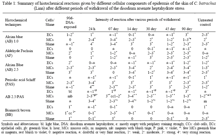
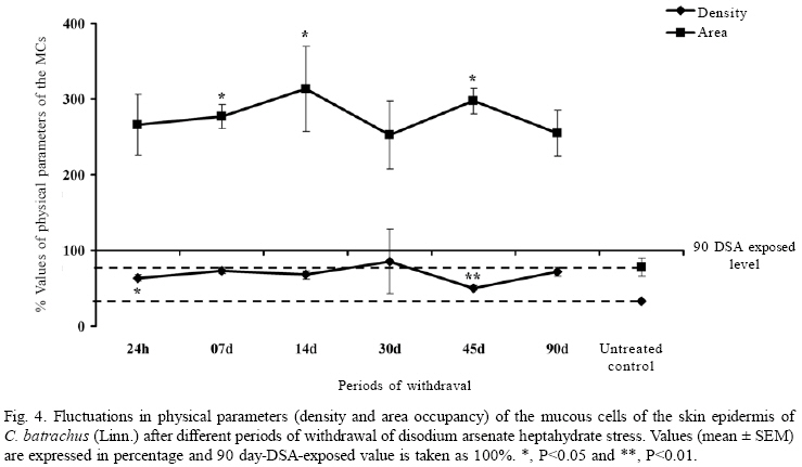
Varanasi-221005, India Received 2 February 2008; revised 15 July 2008; accepted 13 August 2008 Code Number: se08037 ABSTRACT The bottom dwelling air-breathing catfish, Clarias batrachus (Linn.) also respires via its skin (an accessory water-breathing organ). Prolonged (90 days) exposure to disodium arsenate heptahydrate has caused massive damage (e.g. wear and tear of various cellular components including club cells, hypertrophy and hyperplasia of the goblet mucous cells, altered staining and the slimy secretion) to the epidermis of its skin. The present study investigated the recovery in architecture of the damaged epidermis following return of the 90 days disodium arsenate heptahydrate exposed fish to clean water. The significant regeneration of its different cellular components (epithelial cells, Club cells, Mucous cells) took place after 24 h of withdrawal when sloughing; wear and tear and other damages of the epidermis of the skin got substantially reduced. The histopathological alterations which still continued included squeezing out of contents of the Club cells that formed a thin layer on the body surface. Regeneration of the Club cells continued throughout the epidermis even though the newly formed Club cells still showed massive sign of degeneration. Altered staining behaviour and hyperactivity of the Mucous cells continues even after prolonged withdrawal of the stress of the arsenic salt. Similarly the glycoproteins of the slime secreted by the mucous cells retained their sulphate moieties. This indicates that disodium arsenate heptahydrate induces certain permanent non-reversible damages including altered mucogenic activity in the epidermis of the skin of C. batrachus. Key words: Clarias batrachus (Linn), disodium arsenate heptahydrate, histopathology, recovery, skin epidermis, toxicity INTRODUCTION The toxicity of arsenic in the cutaneous and respiratory organs of the mammals and non-mammalian test organisms is well documented. The air-breathing fish Clarias batrachus (Linn) also respires through its skin along with gills and air- breathing organs (Banerjee, 2007). Recently Singh and Banerjee (2008) have analysed the toxic impact of sublethal concentration (1 ppm) of an arsenic salt, disodium arsenate heptahydrate (DSA). The toxicity rendered by the DSA to the skin of C. batrachus L. includes wear and tear, sloughing, mucous cells and club cells hyperplasia along with sever degenerative changes which make the fish literally unsuitable for the human consumption. The exposed fish however continue to survive in toxic solution for several days (90 days). To check if these exposed fish can recover and become suitable for their use as food, efforts have been made in this paper to analyse the degree of recovery following their return to clean water using certain histopathological techniques. MATERIALS AND METHODS Irrespective of the sex, live specimens of Clarias batrachus (Linn) (15±1 cm in length and 45±5 g weight) from a single population were acclimated in the laboratory in 25 L capacity plastic tubs containing tap water (having dissolved 6.3 O2 mg/L, pH=7.2, water hardness 23.2 mg/L and room temperature 28±3 ºC) for 30 days. Regular feeding followed by renewal of water was done at every 24 h interval during acclimation as well as entire experimental period. Ten groups of ten fish each were exposed separately to ten litres of sublethal concentration (1 ppm) of disodium arsenate heptahydrate (DSA) (s. d. fine-chem. Ltd. Mumbai, Min. assay 99.0-102.0%) prepared in tap water for 90 days. After treatment with DSA, the fish were transferred to clean (tap) water (without having arsenic salt). Three fish (n=3) each from untreated control as well as withdrawal fish groups were cold anaesthetized and sacrificed by spinal dislocation after every 24 h, 07 days, 14 days, 30 days, 45 days and 90 days of withdrawal period and the skin pieces (5 x 5 mm2) just below the anterior end of the dorsal fin were fixed in 10% neutral formalin (Lillie and Fullmer, 1976), aqueous Bouin's fluid (Bouin, 1897), 70% alcohol and Helly's fluid (Pearse, 1985) for histopathological analyses. Lugol's iodine was used to remove the mercury from Helly's fluid fixed tissue (Pearse, 1985). Paraffin sections (6 µm thick) were cut using a Lieca semi-motorized rotary microtome (Model Lieca RM 2145, Lieca Microscopy and scientific instruments Group, Switzerland), and stretched on glass slides having distilled water at 40 ºC. Serial sections were stained with Ehrlich's haematoxylin and eosin (H/E) (Ehrlich, 1886) for routine histopathology, periodic acid- Schiff (PAS) (McManus, 1948), alcian blue (AB) pH=1.0 (Lev and Spicer, 1964) and pH=2.5 (Mowry, 1956), alcian blue pH=2.5/periodic acid-Schiff (Mowry, 1956), aldehyde fuchsin (AF) (Pearse, 1985) and Bismarck brown (Gurr, 1958) for certain carbohydrate histochemistry. Observations were made under light microscope with an imaging system (Leica DM, 2000). Mucogenic activity was determined by measuring the density and area occupancy of the mucous cells in the alcian blue pH=2.5/periodic acid-Schiff stained sections with help of the software, Motic Images 2000, version 1.3. One-way ANOVA followed by Dunnett t-test was performed using the software SPSS, version 10, for statistical analyses. Measurements taken from the various control groups (untreated fish) at different time intervals of recovery experiment were almost identical hence averages of all the control groups were taken into consideration. RESULTS Control fish (untreated) The skin epidermis of Clarias batrachus (Linn) is a typical stratified epithelium having an outermost layer (OL), a middle layer (ML) and a basal layer (BL). Its main cellular constituents include polygonal epithelial cells (ECs), goblet mucous cells (MCs) and club cells (CCs) (Fig. 1A). The OL is composed of 2-3 layers of compactly arranged ECs. In between the MCs are regularly distributed. The middle layer is mainly composed of one or two layers of large sized binucleated CCs (Fig. 1E). These cells are somewhat oval in shape. The contents of CCs are slightly eosinophilic in nature and invariably showed some degree of shrinkage in histological preparations. The contents of these cells mostly showed negative PAS and AB 2.5 reactions (Fig. 2A and B). The space between the CCs remains filled by vertically elongated ECs. The MCs of OL stain, almost negatively with BB and AF, weakly to strongly with AB 2.5 (Fig. 2B), moderately to strongly with PAS and AB 1.0 and gave magenta colouration with bluish tinge with AB 2.5/ PAS (Fig. 2A) technique (Table 1). ECs of OL stain faintly with AF and AB 2.5/ PAS (Fig. 2A), weakly with BB and PAS, weakly to moderately with AB 2.5 (Fig. 2B) and showed moderate to strong reaction with AB 1.0 (Table 1). The slime secretion if present on surface also stains moderately to strongly with AB 1.0. Exposed fish (DSA treated) Singh and Banerjee (2008) have described many alterations in the skin epidermis of Clarias batrachus (Linn). The main damage following arsenic stress includes wear and tear, sloughing of the cellular debris on the surface of the skin, hyperplasia and hypertrophy of the epidermal cells (ECs, MCs and CCs) and vacuolization and degeneration of the CCs (Fig. 1A). Initially the CCs showed extensive hyperplasia resulting in crowding out of the other cellular constituents of the epidermis. During later stages of exposure the CCs showed several degenerative changes that included vacuolization due to shrinkage of their eosinophilic contents. They (CCs) also remained confined mainly to the basal layer of the epidermis. The damages and sloughing of the epidermal cells due to immediate contact stress produced by DSA perhaps caused an initial decrease in density as well as area occupancies of the MCs after 03 h (Fig. 3). Both these physical parameters of the MCs although fluctuated periodically increased significantly (P<0.01) at most of the stages of exposure especially during later stages (Fig. 3). Prolonged exposure to DSA stress however disturbed the mucogenic activity resulting the decreased density and area occupancy of the MCs. Sometimes a thick layer of slime containing glycoproteins was also noticed at surface. The MCs and their secretory material showed varied histochemical reactions for different carbohydrate moieties showing more affinities for acidic glycoproteins (Singh and Banerjee, 2008) as observed with AB 2.5 reaction. Recovered fish (withdrawal of DSA stress) Transfer of the fish to clean water induced active regeneration of the epidermis within 24 h when sloughing, wear and tear and other damages of the skin got substantially reduced. Regeneration incorporated all the cellular components of the epidermis. However the squeezed out of the contents of the CCs continued to form a thin crust on the surface of the epidermis (Fig. 1B). The regeneration of the CCs in the middle and lower layers were most prominent. Due to decrease in the density of the CCs in the outermost layers, the structure of the regenerated epidermis differed greatly from that of the control fish. Space vacated by the CCs got occupied by large sized sac like goblet MCs (Figs. 2C and D). However the slimy secretion on the skin surface remained almost unstained with haematoxylin/eosin preparations. The contents of the CCs which occupied mostly the lower layers continued to show great condensation and shrinkage that very often contained minute glaring, black coloured granules (Fig. 1C). The rest of the areas of the CCs often contained a varying amount of fuzzy substance (Fig. 1C). At certain locations the thickness of the epidermis decreased considerably due to appearance of pit like depressions. Regeneration of CCs in the entire epidermis was noticed after 45 days. However the shrinkage and condensation of the contents of these newly developed CCs continued to persist. The MCs very often appeared partially empty especially in AB 1.0 stained properties. The regeneration of the epidermis became more perfect after 90 days of withdrawal (Fig. 1D). However the MCs did not show much alternation. The ECs at the surface layer stained strongly with AB 1.0. The MCs continued to exhibit hyperplasia and hypertrophy even after withdrawal of arsenic stress (Fig. 4). The increased size of the MCs was significantly (P<0.05) more than their increased density at several stages of withdrawal (Fig. 4). The alterations in the density and dimension of the MCs following exposure to clean water were less prominent. However these MCs poured their contents in a common pit which gave the appearance of a lake (Fig. 2E). The intensity of the PAS staining of MCs increased slightly at most of the stages of withdrawal (Table 1). However they continued to show strong AB 1.0 and AB 2.5 reactions even after 24 h of the withdrawal. With AB 2.5/PAS also, most of the MCs stained pinkish violet (Table 1) at certain stages of withdrawal. With AF for sulphated glycoproteins the MCs showed positive reaction with varying intensity at different stages (Table 1). The staining intensity of the MCs with BB mostly remained weak. However the polygonal epithelial cells (ECs) at the superficial layer stained positively with both the AB techniques (Table 1). The condensed contents of the CCs also stained positively with PAS at certain stages (Fig. 2D and Table 1). The slimy secretion on the surface of the skin showed strong AB 2.5 and AB 1.0 reactions at most of the stages of the withdrawal. With BB however the slime stained almost negatively at most stages. DISCUSSION Following exposure to the arsenic salt, the skin of C.batrachus (Linn) shows extensive histopathological damages in its epidermal and dermal components (Singh and Banerjee, 2008). Following withdrawal the intensity of damage and sloughing of the epidermal cells decreases significantly within 24 h of transfer to clean water. Histopathological studies on the withdrawal of toxic stress of metals on the skin of fishes are meagre. Recently, Chatterjee (2008) while studying the withdrawal effects of manganese toxicity reported significant recovery of the epidermis of the air-breathing catfish Heteropneustes fossilis. In the recovery experiment with arsenic exposed C. batrachus (Linn) (present study) the club cells (CCs) especially those at the outermost layers however continue to show several symptoms of damage and degeneration that slough in the early stages of withdrawal. This perhaps helps to get ride of the club cells damaged by the DSA. The CCs at the inner layer of the epidermis continue to show shrinkage and condensation and bear certain black/glaring granules. These cells perhaps continue to retain the toxic arsenic salt (Singh and Banerjee, 2008) within them and get eliminated along with accumulated arsenic salt and/or its different metabolites due to sloughing when reached the skin surface. The goblet mucous cells (MCs) continue to show hypertrophy, hyperplasia and hyperactivity. This may perhaps be due to protection rendered to the skin especially when the regeneration of the epidermis is incomplete and the affected CCs continue to slough from the surface. The shift in the staining properties of the MCs can also be related to the protective role played by the slime. It is also possible that the secretory nature of the slime secreted by the MCs got permanently altered by the prolonged treatment of the DSA. Intensities of AB 2.5 and AB 1.0 staining of the MCs of the present study are less stronger than those noticed in DSA treated C. batrachus L. (Singh and Banerjee, 2008). This indicates the decreased requirement of sulphated and acidic mucopolysaccharides when the toxicity of the DSA is withdrawn. Initial regeneration of the epidermis perhaps helps to protect the fish from the entry of pollutants, microorganisms and also to maintain osmoregulation. Subsequent sloughing from the surface of the epidermis helps to get ride of the damaged cells when they migrate to outer surface from inner layer of the epidermis. The alteration in the configuration of the epidermis at certain stages is due to rapid regeneration of ECs to fill the space vacated by sloughing. A positive PAS reaction shown by the CCs indicates the altered physiology of these cells which otherwise stain negatively for various carbohydrates (Singh and Banerjee, 2008). It was interesting to note that the alteration in the skin following returning of the fish to clean water was not remarkable. This indicates that the DSA has perhaps caused long lasting irreversible toxicolopathological alterations in the skin of C. batrachus L. A survey of Fig. 4 indicates that the density of the MCs remained above the untreated control level throughout the tenure of return of the fish to the clean water. This indicates that the DSA treatment permanently alters the secretory activity of the epidermis and following withdrawal of arsenic stress, the MCs failed to return to their normal secretory physiology, even though prolonged treatment to clean water ameliorated the toxic stress of the arsenic salt to a great extent because the increase in the density of the MCs was not statistically significant. The area of the MCs of the clean water treated fish showed more increase than that of density. This also indicates the increased size of the MCs (hypertrophy) along with their increased density (hyperplasia). All these may perhaps be due to decreased stress of the DSA following return of the fish to plain tap water and in an effort to facilitate the regeneration process because increased mucogenic activity substantially reduces damages of the fish skin (Banerjee, 2007). Because of the permanent histopathological alteration inflicted to the skin of the fish by the arsenic salt, it will still not safe to consume these fishes after prolonged withdrawal of the arsenic stress. ACKNOWLEDGEMENTS Financial support to Ajai Kumar Singh from Council of Scientific and Industrial Research, Govt; of India, New Delhi, India is gratefully acknowledged. REFERENCES
© 2008 Tehran University of Medical Sciences Publications The following images related to this document are available:Photo images[se08037f4.jpg] [se08037f3.jpg] [se08037f2.jpg] [se08037f1.jpg] [se08037t1.jpg] |
| |||||||||