 |
| About Bioline | All Journals | Testimonials | Membership | News |
|
||||||
|
||||||
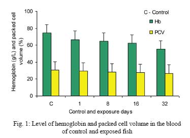
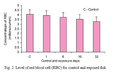
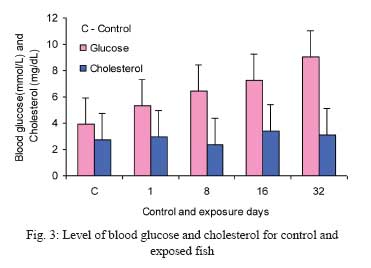
Iranian Journal of Environmental Health, Science and Engineering, 2009, Vol. 6, No. 1, pp. 23-28 The Impact of Toxic Heavy Metals on the Hematological Parameters in Common Carp (Cyprinus Carpio L.) *R. Vinodhini, M. Narayanan
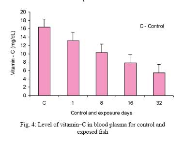
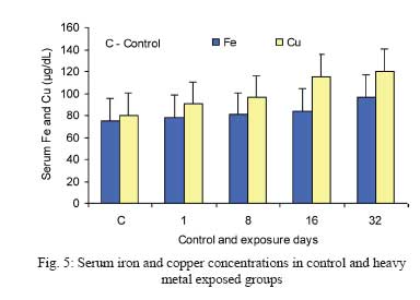
Aquatic Biodiversity Research Centre, Dept. of Advanced Zoology and Biotechnology, St.Xavier’s College, Palayamkottai – 627 002, Tamilnadu, India Code Number: se09005 Received 11 December 2007; revised 23 July 2008; accepted 8 November 2008 ABSTRACT The aim of the present investigation was to determine the effect of heavy metal pollutants such as cadmium, chromium, nickel and lead in aquatic system on common carp (Cyprinus carpio L.) by using a set of biochemical parameters. The experimental group of fish was exposed to a sublethal concentration of 5 mg/L of combined (Cd+Pb+Cr+Ni) metal solution containing 1.25 mg/L of each metal ion (1/10th of LC 50/48 h) for a period of 32 days. The results indicated that the values of the hemoglobin were in the range of 55.30±1.20 g/L to 74.55±1.33 g/L (p<0.001) and the packed cell volume was in the range of 26.72±0.26% to 30.68±0.43% (p<0.01). Concentrations of red blood cells, blood glucose and total cholesterol were significantly elevated. The level of serum iron and copper was increased. The results showed the decreased activity of vitamin C during chronic exposure to toxic heavy metals, which indicates the presence of reactive oxygen species–induced peroxidation. The study suggested that the presence of toxic heavy metals in aquatic environment has strong influence on the hematological parameters in the fresh water fish common carp (Cyprinus carpio L.). Key words: Heavy metals, hematology, blood glucose, common carp, Cyprinus carpio L. INTRODUCTION The tremendous increase in the use of heavy metals over the past few decades has inevitably resulted in an increased flux of metallic substances in the aquatic environment (Yang and Rose, 2003). The metals are of special concern because of their diversified effect and the range of concentration stimulated toxic ill effect to the aquatic life forms. Industrial wastes constitute the major source of metal pollution in natural water (Livingstone, 2001). Aquatic systems are exposed to a number of pollutants that are mainly released from effluents discharged from industries, sewage treatment plants and drainage from urban and agricultural areas. These pollutants cause serious damage to aquatic life (Karbassi et al., 2006; Al–Masri, 2002). A large part of these elements exert their toxic effect by generating reactive oxygen species (ROS), causing oxidative stress. Most of the heavy metal ions are toxic or carcinogenic in nature and pose a threat to human health and the environment (Damien et al., 2004; Farombi et al., 2007). The contamination of fresh water with a wide range of pollutants has become a matter of great concern over the last few decades, not only because of the threat to public water supplies, but also with the damage caused to the aquatic life. The river systems may be excessively contaminated with heavy metals released from domestic, industrial, mining and agricultural effluents (Vander Oost et al., 2003). Cadmium is a nonessential heavy metal; however, it is considered as one of the most toxic water contaminants and could cause toxicity at each level in organisms, from populations and communities to cell elements (Rashed, 2001). The lethal values (96 h LC50) for fish range from 0.5 µg/dm3 to 21.1 mg/dm3 (Lin and Dunson, 1993). Even at sublethal concentration, cadmium has a cumulative polluting effect and could cause serious disturbances in fish metabolism such as abnormal behavior, locomotor anomalies or anorexia (Woo et al., 1994; Bryan et al., 1995; Cicik and Engin, 2005). Cadmium may also affect the blood cells (Witeska, 1998). Chromium exists primarily in Cr (III) and Cr (VI) oxidation states; the later, hexavalent species, being considered as more toxic in the environment due to its higher solubility and mobility. These species are known to be associated with a spectrum of DNA lesions occurring during Cr (VI) exposure (Reynolds et al., 2004). Lead has long been known to alter the hematological system by inhibiting the activities of several enzymes involved in heme biosynthesis. Once absorbed, it is distributed particularly to the liver, kidney, heart and male gonads as well as it affect the immune system (ATSDR, 2005). Nickel may cause some morphological transformations in numerous cellular systems and chromosomal aberrations (Coen et al., 2001). Water born metals may alter the physiological and biochemical parameters in fish blood and tissues. The reaction and survival of aquatic animals depend not only on the biological state of the animals but also on the toxicity, and type and time of exposure to the toxicant (Brungs, 1977). Hematological and biochemical profile in fish is proved to be a sensitive index for the evaluation of fish metabolism under metallic stress. However, there are no studies demonstrating the change in blood biochemistry of common carp exposed to a mixture of heavy metals such as cadmium, chromium, nickel and lead. Henc the aim of this study was to investigate the hematological and biochemical changes in common carp (Cyprinus carpio L.) influenced by selected heavy metals. MATERIALS AND METHODS The fresh water common carp (Cyprinus carpio L.) of (10–13) cm length and 35.70±0.60 g body weight) were collected from the ponds of southern districts of Tamil Nadu, India. They were acclimated to laboratory conditions for a week prior to the experiments. The fish were kept in batches of 20–25 individuals with a photoperiod of 12:12 h light and dark cycle with constant filtration. All the fish were fed with commercially available fish feeds at a daily rate of 3–4% body weight throughout the experiment. Analytical grade cadmium chloride, lead nitrate, potassium chromate and nickel sulphate supplied by BDH (India) were used as metal toxicant throughout the experiments. Fish were divided into two groups, with the first group serving as control and other as experimental group. The experimental group was exposed to a sublethal concentration of 5 mg/L of combined (Cd+Pb+Cr+Ni) metal solution containing 1.25 mg/L of each metal ion (1/10th of LC 50/48 h) for a period of 32 days. The heavy metal concentrations were selected based on preliminary results, shown to be sublethal after a 32 day period of exposure. At the end of exposure period, the control and experimental fish were starved for 24hrs for analyzing the biochemical assays. The blood samples were taken immediately by cardiac puncture method using vacutainer tubes. Blood was sampled from ten fish of each control and metal exposed groups during each exposure period of 1, 8, 16 and 32 days. The blood parameters such as hemoglobin (Drabkin and Austin, 1935), blood glucose by glucose oxidase method (Bergmeyer and Bernt, 1974), total cholesterol by the method of Allain (1974) and Young (1997), red blood cells by Ochei and Kolhatkar (2005) and packed cell volume by Wintrobe’s tube method (Ochei and Kolhatkar, 2005) were determined. The plasma ascorbic acid content was estimated by using -2, 6 dichlorophenol indophenol dye titration method (Harris and Ray, 1935). The concentration of iron (detection limit 0.005 ng/mL) and copper (detection limit 0.002 ng/mL) in serum samples were determined by using graphite atomic absorption spectroscopy method (Monisov, 1992). Data reported in the paper are means of two or more assays. All measurements were performed in triplicate and the results were expressed as mean±S.D. The comparison of the control and experimental groups was statistically analyzed by student’s t test and the validity of investigation was expressed as probability (p) values. Values of p<0.05 as less significant, p<0.01 were considered significant and p<0.001 as highly significant. Values of p<0.05, p<0.01 and p<0.001 were considered as less significant, significant and highly significant, respectively. RESULTS The water quality parameters like temperature, pH, electrical conductivity, total dissolved solids, alkalinity, total hardness, dissolved oxygen, total ammonia and salinity measured in the municipal water used in the experimental ponds and the quality of water during heavy metal induction period are presented in Tables 1 and 2, respectively. The length and body weight of heavy metal exposed and control groups are presented Table 3. The concentration of blood hemoglobin (g/L) was in the range of 55.30±1.20 to 74.55±1.33 (p<0.001), and the packed cell volume (%) of the blood sample was in the range of 26.72±0.26 to 30.68±0.43 (p<0.01). The red blood cell (millions/cu/mm) value was in the range of 3.27±0.16 to4.05±0.18. The values were statistically significant at p<0.01. Fig. 1 shows the level of hemoglobin and packed cell volume in the blood of control and exposed fish. Levels red blood cell (RBC), and blood glucose and cholesterol are presented in Figs. 2 and 3, respectively. Results of the level of vitamin–C in blood plasma and serum iron and copper concentrations are shown in Figs. 4 and 5, respectively. The level of blood glucose (mmol/L) was reported to be in the range of 3.95±0.10 to 9.07±0.15 (p<0.001). Similarly, the level of total cholesterol (mg/dL) in blood sample was in the range of 2.77±0.06 to 3.12±0.06 (p<0.01). The value of vitamin–C (mg/dL) in blood plasma was in the level of 16.36 ± 0.03 to 5.43 ± 0.03 (p<0.001). The serum iron (μg/dL) and copper (μg/dL) values were in the range of 75.58±0.025 to 96.83±0.026 and 80.43±0.030 to 120.12±0.020 respectively. The values were statistically significant at p<0.001. DISCUSSION The present study demonstrated the obvious toxic effect of heavy metals on the biochemical parameters of common carp. The recorded water quality parameters indicate the municipal water was very much suitable for aquaculture. The values of water quality parameters measured in the experimental ponds indicated significant increase during the heavy metal exposure period. Specifically the level of dissolved oxygen was found to be increased in the experimental pond during day and night. This might be due to the effect of heavy metal induced alterations in the respiratory function of fish. The increase in oxygen level could inhibit the respiratory factors. A massive expulsion of mucus and swelling with necrosis around the gill surface of common carp observed in the study might be the impairment in respiratory function. The increase in dissolved oxygen affects the fish by inducing a vulnerable stress around the gills with a drop in metabolism of carp. The concentration of hemoglobin decreased significantly (p<0.001) in the blood of fish exposed to combined heavy metals (Fig. 1). Heavy metals such as cadmium, chromium, Nickel and lead might alter the properties of hemoglobin by decreasing their affinity towards oxygen binding capacity rendering the erythrocytes more fragile and permeable, which probably results in cell swelling deformation and damage (Witeska and Kosciuk, 2003). It was evidenced that cadmium influences the differential blood count (Gill and Epple, 1993). Heavy metals induced a significant decrease in the hematocrit level of the fresh water fish common carp during the exposure period of 1,8,16 and 32 days (Fig. 1). The results are in good agreement with earlier works that reported a significant decrease in RBC’s hemoglobin and packed cell volume of fresh water fish exposed to heavy metals (Vutkuru, 2005; Shalaby, 2001). The perturbation in these blood indices may be attributed to a defense reaction against toxicity through the stimulation of erythropoiesis. The related decrease in hematological indices proved the toxic effect of heavy metals that affect both metabolic and hemopoietic activities of Cyprinus carpio L. The present data demonstrated a significant decrease in red blood cell of common carp exposed to 5 mg/L of combined heavy metals (Fig. 2). Our results are supported by previous research work that various heavy metals and toxins enter into the aquatic system exerted a specific toxic effect on fish blood and tissues (Mousa and Khattab 2003; Vosyliene and Kazlauskiene, 2004). The blood of common carp showed significant increase in glucose during 32 days of heavy metal intoxication. This might be due to the vulnerable stress induced by the heavy metals resulted in hyperglycemia (Fig. 3). Previous investigation proved that, cadmium modulate the metabolism of carbohydrates, causing hyperglycemia by stimulating the glycogenolysis in some marine and fresh water fish species (Zikic et al., 1997; Levesque et al., 2002). Similar trend with characteristic hyperglycemia was observed in common carp throughout our experiment. Heavy metals increase the glucose content in blood, because of intensive glycogenolysis and the synthesis of glucose from extra hepatic tissue proteins and aminoacids (Almeida et al., 2001). Cholesterol is the most important sterol occuring in animal fats. It is equally distributed between plasma and red blood cells, but in adrenal cortex, it occurs in the esterified form. The cholesterol occurs as white (or) faintly yellow almost odorless granules. In the present investigation, the blood cholesterol level was significantly (p<0.01) increased in heavy metal exposed experimental groups (Fig. 3). The increased levels of cholesterol develop weakness in the body and swimming ability of the fish was observed in our study. Ascorbic acid (Vitamin–C) is an essential co-nutrient in aqua-feeds and an indispensable nutrient required to maintain the physiological processes of different animals including fishes. It is closely related to the antioxidant action, protecting the structure and fluidity of biological membrane and control of oxidizing reaction of fatty acids (Brake, 1997). The levels of vitamin–C was found to be decreased in heavy metals exposed experimental fish Cyprinus carpio L. (Fig. 4). High levels of ascorbic acid are efficient in reduction of toxicity, preventing disease and enhancing fish tolerance to environmental stress (Abdel–Tawwab et al., 2001). The decrease level of ascorbic acid might be their detoxifying effect against the toxicity exerted by the metals. The level of iron was found to be increased (Fig. 5) in the heavy metal toxicity that can generate free radicals through the oxidation of ferrous to ferric state by the Fenton’s reaction. It is noticed that long-term effect of heavy metals stimulates lipid peroxidation (Faix et al., 2005). The serum copper was found to be increased on the exposure days with heavy metals (Fig. 5). It was evidenced that copper ions showed a transient effect on calcium homeostasis (Viarengo et al., 1996) but specific inhibition of Na+ uptake in fish gills (Mc Donald and Wood, 1993) and thus caused significant losses of Na+ (Reid and Mc Donald, 1988). This finally disrupts the enzyme sodium potassium ATPase by reproducing membrane damage to the cells. In conclusion, our results confirm the sublethal effect of combined heavy metals on Cyprinus carpio L. by using a set of biochemical parameters. The decreased level of hemoglobin, hematocrit and RBC revealed the hematotoxic effect of heavy metals. The major effect of heavy metals decreased the antioxidant enzyme vitamin–C that indicates the presence of ROS– induced lipid peroxidation in fish. Biochemical constituents such as glucose, cholesterol, iron and copper shows an increasing trend because prolonged metallic stress in fish makes adaptation difficult and creates weakness in fish. These parameters could be effectively used as potential biomarkers of heavy metal toxicity to the freshwater fish in the field of environmental biomonitoring. ACKNOWLEDGEMENTS The authors wish to thank the college Principal Rev. Dr. Alphonse Manickam S.J and Prof. M.Thomas Punithan, Head of the Department of Advanced Zoology and Biotechnology, St. Xavier’s College (Autonomous), Palayamkottai, Tamilnadu, India, for their kind encouragement and their laboratory facilities. REFERENCES
© Copyright 2008 - Tehran University of Medical Sciences Publications The following images related to this document are available:Photo images[se09005f4.jpg] [se09005f3.jpg] [se09005f5.jpg] [se09005f2.jpg] [se09005f1.jpg] |
| |||||||||