 |
| About Bioline | All Journals | Testimonials | Membership | News |
|
||||||
|
||||||
Metallothionein Induction In Two Species Of Pseudomonas Exposed To Cadmium And Copper Contamination 1M. Enshaei,*2A. Khanafari, 2A. Akhavan Sepahey 1 Department of Microbiology, Islamic Azad University,
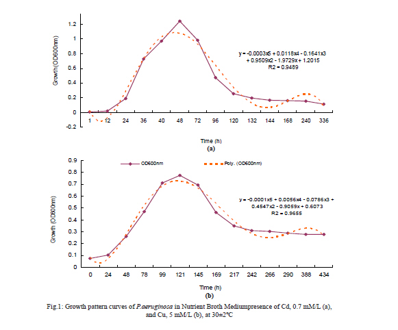
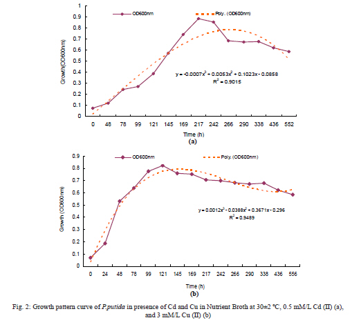
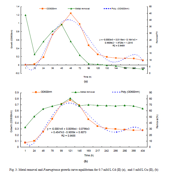
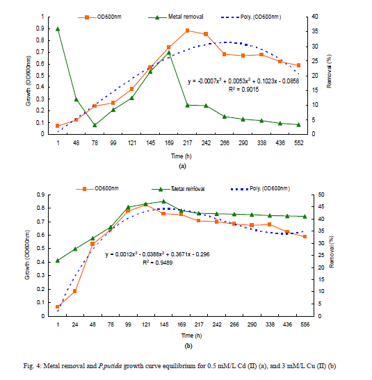
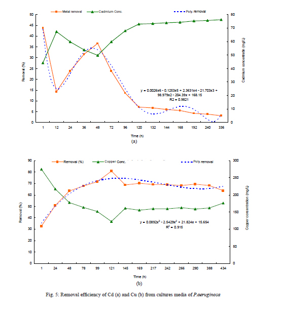
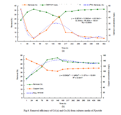
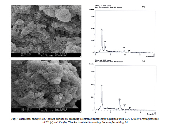
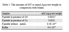
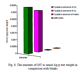
Sciences and Researches Branch, Tehran, Iran, Received 13 January 2010; revised 15 June 2010; accepted 20 June 2010 Code Number: se10033 Abstract The main objective of this study was to investigate the effect of cadmium (II) and copper (II) salts concentrations on uptake, tolerance, growth pattern and metallothionein induction as a biomarker by two bacterial strains including Pseudomonas aeruginosa and Pseudomonas putida PTCC 1694. For this purpose, the minimum inhibition concentration, minimum bactericidal concentration, growth and uptake patterns of Pseudomonas aeruginosa and Pseudomonas putida were determined in culture media with 0.09-10mM/L of Cd and Cu salts in pH7±0.2= at 30±2 ºC. Growth rate and amount of metal uptake were determined by spectrophotometer and atomic absorption assay every 24 hours for 14 to 23 days. Biosorption of the heavy metals on the bacterial cell-wall surfaces after preparation were analyzed by Scanning Electron Microscopy (SEM) equipped with Energy Dispersive Spectroscopy (EDS). Metallothionein production was evaluated by silver saturation methods. The results showed that the growth was directly inhibited at the concentration of 1.5 mM/L Cd(II) and 9 mM/L Cu(II) for P. aeruginosa and 0.95 mM/L Cd(II) and 7.5 mM/L Cu(II) for P. putida. Results of the growth pattern showed that the log phase for Pseudomonas aeruginosa and Pseudomonas putida lasted 48 and 121 hours, 217 and 121 in presence of cadmium and copper, respectively. The stationary phase was very short and very soon after log phase, the microorganisms went into death phase. The maximum biosorption of metal from cultures of two strains was 36.6% and 28% of cadmium and 80% and 47% of copper of final concentration. The result of elemental analysis with SEM-EDS approved surface adsorption of cadmium and copper. Since the exact number of Ag-binding sites per metallothionein molecules is unknown for Pseudomonas putida, results were expressed as nanomoles of Ag-binding site per gram of wet weight as equal to 0.0033 and 0.0031. Key words: Pseudomonas aeruginosa; Pseudomonas putida; Metallothionein; Ag-binding site; Cadmium; Copper Introduction Human activities such as mining and industrial waste disposal have caused accumulation of heavy metals in environment (Xincai et al., 2008). These metals have an important role in different biochemical reactions and are poisonous for cells in high concentrations (Nies, 1999). Unlike organic contaminants which can be converted into non-toxic derivatives, metals are intrinsically persistent in nature (Valls et al., 2000). Metals play an integral role in the life processes of living organisms. Some metals such as Ca, Co, Cr, Cu, Cu, Fe, K, Mg, Mn, Na, Ni and Zn are essential, serve as micronutrients and are used for redox-processes to stabilize molecules through electrostatic interactions, as component of various enzymes and for regulation of osmotic presser (Botidean et al., 2000; Hussein et al., 2005). Many other metals such as Ag, Al, Cd, Au, Pb, and Hg have no biological role and are nonessential and potentially toxic to living organisms, especially microorganisms (Hussein et al., 2005; Xincai et al., 2008). Toxicity of nonessential metals occurs through the displacement of essential metals from their native binding sites or the ligand interaction (Nies, 1999; Hussein et al., 2005). At high concentrations, both essential and nonessential metals could change enzyme specificity disrupt cellular function and damage cell membrane and structure of DNA (Bruins and Kapils Oeheme, 2000). Nonspecific and complex compounds of heavy metals have toxic effects in higher concentrations. Toxic complexes of some of heavy metal ions such as Cd, Ag and Hg are very harmful for cell vital activity, so the internal concentration and increasing of heavy metal ions should be controlled (Neis, 1999). Microbial heavy metal accumulation often comprises of two phases (Gadd, 1990). An initial rapid phase, is a physical adsorption or ion exchange at cell surface and by a subsequent slower phase involving active metabolism-dependent transport of metal into bacterial cells. During the bioaccumulation, many features of a living cell like intracellular sequestration followed by localization within specific organelles, metallothionein (MT) binding, particulate metal accumulation, extra cellular precipitation and complex formation can occur (Braam and Klapwijk, 1981; Aimal et al., 1983; Gadd, 1990; Waara, 1992; Aimal et al., 1996; Madoni et al., 1996). In addition to these varieties of mechanisms, cells can be genetically manipulated to alter morphological and physiological features (Imai and Gloyna, 1990; Dilek and Yetis, 1992; Langley and Beveridge, 1999). Conventional chemical (precipitation/neutralization) or physical (ion exchange, activated carbon sorption and membrane technology) treatment techniques are inherently problematic in their application to metal-bearing waste streams. Chemical treatment methods can prove costly to the user as the active agent cannot be recovered for reuse in successive treatment cycles (Kuyuca, 1997). The sorption of metals is attributed to the bacterial cell wall (Volesky, 1999; Slaveykova et al., 2003; Fein, 2006) and to the exopolymeric substances (EPS) (Silver, 1991; Canstein, 1999; Xiong, 2002; Pal and Paul, 2008), which contain a large number of negatively charged functional groups such as carboxyl, phosphate and sulfate (Wilhelmi and Duncan, 1995; Price et al., 2001). Various microbial species, mainly Pseudomonas, have been shown to be relatively efficient in bioaccumulation of the different heavy metals from polluted effluents (Lovely et al., 1991; Dilek and Yetis, 1992; Hadjispyrous et al., 2001; Price et al., 2001; Slaveykova et al., 2003; Hussein et al., 2005). Metallothionein is a family of cysteine-rich, low molecular weight (MW ranging from 3500 to 14000 Da) proteins which have the capacity to bind both physiological (such as zinc, copper, selenium) and xenobiotic (such as cadmium, mercury, silver, arsenic) heavy metals through the thiol group of its cysteine residues, which represents nearly the 30% of its amino acidic residues (Sigel, 2009). MTs function is not clear, but experimental data suggest may provide protection against metal toxicity, be involved in regulation of physiological metals (Zn and Cu) and against oxidative stress. They are present in a vast range of taxonomic groups, ranging from prokaryotes (such as Syneccococus spp.), protozoa (such as Tetrahymena genera), plants (such as Pisum sativum, Triticum durum, Zea mays, Quercus suber), yeast (such as Saccharomyces cerevisiae, Candida albicans), invertebrates (such as the nematode Caenorhabditis elegans, the insect Drosophila melanogaster, the mollusc Mytilus edulis, or the echinoderm Strongylocentrotus purpuratus) and vertebrates (such as the chicken, Gallus gallus, or the mammalian Homo sapiens or Mus musculus). According to the importance of environment bioremediation and industrial waste treatment, the objective of this research was to study the uptake, tolerance, growth pattern and metallothionein induction as a biomarker in presence of two heavy metals (cadmium and copper) contamination by two bacterial strains Pseudomonas aeruginosa and pseudomonas putida PTCC 1694. Materials and Methods Bacteria strain and growth condition Two bacterial strains including P.aeruginosa (from Mofid Infants’ Hospital, Tehran) and P.putida PTCC 1649 (from Persian Type Culture Collection, PTCC, Tehran-Iran) were employed to study their uptake, tolerance, growth pattern and metallothionein induction in presence of copper and cadmium salts concentrations. The strains were identified according to their morphological and physiological properties and were grown in nutrient broth medium (Merck); pH was adjusted to 7±0.01. Inoculums culture were performed in 100 cm3 Erlenmeyer flasks containing 20 mL of sterile nutrient broth medium and incubated on inductive stirring system at 30±2 ºC and used at the density of 0.8-1 at 600 nm by HACH® DR/2000 spectrophotometer (Hussein et al., 2005; Kasra Kermanshahi et al., 2007). Metal solutions Different metal concentrations were prepared by dissolving 3CdSO4.8H2O (769.51 g/mol; Merck) and CuSO4.5H2O (242.968 g/mol; Merck) salts in deionized water to obtain desired concentration of each metal in mM/L. All glassware was washed with 0.1 M HCl before and after each experiment to avoid binding of the metal (Hussein et al., 2005; Kasra Kermanshahi et al., 2007). Salt-tolerance levels Nutrient broth supplemented with different metal concentrations (0.09-10 mM/L) of 3CdSO4.8H2O and CuSO4.5H2O was provided and autoclaved 15 min at 121ºC for sterilization. Each medium was inoculated with 1 mL of inoculums culture and incubated on inductive stirring system at 30±2 ºC for 24-48 hr. After that, every culture sample was transferred to nutrient agar medium and again incubated at 30±2 ºC for 24-48 hr and minimum inhibitory concentration (MIC) and minimum bactericidal concentration (MBC) were determined (Hussein et al., 2005; Kasra Kermanshahi et al., 2007; Khanafari et al., 2008). Bacteria Growth curve Growth pattern curve for P.aeruginosa was determined at concentrations of 0.7 mM/L and 5 mM/L for cadmium and copper salts separately (Hussein et al., 2005; Kasra Kermanshahi et al., 2007). For P.putida, concentrations of 0.5 mM/L and 3 mM/L of cadmium and copper salts were chosen and incubated on inductive stirring system at 30±2 ºC for 13-23 days. The growth was investigated with determining cultures cell density at 600 nm using HACH® DR/2000 spectrophotometer and compared with blanks (same culture medium without salt concentration) (Hussein et al., 2005; Kasra Kermanshahi et al., 2007). Metal uptake or removal Simultaneously with sampling for determining the growth pattern in presence of metals, samples were taken for investigation of metal uptake or removal from the medium. All samples were centrifuged at 10000×g for 20 min and supernatants were diluted in deionized water with 5% nitric acid; then metal concentration was measured with atomic absorption spectrometry (AAS-200), Variant® (Hussein et al., 2005; Kasra Kermanshahi et al., 2007). Scanning electron microscopy (SEM) analysis Investigation of adsorption of the metals after fixation and dehydration was conducted with SEM equipped with Energy Dispersive Spectroscopy (EDS) (LEO 440i England) (Roane, 1999). Estimation of metallothionein induction by P.putida The silver saturation method was used for evaluation of metallothionein induction by P.putida in presence of 0.5 mM/L Cd salt and 3 mM/L Cu salt and blank sample (including P.putida, cultured in absence of two above metals) and a buffer sample without any bacteria suspension, in order to obtain the Ag complex efficiency of hemoglobin (Schuhamer and Cherian, 1986; Lecoeur et al., 2004; Deng et al., 2007). Extraction of metallothionein 500 µL of equivalent mixtures of dithiolthreitol (DTT), 10 mM/L, phenyl methanes sulfonyl fluoride (PMSF), 5 µM/L, and glycine buffer, 5 M/L, at pH=8.5 were added to 0.5 g of wet weight of above samples (harvested at the end of log phases and centrifuged at 10000 ×g for 10 min at 4 ºC), and then were lysis in sonicator for 10 min and cooled on ice The samples were ultra centrifuged (Hettich) at 10000 ×g at 4 ºC for 1 hr to separate the macromolecules (Schuhamer and Cherian, 1986; Lecoeur et al., 2004). Silver saturation and heat treatment 100 µL of supernatant was transferred in a 1.5 mL Eppendorf tube and 50 µL silver nitrate (20 mg/L silver) was added. After 10 min incubation in room temperature, 350 µL glycine buffer at pH=8.5 and 80 µL of bovine hemoglobin 4% were added. The mixture was heated 1 min at 100 ºC, then cooled on ice and centrifuged for 6 min at 13000×g. Supernatants were transferred to a centrifuged tube; 80µL hemoglobin was added, and samples were heated and then centrifuged. This procedure (from the transfer of supernatant in centrifuge tube to heating and centrifugation) was repeated four times to completely remove the excess silver. The last supernatants were diluted in deionized water with 0.5% nitric acid, and then Ag concentration was determined by Inductively Coupled Plasma (ICP) (JOBIN-YVON® hyultra-CE. Since the exact number of Ag-binding site per MT molecules is unknown for P.putida, in order to compare the effect of Cd and Cu on MT content, results were expressed as nanomoles of Ag-binding sites per gram of wet weight ([ng Ag in samples/mL of supernatant] × [tissue dilution/Ag molar mass]). The presence of low molecular weight proteins was investigated by sodium dodecyl sulfate polyacrylamide gel electrophoresis (SDS-PAGE) and 15% resolving gel (Schuhamer and Cherian, 1986; Lecoeur et al., 2004). Statistical analysis All experiments were carried out as three independent sets and the resulting values represent mean along with the standard errors. Data were analyzed by an analysis of variance (P<0.05). Results MICs and MBCs results for P.aeruginosa were obtained as 1.5 and 2 mM/L of cadmium and 9 and 10 mM/L of copper, respectively. For P.putida, these results determined at 0.9, were 1 mM/L of Cd and 7.5 and 8 mM/L of Cu, respectively. Results also showed that pyoverdin pigment was induced in P.aeruginosa at the concentrations of 0.3-0.9 mM/L after 36 hours and 20 hours delay was observed at the concentration of 1 mM/L. The results of growth pattern of P.aeruginosa in presence of 0.7 mM/L Cd and 5 mM/L Cu during 14 days, showed that the log phases elongated and in presence of Cd and Cu it took 48 and 121 hours, respectively. The stationary phase in presence of Cd was very short and in less than 24 hours after the end of log phase and after 120 hours of inoculation, the system went into the death phase (Figs.1a and 1b). The results of growth pattern of P.putida in presence of 0.5 mM/L Cd and 3 mM/L Cu, during 23 days showed that the log phase elongated and in presence of Cd and Cu it took 217 and 121 hours, respectively. In contrast with P.aeruginosa that showed longer log phase in presence of Cu, the P.putida showed the longer log phase in presence of Cd (Figs. 2a and 2b). The highest density of P.aeruginosa was OD600nm Max=1.24 in presence of Cd and OD600 nm Max=0.824 in presence of Cu. The highest density of P.putida was OD600 nm Max=0.884 in presence of Cd and OD600 nm Max=0.824 in presence of Cu. The highest metal removal for P.aeruginosa in presence of 0.7 mM/L Cd at the end of the log phase was 28.75l mg (equal to 36% of total Cd concentration) and 256.8 mg (equal to 80.8% of total Cu concentration) in presence of 5 mM/L Cu (Figs. 3 and 5). The highest metal removal for P.putida in presence of 0.5 mM/L Cd at the end of the log phase was 15.7 mg (equal to 28% of total Cd concentration) and this amount was 90.1 mg (equal to 46% of total Cu concentration) in presence of 3 mM/L Cu (Figs. 4 and 6). Figures 7a and 7b approved the presence of Cd and Cu in surface of P.putida by SEM-EDS method (Figs. 7a and 7b). The amounts of MT as mM Ag/g wet weight in comparison with blanks are shown in Table1. Results of this part revealed that Cd was the better inducer for MT than copper by P.putida (Fig 9). Two bands of low weight protein (almost 7000 Da) were seen in presence of 0.3 mM/L Cd or Cu by SDS-PAGE method. Discussion In this research the effects of two metals including Cd and Cu on live biomass of two species of Pseudomonas in different phases of growth and metallothionein induction were investigated. The results of growth pattern of P.aeruginosa in presence of different of Cd and Cu concentration showed that the log phase elongated and the stationary phase was very short, as if in less than 24 hours after the end of log phase went into the death phase and after 120 hours of inoculation the OD600 nm declined from 1.24 (at the end of log phase) to 0.255, which was probably due to fast cell lysis (Fig. 1). The results of growth pattern of P.putida in presence of Cd and Cu different concentration, showed that the log phase elongated but the stationary phase in contrast with P.aeruginosa that showed longer the log phase in presence of Cu, the P.putida showed longer the log phase in presence of Cd and in the death phase in comparison with P.aeruginosa didn’t lysis so fast (Fig.2). P.aeruginosa in comparison with P.putida showed faster growth in presence of Cd but in presence of Cu growth, the rate was similar for both strains. Many researches have been done on bacterial resistance and absorption of heavy metals (Olafson, 1986; Lovely and Coates, 1997; Shakibaie and Harati, 2004). Olfson in 1986 showed that Cyenococcus TX20 in presence of heavy metal caused an increase in log phase and also presence of copper had more effects on increasing of lag phase than cadmium and it took about 7, 4 and 10 days in presence of Zn, Cu and Cd, respectively. The steps of growth till the end of log phase in presence of Zn, Cu and Cd took 13, 9 and 15 days, respectively; results for bacterial growth in the blank culture (in absence of metals) were two days and log phase took less than 5 days lasted (Olfson, 1986). Results of growth in presence of different concentrations of metals showed that MIC and MBC of P.aeruginosa were 1.5 and 2 mM/L of Cd and 9 and 10 mM/L of Cu, respectively. Results also showed that pyoverdin pigment was induced in P.aeruginosa at the concentrations of 0.3-0.9 mM/L after 36 hours and at the concentration of 1mM/L after 20 hours delay. MIC and MBC of P.putida were 0.9 and 1 mM/L of Cd and 7.5 and 8 mM/L of Cu, respectively. According to the Mulen et al. studies on a group of soil separated bacteria, the MIC of Cd was in the range of 2-2.5 mM/L (Mulen et al. 1989); this range was 0.5-1.5 mM/L according to the Javadi and Karegaran studies (Javadi and Kargaran, 1997). Sabery et al. in 1997 demonstrated that the MIC of Cu in Eshershia coli was 10 mM/L, in Vibrio colera was 5 mM/L and in Pseudomonas was 7 mM/L (Sabery et al., 1997). Kasra Kermanshahi et al. in 2003 showed that in three groups of heavy metal resistance bacteria, separated from the soil of Esfahan province of Iran, the average of MIC for Cu were 8.3, 7.8 and 7.8 mM/L and for Cd were 4.5, 4.9 and 3.8 mM/L, respectively (Kasra Kermanshahi et al., 2007). Husein et al. showed that MIC of chromium in a strain of P.fluorscence was 3 mM/L. They also showed the MIC of Cu and Ni for P.putida as 4 mM/L (Hussein et al., 2005). Shakibaie et al. in 2009 showed that for Pseudomonas separated from soil and water of Kerman province of Iran after treating with mutagen substances, seven strains of mutated bacteria had the highest MIC to Cu and Zn equal to 10 and 20 mM/L, respectively (Shakibaie et al., 2009). This research also showed that the highest metal removal by P.aeruginosa in presence of 0.7 mM/L Cd at the end of the log phase was 28.75 mg (equal to 36% of total Cd concentration) and this amount was 256.8 mg (equal to 80.8% of total Cu concentration) in presence of 5 mM/L Cu. The highest metal removal for P.putida in presence of 0.5 mM/L Cd in the end of the log phase was 15.7 mg (equal to 28% of total Cd concentration) and this amount was 90.1 mg (equal to 46% of total Cu concentration) in presence of 3 mM/L Cu. The result of SEM-EDS approved the presence of Cd and Cu in surface of P.putida. In a similar research, Roane had shown that P.marginalis separated from soil had surface absorption of lead (Roane, 1999). Shakibaie and Harati have also shown that Cr and Cu could sediment on the surfaces of bacteria as nanoparticles (in size of 20-40 nm) in a form of sulfide of Cr and Cu, and had very little internal absorption (Shakibaie and Harati, 2004). Since the exact numbers of Ag-binding sites per MT molecules are unknown for P.putida, results were expressed as nanomoles of Ag-binding site per gram of wet weight (equal to 0.0033 and 0.0031). The results from this study showed that there is a significant (P<0.05) and early increase of MT biosynthesis in P.putida after exposure to Cd and Cu (0.00337 and 0.0031 mM Ag/g wet weight, respectively). The increase was significantly correlated to Cd and Cu bioaccumulation. According to the result of this research, the MT production induced in presence of both Cd(II) and Cu(II) and hence it could be recommended as a biomarker in exposing of these two metals. The case should be studied for other metals. However, because the form of MT existing in Derissena polymorpha is not Cu-inducible (Lecoeur et al., 2004) and the lack of reliable standard methods for isolation or quantification in the different species makes inter-comparison of results difficult, proposing MT as a biomarker may still be remained as a challenge and further studies are needed to confirm the mixture metals influence or very weakly influenced bioaccumulation of Cd, Cu and MT response to each of the metals, with a long-term exposure. Acknowledgements This research was supported by Islamic Azad University, Science and Research branch, Tehran. The authors would like to express their especial regards to Ms. Omidi for providing laboratory facilities. References
Copyright 2010 - Iran Journal of Environ Health Sci Eng The following images related to this document are available:Photo images[se10033f1.jpg] [se10033t1.jpg] [se10033f8.jpg] [se10033f7.jpg] [se10033f3.jpg] [se10033f2.jpg] [se10033f4.jpg] [se10033f5.jpg] [se10033f6.jpg] |
| |||||||||