 |
| About Bioline | All Journals | Testimonials | Membership | News |
|
||||||
|
||||||
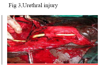
Nigerian Journal of Surgical Research, Vol. 8, No. 3-4, Jul-Dec, 2006, pp. 116-118 Penile fracture a review of management G.O Sanda1, C.F Heyns2, A. Soumana1and S. Rachid1 1Clinic of Urology, Lamordé National Hospital,BP 407 Niamey, Niger Code Number: sr06027 Abstract Objective: To discuss the clinical features and treatment of
penile fracture in the light of the current practice.
Key words: Penis, corpus cavernosum, fracture, diagnosis, management. Introduction Penile fracture isa urological emergency that may occur during coitus when the erect penis strikes against the symphysis pubis or perineum creating a sudden bending of the penis. Fractures may also occur during masturbation and vigorous self manipulation . Other causes described are falling on the erect penis, rolling over in bed or (very rarely), a blow to the flaccid penis. The pathological lesion is a tear in the tunica albuginea of the corpus cavernosum or spongiosum resulting in haematoma formation and penile swelling. Some patients may present in acute urinary retention secondary to urethral injury or periurethral haematoma. Urinary extravasation may be a late complication of unrecognized urethral injury. Characteristically, the patient hears a cracking sound followed by sudden collapse of the erection and intense local pain (1, 2, 3 Patients and Method. During the 3-year period January 2003 to April 2006 .Three patients were managed for penile fracture in our hospital. Their ages were 20, 36 and 42 years, with a mean age of 32,.6 years. The mode of presentation,aetiology of the fracture, intraoperative findings and management were studied. Aetiology. Trauma against the symphysis pubis during coital activity was the etiology in 2 cases, and rough sexual manipulation in one case. All patients accepted hearing a sharp cracking sound with severe pain followed by immediate detumescence, rapid swelling with deformity of the penis. There was acute urinary retention and bleeding per urethra in one patient.. The time to intervention was 3 hours, 7 and 15 days. The rupture was on the right side of the phalus in all the three patients(Fig. 1).The injury involved the both corporal bodies in cavernosum/ spongiosum in one patient and the urethra wasinjured in three sites in this patient(Fig. 3).On admission, all patients were initially sedated with diazepam and pain relieved with ketoprofene . On clinical assessment, the haematoma was confined to the penile shaft in onepatient (Fig. 1) and extended to the scrotum in 2 patients . Diagnosis of penilefracture was based on clinical assessment , in all patients. All surgical intervention was under spinal anaesthesia. An incision was made directly at the curvature Fig. 1; haematoma (Fig. 2) frequently encountered was evacuated. Tunica bleeding was controlled and repair done by application of running absorbable sutures (3/0 polydioxanone-PDS). Theurethral mucosa wasrepaired separately in one patient who had a urethral injury using a running 4/0 polydioxanone-PDS and the corpus spongiosum was repaired with an interrupted 3/0 polydioxanone-PDS (Fig. 3).A suprapubic catheter was placed to divert urine. All patients were however catheterised .The penile wound was not drained and pressure dressings were applied on all patients. Ciprofloxacin antibiotic was given prophylactically in all patients and ketoconazole was used with the purpose of aborting or reducing the frequency and intensity of post-operative erections. The urethral catheter and suprapubic tubes were removed after 5 days in two patients and 28 days in the third patient. Postoperative complications recorded were: wound site infection and penile angulation in one patient. The mean follow-up period was 3 months and routine examination of the phallus was done on each visit. There was no erectile dysfunction and, deformity at 3 months of follow up. Discussion Penile fracture was first reported by Frank in 1807 and there were 550 reported cases in the literature by 1989 4. Fewer than 1500 cases have been reported in the literature from 1935 to 2005 2 Hirasawa 5 reported a series of 231 cases and Zargooshi 6 encountered 172 cases in Iran. The incidence of concomitant urethral injury in reported cases is 10-58% 7,8. Approximately 30% of men with penile fracture demonstrate blood at the meatus. There was urethral injury in one of our three cases. El-Sherif and colleagues 9,10 attributed the low incidence of associated urethral injury in their series to the high incidence of non-coital fractures, whereas Tan11 reported a similarly low incidence despite a high percentage of coital fractures in their series. It is therefore not clear what predisposes to urethral injuries in association with fractured penis. Most cases (75 %) is unilateral, 25 % is bilateral in 10 % the urethra is affected3. The age of patients with penile fracture quoted in literature ranges from 26 to 41 years with a peak incidence at 26-33 years. The proportion of the patients who fall into the peak age range of 26-33 years varies from 9.5% in a Gulf state8 to the 58% reported in Western countries, 9 . The mean age in our series falls within this range. Penile rupture may be diagnosed solely on the history and clinical examination. Classically, patients describe a popping, cracking, or snapping sound with immediate detumescence. They may report minimal to severe sharp pain, depending on the severity of injury. Upon inspection, significant soft tissue swelling due to penile haematoma formation is apparent. The penis is abnormally curved, often in an S shape and deviated away from the site of the injury, due to the mass effect of the haematoma12. At times however, the diagnosis may be difficult if urethral injury is not suspected or if the penile haematoma is not significant for the cavernosal rupture 12 . Sometimes, the physician may appreciate a "rolling sign" which is the palpation of localized blood clot over the site of rupture. If Buck’s fascia has been ruptured, the swelling is contained within Colles’ fascia and a "butterfly-pattern" haematoma may be observed over the perineum, scrotum and lower abdominal wall. Most authors report accurate diagnosis without any imaging studies, but some debate surrounds the usefulness of imaging in diagnosing cavernosal injury 14,15,16 .In our series all three cases were diagnosed clinically and confirmed upon surgical exploration. Imaging studies should be reserved for cases in which the clinical history and physical examination findings conflict or for those in which no injury is apparent. Investigations may include colour Doppler ultrasound 16 , magnetic resonance imaging (MRI) for dorsal vein rupture17 18, urethrography17for urethral injury, and Cavernosography19.Ultrasonography has been suggested as a non-invasive alternative: it is widely available and inexpensive, but is operator-dependent and requires specific expertise. Cavernosography 20 is a useful diagnostic tool that reveals extravasation of contrast material from the corpus cavernosum into the penile soft tissues, indicating an injury of the tunica albuginea. However, false-negative rates are high; risks of tissue reaction to the contrast material and increased corporal fibrosis have been reported. Penile magnetic resonance imaging (MRI) provides excellent delineation of anatomy and can reveal tunical tears and urethral injury. Urethrography should only be done for suspected urethral injury. The only differential diagnosis of penile fracture is rupture of the dorsal veins of the penis 14. Both conditions require surgical exploration and repair, as there is no role for conservative treatment 21,22The "conservative management" advocated by some authors 22 in the past has now been abandoned by most surgeons because of its high complication rate, reaching 25% to 53%.Non-operative management leads to unacceptable deformity, prolonged hospitalization, residual penile mass, pulsatile cavernosal diverticulum, and expanding penile haematoma 7. All our patients were managed by operatively. In these conditions a corporeal albuginea tear is present in 100% of patients. Primary goals of surgical repair are the relief of pain, prevention of erectile dysfunction, restoration of normal voiding and preservation of penile length. Three types of incision are generally used: incision directly over the defect, circumscribing-degloving incision, and inguinal-scrotal incision7 23. An incision directly over the identified defect in the corpus cavernosum allows minimal dissection of the neurovascular bundles, but does not allow complete evaluation of both corpora cavernosa and the corpus spongiosum. Our choice was the direct incision at the site of the tear. Polydioxanone-PDS suture was used in all cases without any difficulty. Circumferential-degloving incisions 7 when applicable usually begin 1 cm proximal to the coronal sulcus and has the advantage of providing excellent exposure. However, decreased penile sensation has been reported with this type of incision. The inguinal-scrotal incision provides excellent exposure of the base, root, and dorsal surfaces of the penis23. In the available literature, surgical therapy has consistently resulted in fewer complications. Recent series from various authors24-28reported good outcomes in 92% of patients treated surgically versus only 59% in those treated conservatively. Another recent series by Shaeer29 revealed that intraoperative injection of methylene blue into the corpora helped reveal the tunical injury and thereby reduced unnecessary tissue dissection, operative time and simplified the repair. Partial or complete urethral transections require primary anastomosis over a catheter. Additionally, urinary diversion via a suprapubic tube may be considered. We closed the urethral defect with 4-0 polydioxanone-PDS sutures in a running fashion, and indwelling urethral catheter for 4 weeks. There is lack of consensus on the need for postoperative suppression of penile erection with diazepam or estrogens routinely used in some studies 24. In our patients, the use of ketoconazole helped in the prevention of early erections. Conclusions Penile fracture is an injury that is easily diagnosable by a proper history and physical examination. If there is doubt, the diagnosis should be confirmed by prompt surgical exploration. Our results show that a direct incision over the injured site is a reliable method to treat unilateral penile fracture with or without urethral injury. Immediate explorationandrepairof the cavernosaltearisessential for a goodoutcome. A urethrogram should be performed on patients with suspected urethral injury. The current policy of early surgical repair of the tunical defect seems to give excellent results in the short term References
Copyright 2006 - Nigerian Journal of Surgical Research The following images related to this document are available:Photo images[sr06027f3.jpg] [sr06027f2.jpg] [sr06027f1.jpg] |
| |||||||||
