 |
| About Bioline | All Journals | Testimonials | Membership | News |
|
||||||
|
||||||
Nigerian Journal of Surgical Research, Vol. 8, No. 3-4, Jul-Dec, 2006, pp. 174-176 Acute abdomen from gossypiboma: A Case series and review of literature M .E Asuquo, N Ogbu, J Udosen, R Ekpo, C Agbor, M Ozinko and K Emelike, Department of Surgery, University of Calabar Teaching Hospital, Calabar.
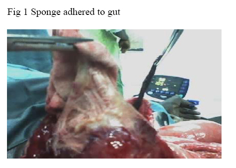
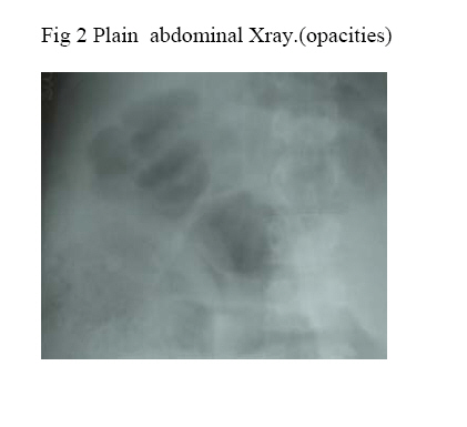
Code Number: sr06043 Abstract Gossypiboma though uncommon is under-reported. It is an infrequent but avoidable surgical error. The retained sponge induces two types of reactions, fibrinous response resulting in granuloma formation and exudative response leading to abscess formation. This serious medical condition may result in significant morbidity and mortality with serious medico legal implications. We present two cases of retained guaze(gossypiboma) seen in a busy surgical unit within three months. The pathogenesis is due to gauze induced adhesions that may cause intestinal obstruction and abscess formation resulting in peritonitis . The plain abdominal radiograph was very valuable in the first line investigation of these patients. It is possible that gossypiboma is underreported and standard protocols are not common except for routine concern for detail while doing laparotomy. Introduction The retention of surgical sponges in the peritoneal cavity otherwise called gossypiboma, is an infrequent but avoidable medical error1. This medical condition can result in significant morbidity and in some occasion’s mortality with serious medico legal implications2. This condition is generally under reported probably for medico legal reasons. The non-absorbable materials of the retained surgical foreign bodies induce principally two types of reactions, an aseptic fibrinous response resulting in adhesions and encapsulation (granuloma). The other is an exudative response leading to abscess formation3. We present two cases of gossypiboma managed in our centre and a review of Literature. Case Report. I A 65-year-old male farmer presented in the accident and emergency unit of the University of Calabar Teaching Hospital, Calabar as a referral with the complaint of progressive abdominal pain/ distension of three weeks duration, vomiting /constipation of five days duration. Prior to this, he had a left inguinal herniorrhaphy in a Peripheral hospital facility and a day after had a laparatomy because of haematuria noticed after the operation. There was no previous history of difficulty in passing urine, or haematuria. Examination revealed an acutely ill-looking elderly man in painful distress, afebrile, dehydrated and pale. Chest was clinically clear. Abdominal examination revealed two surgical scars, a midline sub umbilical and a left inguinal scar. The abdomen was distended with generalized tenderness and guarding in the periumbilical area, bowel sounds were decreased. The rectum was empty on digital rectal examination. A clinical diagnosis of acute intestinal obstruction was made. He was resuscitated with normal saline, Ringers lactate and antibiotics. Investigations revealed a packed cell volume of 32%, urea initially 16.9mmol/l decreased to 5.0mmol/l after resuscitation, the electrolytes and urinalysis were normal. Plain abdominal radiographs showed multiple air/fluid levels, a radio-opaque string as well as a whorl-like opacity, (Fig.1), following this a diagnosis of intestinal obstruction from a retained surgical sponge was made. At laparatomy, about 500mls of greenish stained peritoneal fluid was aspirated. There was a complex mass involving multiple matted loops ileum kinked at 3 points and firmly adherent to the abdominal sponge measuring 25 x 15cm, (Fig. 2). A single perforation of the ileum at the most proximal. point for obstruction was noticed. The large Sponge was morbidly adhered to viscera The retained sponge was dissected out and a resection of about 40cm of ileum with end-to-end anastomosis to establish continuity. Postoperative period was satisfactory and stitches removed on the 10th day. However, patient suddenly died the following day, there was no post mortem to ascertain the cause of death. II A 27-year-old female petty trader referred to the surgical unit with a nine day history of abdominal pain and distension following an emergency Caesarian section due to prolonged labour. She became febrile and developed progressive abdominal pain and distension from the first day post operative, these worsened with time despite the administration of antibiotics and antipyretic. There was associated bilious vomiting which worsened over time. Examination revealed an ill-looking patient, in acute painful and respiratory distress. He was febrile, pale with bilateral pitting oedema. The temperature was 37.8oC, The pulse was 108 /min., respiratory rate 28 /min., blood pressure 110 /80mmHg. Abdomen was grossly distended with a pfannenstiel scar. There was generalized abdominal tenderness with a vague mass in the right flank. Percussion notes were otherwise tympanitic and bowel were absent. Digital rectal examination revealed circumferential-prolapsed haemorrhoids. On investigation, the packed cell volume was 30%, WBC -6.8X10⁹ /l, urea was 6.8mmol/l and electrolytes were normal as well as urinalysis. Plain abdominal X-ray showed a radiopaque shadow in the right side of the abdomen (approx.10 x 15cm.) with mottled appearance, . There were dilated loops of bowel and evidence of free intraperitoneal fluid. Ultrasonography showed multiple calculi in the gallbladder, thick-walled, markedly dilated bowel loops with no significant peristaltic activity and moderate free peritoneal fluid especially in Morrison’s pouch. A diagnosis of peritonitis with a retained surgical sponge was suspected. At laparotomy three litres of pus were evacuated. A retained large surgical sponge was located in the right paracolic gutter measuring 30 x 15cm . It was removed with ease and the peritoneal cavity was lavaged. The patient did well postoperative and was discharged on the 15th day . Fig. 3 shows a large sponge in the abdomen Xray evidence can be seen in Fig. 2 as an oval radiopaque shadow with mottled appearance and dilated loops of small bowel. Discussion Gossypiboma is the nominal expression for a retained surgical sponge. It is derived from the Latin word “ gossypium” meaning cotton and the Swahili word “ boma”, place of concealment. This terminology is not new but many health care workers are just beginning to be familiar with it 3. The actual incidence of retained surgical sponge at operation is difficult to estimate because of underreporting. This may account for the varied reported incidence with some studies reporting an incidence of 1 in 300 – 1 in 1500 cases following laparatomies4 and others 1 in 3000 procedures5.The non-absorbable materials of the retained sponge induce two types of reactions 6,7. One is an aseptic fibrinous response that creates adhesions and encapsulation resulting in foreign body granuloma (pseudotumour) and intestinal obstruction as depicted in Case 1. The other response is exudative in nature and leads to abscess formation with or without secondary bacterial invasion as shown in our second patient. A retained large surgical sponge in the abdomen in contact with multiple loops of the ileum carries a higher morbidity as shown in our first patient. Resection of bowel in may be associated with multiple complications as can be seen in our first patient. . Retained foreign bodies in the abdomen may be associated with several complications due to foreign body reaction. Documented complications include fistula formation, erosion into bowel or vessels, extrusion via rectum and migration into the bladder8 Other foreign bodies that have been forgotten in the abdomen are, towels, artery forceps, pieces of broken instruments or irrigation sets and rubber tubes8..The interval between the operation and the development of symptoms varies from a few days to 28years reported in a female aged 70years5.The presentation may be acute as in our patients or delayed. Acute presentations generally follow a septic course with the formation of an abscess or intestinal obstruction from granuloma formation with adhesions . Delayed presentations may follow months or years after original surgery with adhesion formation and encapsulation depending on the location of the sponge. Most patients will present as a mass or with sub acute intestinal obstruction, although rarely they may result in a fistula, free perforation or even extrusion of the sponge10. The preoperative diagnosis may be difficult and it requires experience in the analysis of the investigations7. The diagnosis is usually made by plain abdominal radiography and a whorl-like pattern of impregnated thread may be seen in early presentation as shown in Case1 (Fig.1), disintegration and fragmentation of the radio-opaque marker may occur over time. The retained guaze may occasionally present as a heterogeneous mass with gas bubbles within a fibroticcapsule as in, and sometimes if the gauze is in contact with the urinary or gastrointestinal tract, a peripheral calcification may be seen 12. These findings further emphasise the importance of conventional abdominal imaging as first line investigation. The Ultrasonographic, CT scan and MRI findings are well documented 13,14. Laparatomy is the treatment of choice, but in some cases if the diagnosis is made early, ultrasound assisted removal with percutaneous abscess drainage or even laparoscopic retrieval may be feasible 15,16. Most reported cases of gossypiboma occur in the presence of a normal pack count17, the best approach to the prevention of this problem is the meticulous implementation of guidelines for operative theatre record keeping. Emergency operation carries a higher risk as depicted in the cases presented and diagnosis requires a high index of suspicion. The conventional abdominal imaging is important as first line investigation; we were able to make a preoperative diagnosis of gossypiboma in the cases where patients came with their plain Abdominal radiographs. Irrespective of the rarity of reports, operating teams should take care to count swabs used in all procedures. Surgeons should develop a habit of performing a brief but thorough routine postoperative wound and cavity exploration prior to wound closure. References
Copyright 2006 - Nigerian Journal of Surgical Research The following images related to this document are available:Photo images[sr06043f1.jpg] [sr06043f2.jpg] |
| |||||||||
