 |
| About Bioline | All Journals | Testimonials | Membership | News |
|
||||||
|
||||||
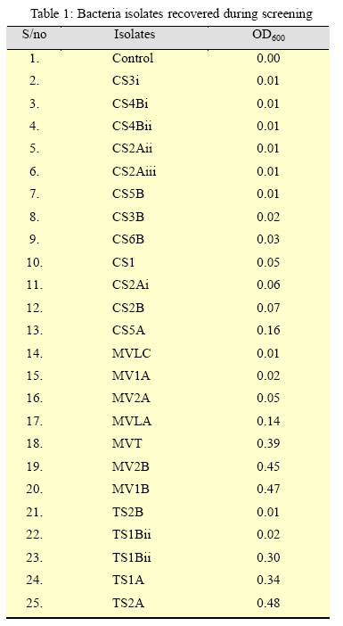
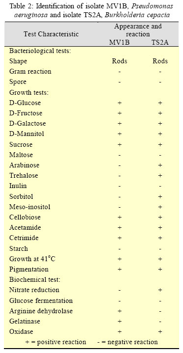
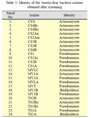
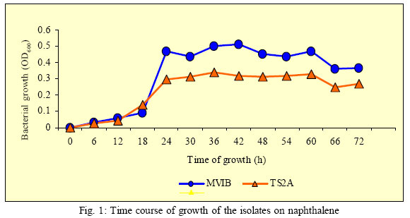
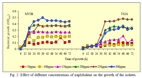
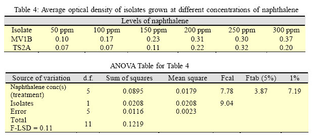
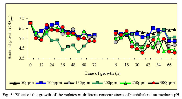
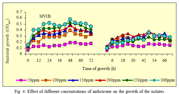
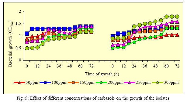
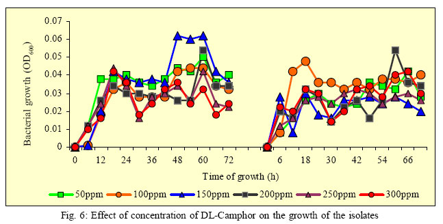
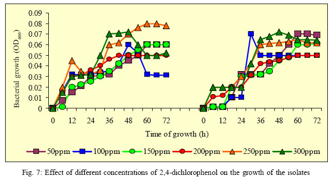
International Journal of Enviornmental Science and Technology, Vol. 3, No. 2, Spring 2006, pp. 181-190 Isolation and characterization of some polycyclic aromatic hydrocarbon degrading bacteria from Nsukka soils in Nigeria *C. I. Nnamchi, J. A. N. Obeta and L. I. Ezeogu 1Department of Microbiology, University of Nigeria, Nsukka, Nigeria Received 18 November 2005; revised 27 February 2006; accepted 3 March 2006; available online 20 April 2006 *Corresponding Author, E-mail:chuksnnamchi@yahoo.com Code Number: st06024 ABSTRACT Twenty-four bacteria capable of utilizing naphthalene, as their sole source of carbon and energy for growth were isolated from three different sites in Nsukka, Nigeria. By standard bacteriological methods, these bacteria were characterized taxonomically as belonging to the genus Pseudomonas, Burkholderia or Actinomycetes. Two of the isolates, which showed the highest growth during screening as demonstrated by an increase in their optical densities (OD600) and identified as Pseudomonas aeruginosa and Burkholderia cepacia respectively, were also able to grow in anthracene and carbazole, but not very much so in 2,4-dichlorophenol and D-camphor. The isolates showed a concentration-dependent growth in all the compounds they grew in. There were visible changes in the colour of the growth medium of the isolates during their incubation, suggesting the production of different metabolites. There were also changes in their medium pH during growth. These studies demonstrate the possession by the bacterial species of novel degradative systems. Key words: polycyclic aromatic hydrocarbons (PAHs), persistence, recalcitrance, biodegradation, bioavailablity INTRODUCTION Polycyclic aromatic hydrocarbons (PAHs) are ubiquitous contaminants of aquatic and terrestrial ecosystems whose presence is attributable to a number of petrogenic and pyrogenic sources, which had increased since the end of the Second World War (Laflamme and Hite, 1978; NAS, 1983; Jonsen et al., 2005). As hydrocarbons, PAHs are composed of carbon and hydrogen; the carbon atoms being arranged in a series of adjoining six-membered benzene rings. Thus, all PAHs have in common a singular feature that is based on two or more fused benzene rings (Chaudry, 1994). Their biochemical persistence in the environment arises from dense clouds of π-electrons on both sides of the ring structures, making them resistant to nucleophilic attack (Jonsen et al., 2005). Environments contaminated with PAHs are considered hazardous as studies using animals have shown the specific carcinogenic, mutagenic and teratogenic effects of some PAHs (Miller and Miller, 1974; Moore et al., 1989; Autrup, 1990). Even though higher molecular weight PAHs such as those containing four or more benzene rings are considered to be responsible for the majority of the potential hazards of these compounds to the environment and human health (EPA, 1984), lower molecular weight types such as naphthalene (the simplest containing two benzene rings), anthracene and phenanthrene (both of which contain three benzene rings) are known to have health effects that though are comparatively mild could be potentially hazardous (Klaasen, 2001). Furthermore, some like phenanthrene is considered as a model substrate in environmental PAHs degradation studies because its structure is found in the nucleus of carcinogenic PAHs such as benzo[a]anthracene and 3-methylcholanthrene (Cerniglia and Yang, 1984). As a result of these hazardous effects of PAHs, there is much interest in their environmental effects. The same is also true of other recalcitrant organic compounds such as carbazole, 2, 4-dichlorophenol, and DL-camphor, which belong to other classification groups. Although some physical processes such as volatilization, leaching, chemical and photo oxidation are often effective in reducing the environmental level of PAHs (Bossert and Bartha, 1984; Heitkamp et al., 1988), biodegradation using microorganisms is usually the preferred and major route of PAH removal from contaminated environments because of some inherent advantages such as its cost effectiveness and more complete cleanup (Pothuluri and Cerniglia, 1994). Moreover, the physical processes are often limited to aquatic environments only. The microorganisms should possess all the necessary enzymes needed to degrade PAHs. It is known that selection or adaptation of PAHdegrading microorganisms as with other chemicals occur as a result of their previous exposure to this substances in the environment (Lewis et al., 1984; Spain et al., 1980). However, these adaptations occur slowly, and usually depend on the recalcitrance or biodegradability of the particular substance involved (Spain et al., 1980). This is especially so considering that PAHs usually have low aqueous solubility and thus, are poorly available (low bioavailability) for microbial utilization. (Jonsen et al., 2005). A lot of isolated microorganisms have been successfully utilized in major hazardous waste clean-up processes, as for example, in industrial process streams and effluents (Levinson et al., 1994). Unfortunately, most of these studies were carried out in Western countries, and to a limited extent in South America and Asia (Kiyohara et al., 1982; Ghoshal et al., 1996; Prantera et al., 2002). In Africa, there is limited information on microbial degradation of polycyclic aromatic hydrocarbons. In this work, we report the isolation and characterization of some PAH (naphthalene)-degrading bacteria from soils in Nsukka environment, and their course of growth in naphthalene and other aromatic compounds. MATERIALS AND METHODS Collection of soil samples About 5 g soil samples were asceptically collected with a sterile scoop from soils in Timber Shed and Mechanic Village, both in Nsukka locality and also from the coal dump behind Oba Akenzua refectory in the University of Nigeria, Nsukka. All samples were placed into sterile polythene bags and stored at 4 oC immediately they were brought to the laboratory. Isolation of bacteria from the soil samples Bacteria were isolated from the soil samples using an enrichment medium containing naphthalene. Themedium consisted of (g /L) NH4SO3, 2.5; Na2HPO4, 1.0; MgSO4, 0.5; Fe2(SO4)3, 0.01; CoCl2, 0.005; CaCl2, 0.001; KH2PO4, 0.0005; MnSO4, 0.0001; (NH4)6Mo7O2.4H2O, 0.0001 and naphthalene, 0.65. The naphthalene was added after autoclaving the medium. The medium was first dispensed in 30 mL volumes into 150 mL Erlenmeyer flasks and autoclaved at 121oC and 15 psi for 15 min.s. Thereafter, 1.0 g of each soil sample was inoculated into each flask of the medium and incubated at 120 rpm at 30 oC in a Gallenkamp orbital shaker (Cat No: IH-460, Made in England) for one week. Afterwards, 1.0 mL sample was taken from each culture and transferred into fresh enrichment medium, followed by incubation as described above for one week. The enrichment procedure was repeated for the third time, before their bacterial contents were isolated using a solid medium containing the enrichment medium and 15.0 g/L of pure agar. Inoculated plates were purified by repeatedly subculturing. Pure cultures obtained by this procedure were stored in slants of enrichment medium with 15.0 g/ L pure agar, and also in nutrient agar, and stored at 4oC. Screening of the isolates for the ability to use Naphthalene as sole source of carbon for growth A loopful of each isolate was inoculated into large test tubes containing 25 mL of screening medium. The screening medium was the same as the enrichment medium, except that 15 mg of naphthalene dissolved in DMSO was added to each tube after autoclaving, as sole source of carbon. Thereafter, the test tubes were statistically incubated by keeping on the laboratory bench at room temperature (23 – 25 oC) for three days. The ability of each isolate to utilize naphthalene was indicated by an increase in turbidity of the medium measured at 600 nm using a Spectronic 21D (Milton Roy, Made in USA) UV spectrophotometer. Identification, characterization and standardization of isolates Two isolates, which gave the highest OD readings, were identified to their species level, while the others were identified to their genus level only, using conventional microbiological and biochemical procedures. The tests were carried out according to the procedures described by Cowan and Steele (1974) and Cheesebrough (1998) and Bergey’s manual of systematic bacteriology (1993). All the isolates were code-named; the two referred to above were named MV1B and TS2A and subsequently used for further studies. Before usage in subsequent works, cells were washed and standardized to the McFarland nephelometer standard of 0.5 (Baron and Finegold, 1990). In all cases, 1o/ov/v of standardized inoculum was used according to the volume of medium used. Determination of the time course of growth of the isolates The two isolates were inoculated into 250 mL Erlenmeyer flasks containing 75mLof sterile enrichment medium (already described) in triplicates. The flasks were then incubated in a Gallenkamp orbital shaker as previously described for three days. At six-hourly intervals, 5.0 mL sample was collected from each flask and assayed for OD at 600 nm in a Spectronic 21D (Milton Roy, Made in USA) UV spectrophotometer. Effect of the concentration of Naphthalene, a polycyclic aromatic hydrocarbon on the growth of the isolates The enrichment medium (3000 mL) was prepared in a 4 L flask and dispensed in 75 mL volumes into thirtytwo 250 mL Erlenmeyer flasks before autoclaving. The flasks were then divided into six sets of six flasks each. Thereafter, the following levels of naphthalene (dissolved as before) were added to each of the six sets of flasks: 50 ppm, 100 ppm, 150 ppm, 200 ppm, 250 ppm and 300 ppm. Three sets of flasks were inoculated with isolate MV1B, and the others with isolate TS2A. Inoculated flasks were then incubated as previously described for three days. Five milliliter sample was asceptically collected from each flask and assayed for the level of microbial growth by measuring the OD as described previously and pH using a Pye Unicam (model 90, MKZ, Cambridge, UK) pH meter. Effect of the concentration of some other aromatic compounds on the growth of the isolates The ability of the isolates to grow on varying amounts of anthracene (a PAH), Carbazole (a heterocyclic aromatic compound), 2,4-dichlorophenol (a chlorophenol), and DL-Camphor was studied. The preparation and sterilization of media as well as the addition of the hydrocarbons were carried out as in the case of naphthalene above. Thereafter, each set of triplicate flasks were inoculated with the two isolates as applicable, and incubated as described before for three days. Samples were also collected at six hourly intervals and measured for optical density. Statistical analysis Statistical analysis was performed using the least significant difference test method (Obi, 1990). RESULTS Screening and characterization of the isolates Twenty-four bacteria isolates were obtained after screening shown in Table 1. Two isolates, MV1B and TS2A, which were found to degrade naphthalene, better than the rest of the isolates were identified as Pseudomonas aeruginosa and Burkholderia cepacia. The characteristics of the two bacteria are shown in Table 2. Results also indicate that most of the isolates with low ODwereActinomycetes as most of them produced aerial mycelia, which bore chains of spores, while the remaining isolates were of the genus Pseudomonas and Burkholderia. This is shown in Table 3. Time course of growth of isolates MV1B and TS2A on Naphthalene The time course of the utilization of naphthalene by the two isolates as their sole source of carbon for growth is shown in Fig. 1. From the Fig., it is seen that both organisms grew relatively well in naphthalene. Of the two, isolate MV1B, Pseudomonas aeruginosa, grew better, peaking at OD value of 0.51 at 600 nm wavelength after 18 hours, whereas isolate TS2A Burkholderia cepacia peaked at an OD value of 0.34 after 30 h. During the course of their growth in naphthalene, the isolates also produced coloured metabolites, which were greenish yellow for isolate MVIB, and faint yellow for isolate TS2A. Effect of naphthalene concentrations on the growth of the isolates There was a correspondingly higher growth of both isolates as the levels of naphthalene were increased from 50ppm to 300ppm (Fig. 2). In isolate MV1B, the isolates showed significant (p < 0.01) differences in the growth of the isolates at the different naphthalene levels. Using the calculated F-LSD value of 0.11, it wasobserved that the differences existed between naphthalene levels of 300ppm against 50ppm, 100ppm and 150ppm;250ppm lowest optimum growth of 0.18 (OD600) was observed when the level of naphthalene was lowest (50 ppm), while the highest optimum growth of 0.50 was recorded when the level of naphthalene was highest (300 ppm). Similarly, the lowest optimum growth in isolate TS2A was 0.10 at naphthalene level of 50 ppm, while the highest was 0.45 when the naphthalene level was 250 ppm (OD600). Table 4 illustrates the average optical density measurements of the two isolates grown at different levels of naphthalene. Analysis of variance (ANOVA) of the effect of the different levels of naphthalene on the growth (mean OD) of the against 50 ppm, 100 ppm and 150 ppm; and between 200 ppm against 50 ppm and 100 ppm. Effect of different concentrations of Naphthalene on medium pH Results of the effect of naphthalene on the growth of the isolates on medium pH illustrated in Fig. 3 showed that there was a fall in pH from a nearly neutral initial medium pH in almost all the concentrations, to acidic and weakly acidic levels (between 5.2 to 6.0 for MV1B, and 4.0 to 6.0 for TS2A) by the end of the experiment. Effect of the concentrations of some aromatic compounds on the growth of the isolates The two isolates grew on anthracene, carbazole and marginally on DL-camphor and 2,4-dichlorophenol, using them as their sole source of carbon for growth as illustrated in Fig.s 4, 5, 6, 7. Of these, the isolates grew well on anthracene and carbazole, and poorly on DLcamphor and 2, 4-dichlorophenol. The growth of the two isolates on anthracene was also concentrationdependent as observed in the case of the utilization of naphthalene. In isolate MV1B, the lowest optimum growth of 0.189 at 600 nm was observed when the level of anthracene was lowest (50 ppm); while the highest optimum growth of 0.578 was recorded when the level of anthracene was highest (300 ppm). Similarly, the lowest optimum growth in isolate TS2A was 0.173 at anthracene level of 50ppm, while the highest was 0.380 when the level of anthracene was 250 ppm (OD600). The growth of the isolates in carbazole only partially followed the concentration-dependent pattern observed in naphthalene and anthracene. In isolate TS2A, the highest growth of 1.80 at 600 nm was given when the level of carbazole was 300 ppm, while the lowest growth of 1.20 was observed when the concentration was 50ppm. On the other hand however, isolate MV1B had the highest growth of 1.40 (OD600) at the two carbazole levels of 100 ppm and 250 ppm, while the lowest growth of 0.45 was observed at the highest level of 300 ppm. The isolates also had slight growth on 2,4-dichlorophenol and DL-camphor. There was no clear-cut effect of their different levels on the growth of the isolates. DISCUSSION AND CONCLUSION Atotal of twenty-four bacterial isolates were obtained and screened for their ability to utilize naphthalene as sole source of carbon and energy. Most of these were identified as Pseudomonas, Burkholderia or Actinomycetes. This conforms to the high degradative ability and ubiquity associated with these bacterial types as it concerns biodegradation of both soil and water environments polluted with petroleum and its many products (Atlas, 1984; Chaudhry, 1994 Jonsen et al., 2005). Of this lot, only two species (P. aeruginosa and B. cepacia) were chosen for further studies as a result of their high growth yield in the relatively short period of three days, as well as other considerations such as their ability to produce coloured metabolites. The two species showed unusually rapid growth rate when cultured in naphthalene – a polycyclic aromatic hydrocarbon. This trait of Pseudomonas and Burkholderia species both of which formerly belonged to the genus Pseudomonas to grow on highly xenobiotic compounds is made possible by the wealth of catabolic enzymes they possess, and more importantly, by their immense capacity for adaptive change. It is believed that this adaptive capacity is promoted by their inherent patterns of regulation, which allows for the coincidental induction of different catabolic pathways, resulting in novel patterns of biodegradation (Ornston and Yeh, 1982). This probably explains why the two isolates not only grew on and hence metabolised naphthalene, which is considered the simplest and hence, the easiest of all polycyclic aromatic hydrocarbons to degrade, but also to some extent degraded other recalcitrant aromatic compounds that belonged to other classification groups. The ability of Pseudomonas and Burkholderia species to degrade naphthalene and other PAHs has been reported by several workers (Catterall et al., 1971; Cerniglia, 1984; Heitkamp et al., 1987; Mueller et al., 1990; Kastner et al., 1994; Mueller et al., 1997; Bosch et al., 2000; Jonsen et al., 2002; Jonsen et al., 2005). In all the cases, the ability of the microorganisms to solely utilize the PAH substrates as sources of both carbon and energy were emphasized. The two bacterial species under consideration showed highly significant (p < 0.01) growth on naphthalene. This is reinforced by the intense colouration noticed during their growth. This colouration or pigmentation is believed to result from the production and accumulation of different metabolites during the course of growth of the bacteria (Mueller et al., 1990), and suggests significant utilization of the substrates. This probably accounts for the slight fall in the pH of the medium during the growth course, suggesting the possible production of acidic metabolites (Fig. 3). No doubt, this is because of the availability of nutrients (the PAHs), and the ability of the bacteria to break them down to simpler utilizable materials, as well as the intense mixing and aeration in the experimental set-up. In natural oligotropic environments such as soils, this is usually not the case because of the low nutrient concentrations and lack of aeration in such heterogeneous environments (Jonsen et al., 2005) The two isolates also showed very fast growth rate and high growth capacity as evidenced in the fact that: (1), they showed very short lag phase of between 12 – 18 h. (Fig.s 1 and 2) though isolate TS2A went beyond that in Fig. 2, and (2), they achieved peak growth within the first 48 h. of growth. The importance of this observation becomes evident when it is recognized that on the average, naphthalene usually has a complete biodegradation half-life that runs into several months (Lee, 1998). This capacity may be attributed to the peculiar genetic make-up of the bacterial species, even though their high exposure to the substrates arising from the many preliminary subculturings and prior exposure to the substrates in the polluted soils from which they were isolated may have contributed. The results also showed that increases in naphthalene concentration proportionately increased the growth of microorganisms (Fig. 2). This agrees with the findings of Bauer and Capone (1985) and others that PAH degradation generally increases with increases in the concentration of PAHs. According to them, the rates of biodegradation of PAHs are concentrationdependent and conform to Monod’s kinetics and first order concentration models (Fu et al., 1996; Ghoshal et al., 1996). Incidentally, it was observed that isolate TS2A (Burkholderia cepacia) usually reached maximum growth at 250 ppm (which was not the highest naphthalene concentration used during the experiment), suggesting that though higher concentrations of naphthalene usually gave higher bacterial growth, this ceased when a threshold concentration was reached. The further experimental results which showed good growth of the bacterial species in both anthracene and other recalcitrant aromatic compounds, most of which proceeded with minimal acclimation or lag period (Fig.s 4 and 5) could be explained by the fact that by virtue of their acclimation and adjustment to naphthalene, the isolates might have adjusted to them. This agrees with the statement ofAlexander (1999) that the acclimation of a microbial community to one substrate frequently results in the simultaneous acclimation to some, but not all structurally related molecules. Also, individual microbial species have the ability to act on several structurally similar substrates, and therefore more easily act on their analogues after the first addition (Bauer and Capone, 1985; Soulas et al., 1983; Obrigawitch et al., 1983; Mitchell and Cain, 1996). It is therefore not surprising that the two isolates grew on these other organic aromatic compounds, considering that they are all commonly composed of benzene rings as naphthalene. REFERENCES
© 2006 Center for Environment and Energy Research and Studies (CEERS) The following images related to this document are available:Photo images[st06024f6.jpg] [st06024f3.jpg] [st06024f5.jpg] [st06024t3.jpg] [st06024t2.jpg] [st06024t1.jpg] [st06024t4.jpg] [st06024f1.jpg] [st06024f2.jpg] [st06024f4.jpg] [st06024f7.jpg] |
| |||||||||