 |
| About Bioline | All Journals | Testimonials | Membership | News |
|
||||||
|
||||||
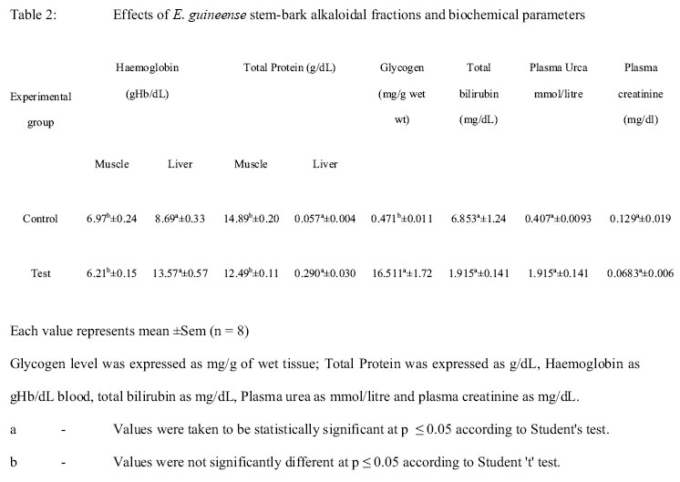
African Journal. Traditional, Complementary and Alternative Medicines Vol. 1, Num. 1, 2004, pp. 45- 54 Research Paper TOXICITY OF ERYTHROPHLEUM GUINEENSE STEM-BARK: ROLE OF ALKALOIDAL FRACTION Adeoye, B.A 1and Oyedapo, O.O.1* Department of Biochemistry, Obafemi Awolowo University Ile-Ife, Nigeria 220005 e-mail:ooyedapo@yahoo.co.uk* Code Number: tc04005Abstract The effect of the in vivo administration of the total alkaloidal fractions of the stem-bark of the Erythropleum guineense on certain biological parameters of Sprague-Dawley rats was investigated. The results revealed that plasma alanine aminotransferase activity, liver aspartate aminotransferase activity, liver glycogen, liver protein, creatinine and haemoglobin concentrations were reduced while plasma aspartate aminotransferase activity, liver alanine aminotransferase activity, plasma urea concentration, bilirubin concentration and alkaline phosphatase activity were elevated in Sprague -Dawley rats treated with total alkaloidal fractions of E. guineense stem-bark. The phytochemical assays revealed that the chemical composition of E. guineense include alkaloids, saponins, cardiac glycosides and tannins. The toxicity of the stem-bark of E. guineense could be attributed to the combined toxicity of other constituents such as tannins, saponins and glycosides with the alkaloids as earlier speculated. Keywords: Toxicity, hepatic, Erythrophelum guineese, alkaloidal fraction, aminotransferases, alkaline phosphatase. IntroductionStudies have revealed that Erythrophleum spp ( family Leguminoseae ) are extremely toxic to livestock all over the world especially to goats, sheep and cows. Several species of Erythrophleum which include E. guineense, E. invorense, E. lasicanthum, E. chlorostachys and E. africanum are known to be poisonous (Watt and Bayer-Brnadwyle 1962, Dalziel 1959, Griffin et al., 1971, Loder et al., 1974). In the Savannah regions of Nigeria, the cattle herders always prevent their animals from grazing along the routes where these species are known to grow (Nwude 1981, Nwude and Chineme, 1981). Further investigations have reported cases of accidental human poisoning after the use of the bark of these plants for traditional uses. E. gunineense is a widely distributed handsome tree with a number of undefined varieties or sub-species which have been the cause of some confusion. The plant is also reputed for its uses as an ordeal poisons for executing capital punishments for witches, to kill or scare away stubborn pests from cultivated farms (Dalziel 1959, Dalma 1970, Loder et al.; 1974). Investigators have implicated alkaloids to be directly involved in eliciting diverse biochemical and pharmaceutical characteristics on biological systems (Clarke 1970). Some alkaloids (pyrrolizidine alkaloids, PAs) have been implicated to be gastro-intestinal tract-irritants and cholinesterase inhibitors and thus affect the nervous systems by causing drowsiness, salivation, laboured breathing, trembling, loss of consciousness, coma and death due to paralysis (Ahmad et al., 1994; Mattocks, 1986; Roberts and Wink, 1998). Presently, there are mere speculations on the nature and activity of the toxicity of E. guineense. It was thought worthwhile to initiate series of studies into the nature of the toxic principles of the stem-bark of E. guineese and their modes of action. As such, the study was designed to investigate the phytochemical constituents of the stem-bark of E. guineese and isolate its total alkaloid content with a view to studying the biological effects of stem alkaloid on certain biochemical parameters of Sprague-Dawley rats so as to have an insight into its role in the overall toxicity of E. guineense. Materials and Methods Plants MaterialsDried stem-barks of E. guineense (G.Don) were purchased from a local herb seller at Ile-Ife, Nigeria. The plant was identified and authenticated by Dr. J. O. Faluyi, Department of Botany, Obafemi Awolowo University Ile-Ife, Nigeria. The stem-barks were cleansed, cut into tiny bits and later ground to powder. Reagents and ChemicalsAll the reagents used were of analytical grades and were obtained from various sources. The solvents were re-distilled before use. Solutions, buffers and reagents were prepared with glass-distilled water. Isolation and Extraction of stem-bark total alkaloidsEthanolic extract of the stem-bark of E. guineense was prepared according to a procedure as earlier described (Oyedapo and Amos, 1997). 750g of powdered stem-bark were suspended in 1.5L of 80%(v/v) ethanol over a period of 15days at room temperature. The suspension was filtered through two layers of cheese-cloth followed by centrifugation at 5000rpm for 10min. The solvent from the supernatant was removed under reduced pressure at 40oC. The brown residue was dried by repeated additions and evaporations of 80%(v/v) ethanol to make scrapping of the residue from the flask easy. The yield was 315g representing 42% of the starting material and was termed ethanolic extract (EE). Isolation and extraction of alkaloids were performed according to a procedure that was based on those earlier described (Kam et al., 1999; Yang et al., 1999). 250g of the extract was dissolved in 600ml of 5% (v/v) HCl solution followed by successive partitioning of acidic filtrate with benzene (l00ml x 3), chloroform (100ml x 3) and ethyl acetate (100ml x 3) to remove non-alkaloid materials. All the organic fractions gave negative tests for alkaloids detecting reagents. The acidic aqueous solution was basified with ammonia solution to pH 12, followed by the extraction of released alkaloids with chloroform (150mlx5) until the basic solution gave negative tests with alkaloid detecting reagents. The chloroform extracts were combined followed by evaporation at 35°C to dryness under reduced pressure to give total alkaloid fraction. The residue was finally taken up in a mixture of ethanol/water (1:1v/v) and was diluted to appropriate concentrations. Phytochemical ScreeningThe ethanolic extract and alkaloid fraction were assayed for the presence of the secondary metabolites using standard procedures (Oyedapo et al., 1999; Sofowora, 1993). (a) For alkaloids, 0.1g of the extract was stirred in 10%(v/v) HCl on a steam bath followed by filtration. The filtrate (1ml) was mixed with a few drops of Meyer's reagent. To another 1ml of the filtrate was added few drops of Wagner's reagent and a few drops of Drangendorff reagent was added to another 1ml of the filtrate. The mixtures were observed for turbidity or formation of precipitate. (b) Saponins were screened by dissolving 0.1g of the extract in 2ml of distilled water, with vigorous shaking until froth appeared. The tubes were warned for 10 min.in a water bath. The presence or absence of frothing was noted after warming. (c) For tannins, 0.1g of the extract was taken up in 10ml-distilled water, and filtered. Then, a few drops of ferric chloride reagent were added to 1ml of the filtrate. The mixture was observed for the formation of blue, blue-black, green or green-black colouration or precipitate. (d) Tests for flavonoids involved (i) suspending 0.1g of the extract in 5ml ethanol, followed by shaking and filtering. To 1ml of the filtrate was added a few drops of 0.5N alcoholic KOH. The mixture was observed for yellowish suspension or precipitate. (ii) (0.1g) of the extract was suspended in 5ml of ethylacetate, shaking vigorously and filtered. To 1ml of the filtrate was added few drops of dilute ammonia solution. The alkaline layer was observed for turning light or deep brown. (e) Cardiac glycosides were screened by dissolving (0.1g) of the extract in 5ml chloroform followed by filtration. Concentrated sulphuric acid was carefully layered at the bottom of the tube without disturbing the solution. It was observed for the formation of a sharp brown ring at the chloroform/sulphuric acid interface. AnimalsHealthy Sprague-Dawley rats of both sexes with an average weight 190g were acclimatised for about four weeks in the laboratory .The animals had free access to Standard pellet diet (Guinea Feeds, Benin, Nigeria) and were watered ad libitum. The rats were randomly divided into two groups of 5 rats each (control group and experimental group) Estimation of acute toxicityForty white albino mice in eight groups (5 rats/group) were given orally isolated alkaloid 0,25,50,75,100,125,150 and175mg/kg bwt dissolved in a mixture of ethanol/water (1:1v/v). The control group received ethanol/water (1:1v/v). All the groups were kept under observation for the next 24-96hr.The minimum and maximum concentrations that produced 0% death and 100% death were obtained. The LD50 value was estimated from the plot of % mortality versus concentration. Treatment of AnimalThe animals were treated as follows: the control group (group 1) received vehicle (ethanol/water 1:1) while the experimental group (group 2) received the drug at 40mg/kg bwt twice as initial dosage and 15mg/kg bwt twice daily for seven consecutive days. The mode of administration was orally with feeding syringe as described by Chang et al., (1992) and Waynforth, (1969). On day 8, the rats were sacrificed, the blood was collected by cardiac puncture into an anticoagulant while the muscles and the livers were removed aseptically. Blood plasma was prepared from the collected blood samples as earlier described by Oyedapo et al., 1999. The blood samples were centrifuged at 4000 rpm for 10 min at room temperature on Galemkamp Junior Table centrifuge. The plasma was collected, transferred into sterile vials and stored in the deep-freezer for further analyses. Biochemical AssaysBiochemical assays were carried out on the plasma, whole blood, livers and muscles as follow, estimation of total protein concentrations as described by Gornallet et al., (1949) with bovine albumin as standard. Extraction, isolation and quantitation of muscle and liver glycogen were performed according to the procedures described by Cowgill and Pardee (1966), Dubois et al., (1956) and Jermyn 1975. The aminotransferases (L-alanine and L-aspartate) were assayed according to the method of Reitman and Frankel (1957) using a Randox Diagnositic kit. The assay mixture consisted of blood plasma (10µl) and alanine aminotransferase (ALT) substrate (50µl) followed by incubation at 37°C for 30 min. The absorbance was read against the reagent blank after 5min at 550nm. One unit of alanine aminotransferase activity was defined as the amount of protein that liberated one µmole of pyruvate/ml per min under experimental condition. Aspartate aminotransferase (AST) was assayed as described above using AST substrate. One unit of AST activity was defined as the amount of protein that liberated one mole oxaloacetate/ml/per min under experimental condition. Plasma alkaline phosphatase activity was assayed as previously described (Oyedapo, 1996) with p-nitrophenyl phosphate (disodium salt) as substrate. The released product phenolate ion was measured at 410nm on CamSpec Visible Spectrophotometer. The blood haemoglobin was quantified as cyanomethemyoglobin as described by Mattentheiner (1971). The absorbance was read against the blank at 540nm. Plasma creatinine was assayed using Jaffe's alkaline-picrate method as described by Chawla (1999). The plasma protein was precipitated by the addition of 2ml of 10% (w/v) sodium tungstate and 2ml of 0.67M sulphuric acid to 2ml of plasma. The supernatant was used for creatinine determination. The test consisted of 3m1 supernatant, 2.5M NaOH (1m1) and 0.04M picric acid (l ml). The reaction mixtures were mixed thoroughly and allowed to stand for 5min. The absorbance was read at 520nm against the blank that consisted of distilled water and other reagents as above. The total plasma bilirubin was assayed according to Sigma Diagnostic assay procedures 1988 and plasma urea as described in Randox laboratory limited handbook with kits. Statistical Analysis Data were analysed statistically using Students' 't' test. The results were expressed as the mean ±SEM. The significance of the differences between control and test groups were determined by the Students 't'test and values of P < 0.05 were taken to imply statistical significance (Parker, 1979). Results and discussion The phytochemical tests revealed that the chemical composition of E. guineense included alkaloids, saponins, tannins, glycosides and flavonoids that may be present in small quantity amongst others. The isolated alkaloid fraction also gave positive tests for alkaloid detecting reagents-Mayer's, Wagner's, Picric acid and Drangendorff. The toxicity of the isolated alkaloid administered orally was estimated to be 62 mg/kg bwt Studies have revealed that L-alanine aminotransferase, L-aspartate aminotransferase, alkaline phosphatase and bilirubin are the most sensitive tests employed in the diagnosis of hepatic diseases (Herper, 1961; Adolph and Lorenz, 1982). The effect of the total alkaloid fraction of the stem-bark of E. guineense on hepatic marker enzymes was presented in Table 1. The results revealed that, the activity of plasma alkaline phosphatase increased in the alkaloid treated rats by about 132.7% when compared with the control rats. Also, plasma L-aspartate aminotransferase and liver L-alanine aminotransferase activities increased significantly by 28% and 140.0% respectively. Moreover, there were slight decrease in the activities of liver L-aspartate aminotransferase and plasma L-alanine aminotransferase by 4.6% and 7.7% respectively. It can then be surmised that the E. guineense stem-bark total alkaloidal fraction was exhibiting dual activity on the enzymes. It was observed that on one hand, it was activating the activities of plasma L-aspartate aminotransferase and liver L-alanine aminotransferase; at the same time causing slight inhibition of the activities of liver-L-asparatate aminotransferase and plasma L-alanine aminotransferase. The results further revealed a significant increase in the plasma bilirubin of alkaloidal fraction treated rats an increase of 140.9% (Table 2). The increase in plasma bilirubin is a suggestive of a possible damage to the liver occasioned by the treatment with the E. guineense stem-bark total alkaloidal fraction. It may be due to impairment of the liver cells and malfunctioning of the kidney eliminating the excess bilirubin or over production of bilirubin and failure of the liver to effectively conjugate it for excretion. Earlier investigators (Oluwole and Bolarinwa, 1997; Oyedapo, 2001) have observed that extracts of leaves of Jatropa curcas and Phyllanthus amarus caused drastic effect in the haematological parameters of treated rats. The plasma urea level was elevated in the alkaloidal fraction treated rats from 0.407±0.093 mmole/litre to 1.915±0.141mmole/litre. The elevation in the level of plasma urea may probably be due to reduction in liver proteins as a result of degradation of proteins and impairment in the excretory function of the kidney possibly due to the treatment with alkaloidal fraction of this plant. Moreover, it was observed that muscle glycogen level increased in alkaloid fraction treated rats and a concomitant decrease in the liver glycogen in the treated rats when compared with control animals (Table 2). The observation that glycogen accumulates in the muscles but decreases in the liver after the treatment may indicate activation of glycogen phosphorylase by the alkaloid fraction a situation that may be organ specific. While the enzyme is being activated in the liver, it is inactivated in the muscle. Moreover there was a decrease in haemoglobin concentration in the alkaloidal fraction treated rats from 6.97±034 gHb/dL to 6.21±0.15 gHb/dL a reduction of about 10.9% .The end product of haem degradation is the bilirubin which is normally transported entirely by albumin through the blood stream and is excreted in the faeces as a water soluble compound (Dow et al., 1997). On the muscle and liver proteins, it was observed that there was an increase from 8.69 ±0.33g/d to 13.57±0.57g/dL in the muscle protein while liver protein decreased slightly from 14.89±0.20 g/dL to 12.49±0.11g/dL (16.12%). It can also be surmised that the action of alkaloidal fraction is organ or tissue specific. In the muscle, the alkaloidal fraction may be promoting protein biosynthesis as observed in the hepato-protective activity of malotilate on galactosamine induced liver damage (Imazumi et al., 1982) .The alkaloidal fraction may be accelerating the degradation of liver proteins as well as increase in amino acid metabolism a situation that might have led to the high elevation of plasma urea (Table 2.0). The plasma creatinine level decreased significantly from 0.129±0.019 mg/dL in the control rats to 0.083±. 0.006 mg/dL in alkaloidal fraction treated rats, by about 35.66%. Creatinine, a metabolic product that is an important indicator of muscle integrity (Adolph and Lorenz 1982). It can be deduced that alkaloidal fraction may be acting by promoting the uptake of creatinine from circulation in the treated rats, which implies that the integrity of the skeletal muscles in the treated rats is adequately protected. Though in conclusive, it is apparent from the results that treatments of the animals with alkaloidal fraction of the stem-bark of E. guineense alter biochemical parameters and alteration in physiological activity in treated rats, which may imply toxicity (Table 2). It may then be surmised that the total alkaloidal fraction was affecting both the liver and the kidney cells, since it became apparent that these two organs are not functioning properly as they ought to be. As such, further investigations need to be carried out to ascertain the roles and contributions of other phytochemicals to the overall toxicity of E. guineese as well as their individual toxicity. AcknowledgementThe authors wish to express our profound gratitude to Prof. Abdul Malik, Co-Director, H. E. J. Research, Institute of Chemistry, University of Karachi, Karachi -75270, Pakistan for making the facilities in his laboratory available to us; for some parts of this investigation. The Third World Academy of Science (TWAS) for the award of a three-month travel grants to H.E.J. Research Institute of Chemistry, University of Karachi, Karachi-75270 Pakistan. References
© Copyright 2004 -African Journal. Traditional, Complementary and Alternative Medicines The following images related to this document are available:Photo images[tc04005t2.jpg] [tc04005t1.jpg] |
| |||||||||
