 |
| About Bioline | All Journals | Testimonials | Membership | News |
|
||||||
|
||||||
African Journal. Traditional, Complementary and Alternative Medicines Vol. 2, Num. 1, 2005, pp. 70-85 Research Paper ANTI-INFLAMMATORY AND ANTIOXIDANT ACTIVITIES OF CASSIA FISTULA LINN BARK EXTRACTS Raju Ilavarasana*, Moni Mallikab and Subramanian Venkataramanc a Department
of Pharmacology, C. L. Baid Metha College of Pharmacy, Old Mahabalipuram
Road, Jyothi Nagar, Thorapakkam, Chennai 600 096, 2Department
of Microbiology, Sri Ramachandra Medical College and Research Institute
(Deemed University), Porur, Chennai-600 116, cDepartment
of Pharmacology and Environment Toxicology, Dr.A.L.Mudiliar P.G. Institute
of Medical Sciences, University of Madras, Taramani, Chennai-600 113, India.
Code Number: tc05009 Abstract Anti-inflammatory and Antioxidant activities of the aqueous (CFA) and methanolic extracts (CFM) of the Cassia fistula Linn. bark were assayed in wistar albino rats. The extracts were found to posses significant anti-inflammatory effect in both acute and chronic models. Cassia fistula bark extracts showed significant radical scavenging by inhibiting lipid peroxidation initiated by CCl4 and FeSO4 in rat liver and kidney homogenates. Both extracts exhibited significant antioxidant activity in DPPH, Nitric oxide and Hydroxyl radical induced invitro assay methods. Both extracts showed Dose-Dependent protective effect against lipid peroxidation and free radical generation in liver and kidney homogenates. Further, the acute toxicity study with the extracts showed no sign of toxicity up to a dose level of 2000 mg/ po. Thus it could be concluded that cassia fistula bark extracts (CFA & CFM) possess significant anti-inflammatory and anti oxidant properties. Key words: Cassia fistula, Anti-inflammatory, Antioxidant activity. Introduction Cassia fistula linn (Caesalpinaceae) tree is one of the most widespread in the forests of India, usually occurring in deciduous forests The whole plant possesses medicinal properties useful in the treatment of skin diseases, inflammatory diseases, rheumatism, anorexia and jaundice (Anonymous,1992, Kirtikar and Basu 1991). A new bioactive flavone glycoside 5,3',4'-tri-hydroxy-6-methoxy-7-O-alpha-L-rhamnopyranosyl-(1 --> 2)-O-beta-D-galactopyranoside with antimicrobial activity was reported by (Yadava and Verma, 2003). Four new compounds, 5-(2-hydroxyphenoxymethyl)furfural, (2'S)-7-hydroxy-5-hydroxymethyl-2-(2'-hydroxypropyl)chromone, benzyl 2-hydroxy-3,6-dimethoxybenzoate, and benzyl 2beta-O-D-glucopyranosyl-3,6-dimethoxybenzoate, together with four known compounds, 5-hydroxymethylfurfural, (2'S)-7-hydroxy-2-(2'-hydroxypropyl)-5-methylchromone, and two oxyanthraquinones, chrysophanol and chrysophanein, were also isolated from the seeds of Cassia fistula by Kuo et al., (2002). Trolox equivalent antioxidant capacity and ferric-reducing antioxidant power assays showed that the antioxidant activities were strongly correlated with total phenols (Luximon-Ramma et al., (2002). The heaptoprotective activity (Bhakta et al., 1999; Bhakta et al., 2001) and the hypoglycaemic activity (Esposito Avella et al.,1991) have been reported. However, a detailed pharmacological screening of the Cassia fistula bark extracts have not been reported. The present study reports the anti-inflammatory and free radical scavenging activity of Cassia fistula bark extracts.
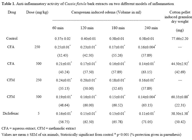
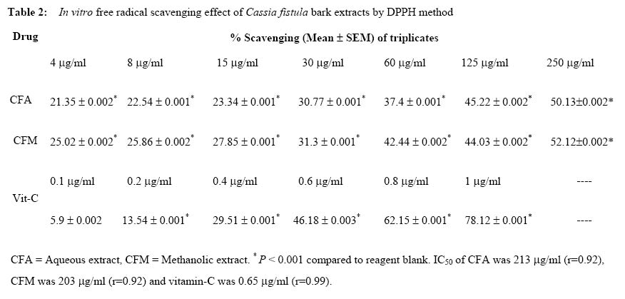
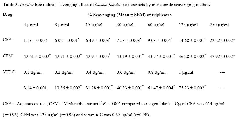
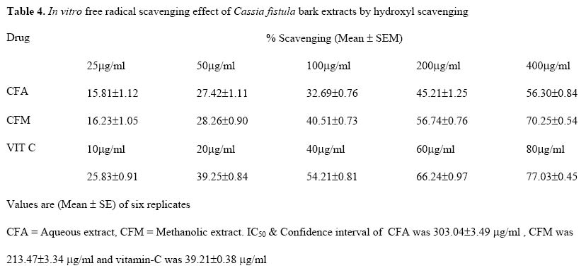
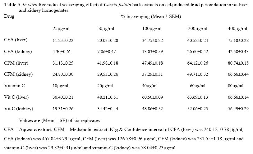
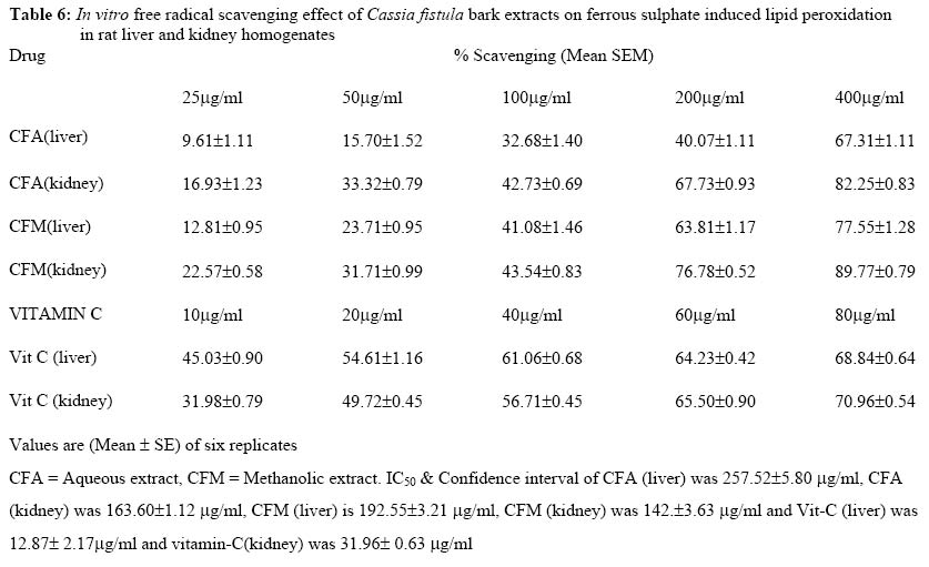
Materials and Methods Plant material The fresh bark of the Cassia fistula was collected from Ariyalur District,Tamilnadu, India in the month of July to August and the plant was identified and authenticated by the Research Officer (Botany), Central Research Institute (Siddha), (Ministry of Health and Family welfare, Govt. of India),Chennai. The voucher specimen of the plant (no: 02/2000) has been kept in the Department of Pharmacology, C. L. Baid Metha College of Pharmacy, Chennai-96, for the future references. Preparation of aqueous and methanolic extracts The freshly collected bark of this plant was chopped, shad dried and coarsely powdered. The powder was defatted with petroleum ether (60-80oc) then successively extracted with methanol and double distilled water using soxhlet extractor. The aqueous and methanolic extracts were dried under reduced pressure using a rotary vacuum evaporator. The percentage yield was 9% w/w for methanol extract (CFM) and 7% w/w for aqueous extract (CFA). The extracts were kept in refrigerator for further use. Chemicals and Instruments Carrageenan (Sigma), 1, 1-diphenyl-2-picrylhydrazyl (DPPH) (Aldrich), Naphthylethylenediamine dihydrochloride (Loba chemie), Thiobarbituric acid (TBA) and Trichloro acetic acid(Sd fine chemicals Ltd). All other reagents used were of analytical grade. UV spectra were recorded in Shimadzu 1601 UV-Visible spectrophotometer. Animals Inbred colony of adult wistar albino rats (170 -200 g) of either sex were used for the pharmacological activities. They were kept in polypropylene cages at 25 ± 2° C, with relative humidity 45-55% under 12h light and dark cycles. All the animals were acclimatized to the laboratory conditions for a week before use. They were fed with standard animal feed (Poultry Research Station Tamilnadu Veterinary and animal sciences University, Chennai, India.) and water ad libitum. The test extracts and the standard drugs were administered in the form of a suspension in water using 1% carboxymethylcellulose as suspending agent. All the pharmacological experimental protocols were approved by the Institutional animal ethics committee (Sanction No: CPCSEA/ CH/ ORG/2001/1910/3 dtd 18.04.2001). Acute toxicity Acute oral toxicity study was performed as per OECD-423 guidelines (acute toxic class method), (Ecobichon, 1997). Wistar rats (n = 6) of either sex selected by random sampling technique were used for acute toxicity study. The animals were kept fasting for overnight providing only water, after which the extracts were administered orally at the dose level of 5 mg / kg body weight by gastric intubation and observed for 14 days. If mortality was observed in 2 out of 3 animals, then the dose administered was assigned as toxic dose. If mortality was observed in 1 animal, then the same dose was repeated again to confirm the toxic dose. If mortality was not observed, the procedure was repeated for further higher doses such as 50, 300 and 2000 mg / kg body weight. Anti-inflammatory activity Carrageenan -induced oedema Paw oedema was induced by injecting 0.1ml of 1% Carrageenan in physiological saline into the subplantar tissues of the left hind paw of each rat (Winter et al., 1962). The extracts CFA (250 & 500 mg/kg) CFM(250mg/kg and 500mg/kg) were administered orally 30 min prior to Carrageenan administration. The paw volume was measured at 60, 120, 180, 240 minutes by the mercury displacement method using a plethysmograph. The percentage inhibition of paw volume in drug treated group was compared with the control group. Diclofenac sodium (5 mg / kg p.o. ) was used as reference standard. Cotton pellet induced granuloma Wistar albino rats (170 -200 gm) of either sex were divided into 4 groups of 6 animals in each group. Cotton pellets weighing 30±1mg were autoclaved and implanted subcutaneously into both sides of the groin region of each rat (D'Arcy etal.,1960), Group I served as control and received the vehicle. The Extracts CFA and CFM at concentration 500mg/kg was administered orally for Group II and III animals for 7 days. Group IV animals received diclofenac at a dose of 5 mg/kg orally for same period. On the 8th day the animals were sacrificed and the pellets together with the granuloma tissues were carefully removed, dried in an oven at 60oC, weighed and compared with control. Diclofenac sodium (5 mg / kg / p.o.) was used as reference standard. Evaluation of Antioxidant activity Scavenging of DPPH radical This assay is based on the measurement of the scavenging ability of antioxidant test substances towards the stable radical. The free radical scavenging activity (Yokazawa et al., 1998) of the extracts (CFA and CFM) were examined in vitro using DPPH radical. The test extracts were treated with different concentrations from a maximum of 250 µg/ml to minimum of 4 µg/ml . The reaction mixture consisted of 1 ml of 0.1mM DPPH in ethanol, 0.95 ml of 0.05 M Tris-HCl buffer (pH 7.4), 1 ml of ethanol and 0.05ml of the herbal extract. The absorbance of the mixture was measured at 517 nm exactly 30 sec after adding extract. The experiment was performed (in triplicate) and % of scavenging activity was calculated using the formula 100 - [100/blank absorbance × sample absorbance]. Scavenging of nitric oxide Sodium nitroprusside (Sreejayan Rao,1997) (5µM) in standard phosphate buffer solution was incubated with different concentration of the test extracts dissolved in standard phosphate buffer (0.025M, pH 7.4) and the tubes were incubated at 25 °C for 5 hr. After 5 h, 0.5 ml of incubation solution was removed and diluted with 0.5 ml Griess reagent (prepared by mixing equal volume of 1% sulphanilamide in 2% phosphoric acid and 0.1% naphthylethylene diamine dihydrochloride in water). The absorbance of chromophore formed was read at 546 nm. The control experiment was also carried out in similar manner, using distilled water in the place of extracts. The experiment was performed (in triplicate) and % scavenging activity was calculated using the formula 100 - [100/blank absorbance × sample absorbance]. The activity was compared with ascorbic acid, which was used as a standard antioxidant. Hydroxyl Radical Scavenging activity
The Hydroxyl Radical Scavenging activity was measured by studying the competition between deoxyribose and the extract for hydroxyl radicals generated from the Fe3+ / ascorbate/ EDTA/ H2o2system . The reaction mixture contained deoxy ribose (2-8mM), Fecl3 (0.1mM), EDTA (0.1mM), H2o2 (lmM), ascorbate (0.1mM), KH2PO4 - KOH buffer (20mM, pH7.4) and various concentrations (25-400 µm of extracts and std 10 to 80 µm /ml) of standard drug in a final volume of 1 ml. The reaction mixture was incubated for 1hr at 37°C, deoxyribose degradation was measured at 532 nm (Mary et al.,2002). CCl4 induced lipid peroxidation Rat liver and kidney ( 30% w/v) homogenate in ice cold 0.15m Potassium chloride was prepared in a homogenizer. Aliquots of 0.5 ml of homogenate were taken in different small conical flasks. These flasks were incubated at 37°C in a constant shaker bath (150 cycles/min) for 45 min with 1.5 ml of Potassium sulphate buffer (pH 7.4), 2 ml of 0.15m Potassium chloride CFA and CFM at (25-400 µg /ml and Vitamin -C 10 to 80 µg /ml) in different flasks and finally 10µl of carbon tetra chloride(CCl4) was added. In case of control, only drug was excluded. The reaction was stopped by the addition of 4 ml of 10% (w/v) trichloro acetic acid and after incubation, the contents were centrifuged at 4000 rpm for 10min and about 2 ml of clear supernatant was transferred to a graduated tube and 2 ml of 0.67% w/v of thiobarbituric acid was added and heated in a boiling water bath for 15 min. The tubes were cooled, bringing the mixture to pH 12-12.5 with Potassium hydroxide, stabilized the colour developed and the absorbance was measured at 543nm (Comporti., 1989). Ferrous sulphate induced lipid peroxidation scavenging The degree of lipid peroxidation was assayed by estimating the thiobarbituric acid-reactive substances (TBARS) by using the standard method (Okhawa et al., 1979) with minor modifications (Tripathi and Sharma, 1998). Briefly, different concentrations of extracts (25-400µgm/ml) were added to the 10% liver and kidney homogenate. Lipid peroxidation was initiated by adding 100 µl of 15mM FeSo4 solution to 3ml of liver homogenate (final concentration was 0.5 mM). After 30min, 100 µl of this reaction mixture was taken in a tube containing 1.5ml of 0.67% TBA in 50% acetic acid. The mixture was heated in a water bath at 850C for 30 min and in a boiling water bath to complete the reaction. The intensity of pink coloured complex formed was measured at 535 nm in a spectrophotometer. The percentage inhibition of lipid peroxidation was calculated, as per the following formula, Inhibition (%) = (control -test) X 100/ control. Statistical analysis The statistical analysis was carried out using one way analysis of variance (ANOVA) followed by Dunnet,s t -test, P- values < 0.05 were considered as significant. Results Acute toxicity study The extracts CFA and CFM did not cause any mortality up to 2000 mg/kg and were considered as safe (X-unclassified),(OECD, 1996). Anti-inflammatory study Carrageenan paw oedema Both extracts CFA (250 and 500mg/ kg) and CFM (250 and 500mg/ kg) exhibited significant (P < 0.01) reduction in paw odema volume of rats. The percentage inhibition of extracts is shown in Table 1. Cotton pellet granuloma Aqueous (CFA) and methanolic (CFM) extracts of Cassia fistula bark at the dose level of 500mg/kg/p.o., significantly (P < 0.001) reduced the weight of the cotton pellet granuloma in rats. The percentage inhibition of CFA was 42.69%, CFM 22.31% and Diclofenac, the reference standard 50.42% as shown in Table 1. Antioxidant effect DPPH scavenging The aqueous (CFA) and methanolic extracts (CFM) of the Cassia fistula bark showed promising free radical scavenging effect of DPPH in a concentration dependant manner up to a concentration of 250 µg / ml. The CFM showed more scavenging activity than the CFA. The reference standard ascorbic acid also demonstrated a significant radical scavenging potential in the concentration of 1µg / ml. The DPPH radical inhibition (%) was 50.13, 52.12 and 78.12 for CFA, CFM and ascorbic acid, respectively (Table 2). Nitric oxide scavenging The Cassia fistula bark extracts (CFA and CFM) showed significant free radical scavenging action against nitric oxide (NO) induced release of free radicals at the concentration 250 µg / ml, showing 22.22% and 47.92% of NO inhibition, respectively. Ascorbic acid was used as reference standard. The % inhibition is shown in Table 3. OH Radical Scavenging The CFA and CFM extracts (25-400µg/ml) significantly scavenged the hydroxyl radicals generated by the EDTA/H2O2 system, when compared with that of Ascorbic acid. The percentage scavenging of OH radicals by CFA and CFM was increased in a dose dependant manner. The standard vitamin-C (10-80µg/ml), also showed scavenging effect (Table 4). Ic50 value of CFA was 303.04±3.49 µg/ml, CFM was 213.47±3.34 µg/ml and vitamin-C was 39.21± 8.38 µg/ml. Lipid peroxidation induced by CCl4 Lipid peroxide formation by CCl4 was inhibited by CFA and CFM at all tested doses (25-400µg/ml). The percentage inhibition of peroxide formation was increased in a dose dependant manner. Vitamin-C (10-80µg/ml) also showed significant reduction in LPO. Results are shown in table 5. The Ic50 confidence interval of CFA in liver and kidney homogenates was 240.12±0.78 µg/ml and 457.84±3.79 µg/ml, respectively. The IC50 value of CFM in liver and kidney homogenate was 126.78 ±0.96 µg/ml and 251.55±1.18 µg/ml, respectively. The IC50s of Vitamin-C were 29.32±0.31 µg/ml and 58.04±0.23 µg/ml in liver and kidney, respectively. Effect on FeSO4 induced lipid peroxidation The Cassia fistula bark extracts (CFA and CFM) showed a concentration dependent inhibition of FeSO4 induced lipid peroxidation in rat liver and kidney homogenates (Table 6). The Ic50 confidence intervals of CFA extract were found to be 257.52± 5.80 µg/ml and 163.60± 1.12µg/ml in liver and kidney homogenate, respectively. The IC50S of CFM and confidence interval of liver and kidney homogenate were found to be 192.55±3.21µg/ml and 142±3.63µg/ml, respectively. The IC50S were found to be 12.87±2.17 µg/ml and 31.96±0.63µg/ml for liver and kidney, respectively for reference standard (Vitamin-C). Discussion In Indian system of medicine, certain herbs are claimed to provide relief of pain and inflammation. The claimed therapeutic reputation has to be verified in a scientific manner. In the present study one such drug Cassia fistula bark was taken for the study. The bark extracts of Cassia fistula possess significant anti-inflammatory effect in the acute and chronic anti-inflammatory model of inflammation in rats. Reactive Oxygen species (ROS) generated endogenously or exogenously are associated with the pathogenesis of various diseases such as atherosclerosis, diabetes, cancer, arthritis and aging process (Guyton et al., 1997, Halliwell and Gutteridge, 1999). Inflammation is a complex process and ROS play an important role in the pathogenesis of inflammatory diseases. (Conner and Grisham, 1996). Thus antioxidants which can scavenge ROS are expected to improve these disorders. Carageenan induced inflammation is a useful model to detect oral action of anti-inflammatory agents (Di Rosa et al, 1971). The development of oedema in the paw of the rat after the injection of Carageenan is due to release of histamine, serotonin and prostaglandin like substances (Vinegar et al., 1969). The significant ameliorative activity of the extracts (CFA and CFM) and standard drug observed in the present study may be due to inhibition of the mediators of inflammation such as histamine, serotonin and prostaglandin. The Carageenan assay is a good method for the comparative bioassay of anti-inflammatory agents. The present results indicate the efficacy of Cassia fistula bark as an efficient therapeutic agent in acute anti-inflammatory conditions. The cotton pellet granuloma method (D'Arcy etal.,1960), has been widely employed to assess the transudative, exudative and proliferative components of chronic inflammation. The fluid absorbed by the pellet greatly influences the wet weight of the granuloma. The results indicate that CFA has more anti-transudative and anti-proliferative activity than CFM. The free radical scavenging activity of the extracts was evaluated based on the ability to scavenge the synthetic DPPH. This assay provided useful information on the reactivity of the compounds with stable free radicals, because of the odd number of electrons. DPPH shows a strong absorption band at 517 nm in visible spectrum (deep violet colour). As the electron became paired of in the presence of free radical scavenging, the absorption vanishes and the resulting discoloration stoichiometrically coincides with respect to the number of electrons taken up. The bleaching of DPPH absorption is representative of the capacity of the test drugs to scavenge free radicals independently. Hydroxyl radical is the principal contributor for tissue injury. The formation of Hydroxyl radical from fenton reaction was quantified using 2, deoxy-D-ribose degradation. The extracts CFA and CFM inhibited hydroxyl scavenging activity. Sodium nitroprusside serves as a chief source of free radicals. The absorbance of the chromophore formed during diazotization of the nitrite with sulphanilamide and subsequent coupling with napthylethylene diamine is used as the marker for NO scavenging activity (Mukherjee, 1989). The chromophore formation was not complete in the presence of Cassia fistula bark extract (CFA and CFM), which scavenges the NO thus formed from the sodium nitroprusside and hence the absorbance decreases as the concentration of the extracts (CFA and CFM) increases in a dose dependent manner Lipid peroxidation has been implicated in the pathogenesis of various diseases including arthritis. It is well established that bioenzymes are very much susceptible to LPO, which is considered to be the starting point of many toxic as well as degenerative processes. The extracts (CFA and CFM) exhibited protection against lipid peroxidation induced by CCl4.. Initiation of lipid peroxidation by ferrous sulphate takes place through ferryl perferryl complex (Gutteridge, 1985). The extracts (CFA and CFM) inhibited the FeSO4 induced lipid peroxidation in a dose dependant manner. The inhibition could be caused by the inhibition of formation of Ferryl perferryl complex. The presence of flavonoids in Cassia fistula ((Yadava and Verma, 2003) may be responsible for the anti inflammatory and antioxidant effects. Alkaloid and flavanoid constituents have already been reported in the barks (Gupta et al., 1989). The results of present study with Cassia fistula bark extracts have good correlations with the therapeutic use of Cassia fistula bark in the treatment of inflammatory conditions by practitioners of Ayurvedic system of medicine. Plants which belong to Caesalpinaceae family are rich in flavonoids and bio flavonoids are known for their anti-inflammatory and antioxidant activities. Further research is in progress to identify the biomolecules responsible for the anti-inflammatory and antioxidant activities. Acknowledgement This work is carried at the Department of Pharmacology, C.L. Baid Metha College of Pharmacy, Chennai-96. Authors are highly thankful to the Secretary and the Principal for providing the facilities. I also thank Dr.Sasikala, Research Officer (Botany), Central Research Institute (Siddha), Chennai -106 for identifying and authenticating the plant. References
The following images related to this document are available:Photo images[tc05009t5.jpg] [tc05009t6.jpg] [tc05009t2.jpg] [tc05009t1.jpg] [tc05009t3.jpg] [tc05009t4.jpg] |
| |||||||||
