 |
| About Bioline | All Journals | Testimonials | Membership | News |
|
||||||
|
||||||
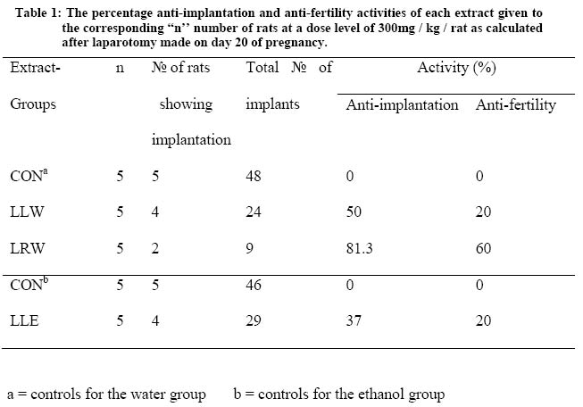
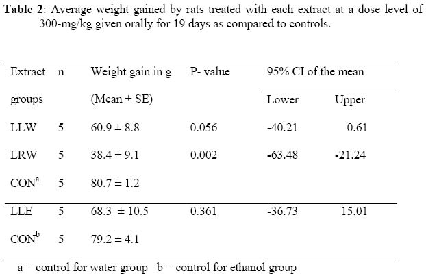
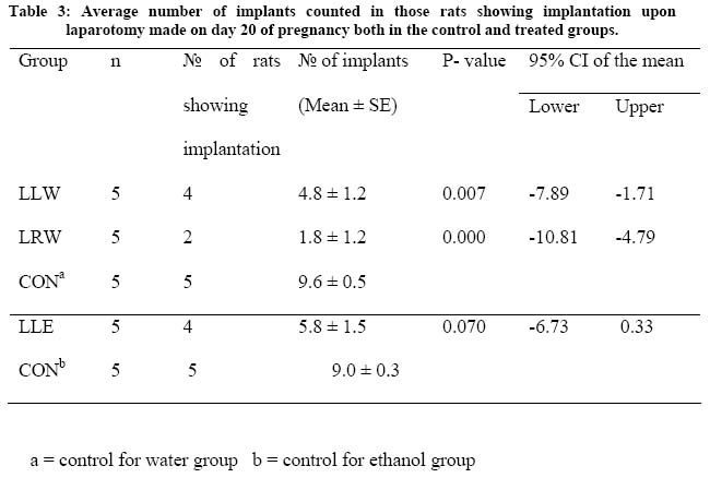
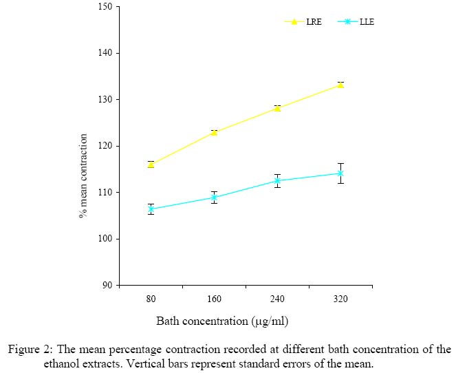
African Journal. Traditional, Complementary and Alternative Medicines Vol. 2, Num. 2, 2005, pp. 103-112 IN VIVO AND IN VITRO ANTI-FERTILITY AND ANTI-IMPLANTATION PROPERTIES OF LEONOTIS OCYMIFOLIA IN RATS Geremew Tafesse1, Yalemtsehay Mekonnen 2, Eyasu Makonnen3 1 Department of Biology, Faculty of Education, Dilla College of Teachers'Education and Health Science, Debub University, Dilla, Ethiopia, 2Department of Biology, Faculty of Science, Addis Ababa University, P.O.Box 1176, Addis Ababa, Ethiopia, 3Department of Pharmacology, Faculty of Medicine, Addis Ababa University, P.O.Box 9086, Addis Ababa, Ethiopia Email: yalemtsehay@yahoo.comCode Number: tc05012 AbstractThe anti-fertility effect of the aqueous and ethanol extracts of the leaves and roots of Leonotis ocymifolia were studied both in vivo and in vitro. The anti-implantation and anti-fertility activities of the ethanol leaves extract were 37% and 20%, respectively. The body weight recorded for 19 days starting from day 1 of pregnancy showed that all rats of the control groups showed considerable weight gains over the period of 19 days. The weight gained by the rats treated with aqueous root extract was significantly less as compared to the controls. On the contrary, those treated with both aqueous and ethanol leaf extract did not show a significant difference in their weight gains over the same period. Aqueous root and leaf extracts reduced the number of implants significantly. No significant difference was, however, observed between the average number of implants counted in rats treated with ethanol leaf extract and the control group. All extracts were observed to increase acetylcholine induced uterine contraction. The mean percentage contractions showed by these extracts in the presence of acetylcholine were significantly different from that of acetylcholine alone (P < 0.05). The results of this study suggest that the leaves and roots of this plant may possess hormonal properties that can modulate the reproductive function of the rats. Key words: Anti-fertility, Anti-implantation, Leonotis ocymifolia, aqueous extract, ethanol extract, leaves, roots, Introduction Among those plants with claimed anti-fertility properties in traditional medicine in Ethiopia Leonotis ocymifolia (Burm. F), Family Labiatae (Minassie, 1991) locally known, as Ras-kimir or Yeferes Zeng is one. Its leaves are used in the treatment of hookworm; while the flowers and roots in gout and leishmaniasis (Mungai, 1997). People living around Bale area in southern Ethiopia use the leaves of this plant to expel intestinal parasites. Other species, L. africana and L. nepetaefolia, are known to possess anti-implantation and/or uterotonic effects (Fransworth et al., 1975; Desta, 1994 ). In addition, an in vitro test showed that L. nepetaefolia induced uterine contraction in rats (Calixto et al., 1991). The objective of the present work was to investigate the antifertility and antiimplantation effect of the crude extracts of the leaves and roots of Leonotis ocymifolia in an attempt to establish the traditional use. Materials and methods Plant material collection Leaves and roots of Leonotis ocymifolia were collected from Bale Mountains National Park (BMNP) around the entrance of the head quarter (Dinsho) 400 km south of Addis Ababa in May 2001. Samples were kept after botanical identification in the National Herbarium of the Addis Ababa University under voucher number 2/2001. Extracts preparation The leaves and roots were dried in a shade and ground into powder. The powder of the leaves and roots was then macerated in distilled water and 90% (v/v) ethanol in a ratio of 1 to 6 (w/v) and 1 to 4 (w/v) respectively. The suspensions of the samples were rotated on a shaker for 24 h at room temperature. Then each sample was filtered using cotton wool and Whatman filter paper No1. The water extracts were lyophilized while the ethanol extracts were evaporated to dryness using Rotavapour at 40oC. The resulting residues were stored at -200C until used for the experiments. The produced extract samples were named as Leonotis root water extract (LRW), Leonotis leaf water extract (LLW), Leonotis root ethanol extract (LRE), and Leonotis leaf ethanol extract (LLE). Experimental animals Rats were bred in the animal house of the Department of Biology, Addis Ababa University. Newly borne female rats 10 to 14 weeks of age ready for the first mating were used for the test (Donnelly, 1987). The newly borne rats were separated from their mothers at their weaning age, and females were separated from males at the age of four weeks in order to prevent uncontrolled mating. Male rats were used for the test after checking their proven fertility through a preliminary mating system with selected rats. They all fed on pellet diet and watered ad libitum In vivo anti-fertility and anti-implantation testThe method described by Wiliamson et al (1996) was employed for the experiment Virgin female rats of age between 10 and 11 weeks weighing between 175 and 200g were divided into control and experimental groups of 5 rats/group. One group was used for the water extracts and the other for the ethanol extracts. The control groups were named as CONa for the water extract and CONb for the ethanol extract groups respectively. Each rat was kept singly in a cage to acclimatize without dosing for one week. The experimental groups were treated with the specific sample orally at a dose of 300mg/kg-body weight/rat, while the control groups received the vehicle (aqueous ethanol (70%) v/v in 0.5 ml volume. The water extract was dissolved in distilled water, while the ethanol extract in 70 % v/v ethanol and distilled water at a ratio of 1:1.5. Males of proven breeding ability were introduced into each cage on the ninth day of treatment. Then vaginal smear and/or vaginal plug were checked each morning to examine the existence of sperm in order to confirm mating. The first morning mating observed was considered day-1 of pregnancy according to the methods employed by Desta (1994) and Uguru et al. (1995). The treatment of animals continued until day-19 of pregnancy, and the weights of the animals were taken daily. The control and experimental rats were sacrificed on day-20 to determine the number of implants. The weights of the animals and numbers of implants were compared with those of controls. The experiment was carried out twice for each sample except Leonotis root ethanol extract because the yield of the crude extract was small. The anti-implantation and anti-fertility activities of each sample were calculated using the following formula (Williamson et al., 1996). Anti-implantation Activity = № implants in control -№ of implants in test group x 100 № of implants in control group
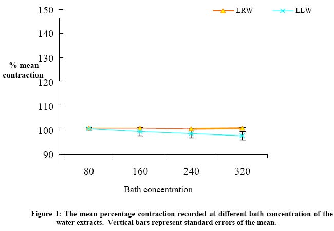
Anti-fertility activity = № of animals showing no implantation x 100 Total № of animals In vitro uterine contraction test Non-pregnant rats were used for the in-vitro (uterine preparation) test. A gentle blow on the head killed each rat. The abdomen was opened and the uterine horns served at their junctions with the fallopian tubes and placed in a dish containing De Jalon´s solution. For each experiment an approximate 5 cm of the uterine strip was set up in a thermostatically regulated 25ml organ bath that contains the solution maintained at 370 C and gassed with air. The uterine strip was tied with a string to a transducer (Grass FT.03) that was connected to Grass Polygraph model 07 to record contractions. A tension of 1gm was applied to the tissue and was allowed to equilibrate for at least 30 min before starting the test. Acetylcholine (Ach) was used as a stimulant to record contractions. A response curve was constructed using ACh at a final bath concentration each of 40ng/ml, 80ng/ml, 160ng/ml and 320ng/ml with a time interval of 3min. Each time, the added ACh was left in contact with the tissue for 30 sec and then was washed with De Jalon' s solution. It was then left to resume its normal contraction. ACh of 80ng/ml was selected as the control concentration that induced sub-maximal contraction of the uterine tissue. Extract sample of the given concentration was added in the organ bath and left in contact with the tissue for 5min. The control ACh (80ng/ml) was added at the end of the 5 min in the presence of the sample and then washed after 30 sec. After rhythmic contraction of the tissue resumed, the control ACh was added in order to establish the reversible contraction capacity of the tissue and also to test the extent the extract acted upon the uterine tissue. The same procedure was repeated for different samples at different concentration. Each sample was tested at 80µg/ml, 160µg/ml, 240µg/ml and 320µg/ml, as final bath concentration. Contraction peaks recorded by the polygraph were measured in cm. The contraction peak of the control ACh (80ng/ml) was taken as a reference (100%). The peak produced due to ACh (80ng/ml) was compared with that produced due to the extract and ACh when added together. The effect of the sample at a given bath concentration was then recorded by measuring the length of the peak due to the extract + ACh. Each value was converted to a percentage contraction by considering a 100% ACh contraction. Then the mean percentage contraction was taken to compare the result (Mekonnen, 1999). Seven uterine strip preparations were used for each sample tested; the average differences were recorded. Statistical analysis For the in vivo anti-implantation test, the mean ± SEM weight gain as well as the mean ± SEM number of implants in each test group was compared with the respective control groups. Independent Student's t-test was used to analyze the result. In the in vitro test the mean ± SEM percentage tissue contraction due to ACh was also compared with the mean ± SEM percentage tissue contraction due to ACh with the extract at each bath concentration. One-way ANOVA test was used to analyze the results. In each test a 95% confident interval was used. Results In vivo anti-implantation/anti-fertility activities Table 1 summarizes the number of rats with implantation and the total number of implants in each group. It shows the percentage anti-implantation and anti-fertility activities exhibited by each extract in comparison to the corresponding controls. It was noted that some of the rats in test groups did not show implantation. As shown in Table 1, the highest rate of anti-implantation activity of LRW was 81.3% while the anti-implantation and anti-fertility activities of LLE extract were 37% and 20% respectively. The body weight recorded for 19 days starting from day 1 of pregnancy showed that all rats of the control groups have considerable weight gains. The body weight gained by rats in most test groups showed a significant difference (P< 0.05) with their corresponding controls (Table 2). The weight gained by the rat groups LRW (t = - 4.624, P = 0.002) was significantly less than the controls. However, when compared with the controls, LLW and LLE treated rats there was no significant difference in their weight gains (t = - 2.238, P = 0.056 and t = - 0.968, P = 0.361 respectively). The number of implants in the LRW treated rats significantly less (t = - 5.982 and P 0.000) than the average number of implants in the control group. The rat treated with LLW (t = -3.578, P = 0.007), showed significant differences with their respective control group. No significant difference was, however, observed between the average number of implants in rats treated with LLE (t = - 2. 09, P = 0.070) and the control group.(Table 3) In vitro uterine contraction effect The contractions of the rat uterus evoke byLRW were significant. They increashed Ach induced uterine contraction. The mean percentage contractions exhitited by these extracts in the presence of Ach were significantly different from that of ACh alone (P < 0.05, F= 46.8, 49.8, and 63.1 respectively, Figure 1). However, extract LLW did not show significant difference in percentage contractions from that of the control (Figure 1). LLE and LRE extracts also showed uterine contractions, which were significantly different from that of the control (P < 0.05, F = 43.1, 31.1and 88.1) respectively. The dose dependent increase in the mean precentage tissue contractions is shown in Figure 2. Discussion The results demonstrated that L. ocymifolia has anti-fertility and anti-implantation activities. The anti-implantation activity observed by LRW might indicate the presence of one or more active ingredients in the root of Leonotis ocymifolia. The root extracts of its related species L. africana was previously reported to have similar effect (Desta, 1994). During the preliminary investigation with L. ocymifolia on mice, the anti-implantation / anti-fertility activity was observed in the leaves in contrast to the present observation. This might be explained in terms of species variability (Uguru et al., 1995). Substances with anti-fertility properties may exert their effects at the ovarian level by inhibiting ovulation and or steroidogenesis. This was also supported by experimental evidence, which suggest that contraceptive steroids act directly on the ovary to inhibit ovulation and/or some aspects of steroidogenesis (Goldfien 1995). It was previously reported that the anti-fertility/ anti-implantation effect of such agents could be due to their estrogenic nature (Waller 1987, Debella et al. 1999). Although it is not conclusive, the absence of implantation sites in some group of rats in the present study might be due to the effect of the extracts. The administration of low concentrations of compounds with estrogenic activity to many species during early pregnancy has been reported to result in rapid passage of ova through the oviducts and expulsion of the ova from the uterus (Fransworth et al., 1975). The decreased fertility due to accelerated transport of ova might be not only because of the expulsion of the fertilized ova from the reproductive tract but also because of the degeneration of the fertilized ova while transported into the uterus too early (Fransworth et al., 1975). The reduction in the number of implants observed in some group of rats in the present study might be due to such properties of the extract. But this is not conclusive since decreasing in the fertility or implantation could be due to uterine environment rather than the rapid transport. It is possible that extracts can produce anti-fertility effect through their ability to alter the estrus cycle of the animal (Makonnen et al., 1997). Therefore, the number of rats showing no implantation in the present study might be due to a prolonged diestrus phase, which gives no chance for fertilization. The ethanol extracts exhibited significantly greater uterine contractions than the water extracts in the present study, which could be explained due to the difference in polarity of the molecules extracted. Uterine contractility produced by the extracts could be due to to their oxytocin-like property or estrogenic activity (Uguru et al. 1995; Mekonnen, 1999). It has been reported that estrogen possesses some oxytocic effect and hence may be involved in increasing uterine contractility (Batra, 1986). Therefore, uterine contractions demonstrated by the extracts in the present study might be partly due to these agents. Those extracts that showed anti-implantation activities in vivo were also capable of showing uterine contraction in vitro. However there was no sign of abortion such as vaginal bleeding or weight loss observed during the in vivo test. This might be because (on one hand) the extract concentration used for in vivo was too low to induce abortion or on the other hand the uterine contracting effect of the extracts could be due to other pharmacological mechanisms that were not sufficient to cause abortion. The exact mechanism(s), of anti-implantation or uterine contraction observed in this study requires further investigations. Moreover, fractionation of the extract is a worthwhile effor to isolate the active compounds responsible for the observed activity. References
© Copyright 2005 -African. Journal. Traditional, Complementary and Alternative Medicines The following images related to this document are available:Photo images[tc05012f2.jpg] [tc05012t1.jpg] [tc05012t3.jpg] [tc05012t2.jpg] [tc05012f1.jpg] |
| |||||||||
