 |
| About Bioline | All Journals | Testimonials | Membership | News |
|
||||||
|
||||||
African Journal. Traditional, Complementary and Alternative Medicines Vol. 2, Num. 3, 2005, pp. 312-325 Research Paper HEALING EFFECT ON CHRONIC GASTRIC ULCERS AND SHORT-TERM TOXICITY PROFILE OF THE LEAF METHANOL EXTRACT OF OCIMUM SUAVE WILD (LAMIACEAE) IN RATS Paul V. Tan1, 4, Christopher Mezui1, George E. Enow-Orock3, Theophile Dimo1, Barthelemy Nyasse2 1Department of Animal Biology and Physiology, Faculty
of Science, P.O. Box 812, University of Yaounde I, Cameroon., 2Department
of Organic Chemistry, Faculty of Science, P.O. Box 812, University of Yaounde
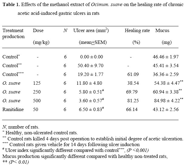
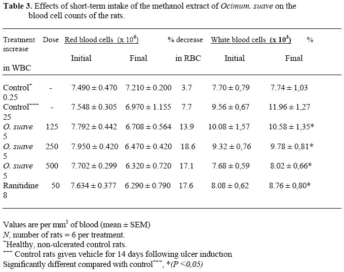
I, Cameroon.,3Pathology Unit, Yaoundé General Hospital, Cameroon., 4 Code Number: tc05034 AbstractThe gastric cytoprotective actions of the extract of Ocimum suave wild (lamiaceae) have previously been demonstrated. We have investigated here the healing effect of the leaf methanol extract of Ocimum suave against chronic gastric ulcers induced in experimental rats. Chronic gastric ulcers were induced using acetic acid and the induced ulcers treated over a period of two weeks using daily oral doses (125 – 500 mg/kg) of the extract. Possible toxic effects of the extract given in the short term were also investigated. The extract reduced ulcer indices from 50.40 in the 4-day controls to 11.8, 5.8, and 3.6, respectively, for the rats receiving 125, 250 and 500 mg/kg of the extract. The highest dose of the extract (500 mg/kg) showed a highly significant (P < 0.001) reduction of ulceration with a corresponding healing rate of 81.25 per cent. Treatment with the plant extract was also associated with a significant increase in mucus production up to 57 per cent (P < 0.01) for the 500 mg/kg dose. A similar increase in mucus production was not observed with ranitidine although it generated a healing rate of 66 per cent. No apparent toxicity signs were observed through food and fluid intakes, vital organ weights, animal behaviour, stool texture, red and white blood cell counts and histopathological evaluation. The results of the present study show that in addition to the previously demonstrated cytoprotective antiulcer actions of the leaf methanol extract of O. suave, the extract also possesses potent healing effects against chronic gastric ulcers. Enhanced mucus production appears to play a significant role in the mode of action of the extract Key words: Ocimum suave; healing effects; chronic gastric ulcers, short-term toxicity Introduction. The search for novel non-toxic, antiulcer preparations from medicinal plants is currently in vogue in order to obtain alternative sources of medicine for the management of gastric hypersecretion and gastroduodenal ulcers. In the developing nations, this turn of events has been prompted in part by the high cost of modern antiulcer medication, as well as the multiple side effects that result from their prolonged use. In Cameroon, a nation-wide OAU/STRC-sponsored ethnobotanical survey (Adjanohoun et al., 1996) revealed the presence of many plants purported by traditional practitioners to be efficient for the management of complaints symptomatic of peptic ulcer disease. We embarked on screening studies (Tan et al., 1997), which have shown that several of these plants do contain useful cytoprotective antiulcer effects. Many species of the genus Ocimum, namely, Ocimum viride Linn, Ocimum gratissimum Linn, Ocimum basillicum Linn and Ocimum canum Sims, have been cited with various medicinal uses (Adjanohoun et al., 1996; Mshana et al., 2000), but antiulcer actions have been attributed to none of them. Ocimum suave has however been shown to posses mosquito repellent, analgesic, antipyretic, acaricidal and antibiotic activities ( Seyoum et al., 2003; Makonnen et al., 2003a&b; Mwangi et al., 1995; Janssen et al., 1989). Studies carried out on the methanol extract of Ocimum suave wild (Lamiaceae, syn.: Labiatae (Thonner, 1915), syn.: Menthaceae (Burger, 1967)) showed that it contained cytoprotective antiulcer effects that are not linked to an antisecretory potential (Tan et al., 2002). This seasonal, ruderal and anthropophilic plant is found in tropical Asia and in West and East Africa, where its geographical distribution is limited to mountainous areas (Watt et al., 1962; Raynal et al., 1979; Hutchinson and Dalziel, 1963). Previous studies showed that the oil obtained from the leaves of O. suave containsphenols (Watt et al., 1962), and recent work shows that the major components of the oil are p-cymene (59 %), alpha-thujene (10 %), myrcene (7 %) and thymol (7 %) (Keita et al., 2000). The gastric mucosal protective action of the methanol extract was essentially exhibited through increased gastric mucus production, and we suggested that such results may pave the way for the establishment of a new anti-ulcer therapy regimen that will not require the use of antacids and anti-secretory agents (Tan et al, 2002). However, although experimental ulcers of the type used in our previous study do provide information about the cytoprotective potency of the extract against selected gastric mucosal irritants, it cannot be concluded that gastric ulcer healing effects will automatically be obtained when the extract is used to treat well-established chronic gastric ulcers of the type seen in many patients. The aim of the present work was therefore to study the healing effect of the extract on chronic experimental gastric ulcers induced in rats using glacial acetic acid. Because some drugs can show mild-to-severe side effects even after short-term intake, we also evaluated the possible toxic effects of the extract on blood cell count, histology of vital body organs, as well as on some aspects of animal behavior. Materials and methods AnimalsMale Wistar rats (150-200 grams) raised in the animal house of the Faculty of Science, University of Yaoundé I, were used. They were fed a standard laboratory diet (S.P.C. Ltd, Bafoussam, Cameroon) and given fresh water ad libitum. Unlike for the prophylactic tests, the animals were deprived of food only for 24 h prior to the induction of acetic acid ulcers but free access to water was allowed. Prior authorization for the use of laboratory animals in this study was obtained from the Cameroon National Ethics Committee (Reg. No. FWA-IRB00001954). Preparation of the plant extract. The leaves of O. suave were harvested in August 2003 from the Sabga and Wainama hills of Bamenda and Jakiri, respectively, in the North West province of Cameroon. Botanical identification was done in the National herbariun, Yaounde by Jean Michel Chrana, by comparison with existing herbarium specimen No. HNC:6077/6914 (R. Letouzey). The dried leaves of the plant were crushed to a powder form and 1 kg of the powder was subjected to cold methanol extraction (2 L) for 48 h. The solution obtained was evaporated using a rota vapor to obtain 87 g of extract. The extract was re-dissolved in water and the solution filtered to obtain the water-soluble portion (pH 4.39). The water insoluble part of the methanol extract was discarded and water soluble portion evaporated to dryness at 50oC using a convection air oven. The resulting brownish solid (20 g) was used for the ulcer healing tests. Ulcer healing test. The method described by Pillai and Santhakumari (1984) was used. Briefly, laparotomy was performed under light ether anesthesia on experimental rats that were deprived of food during the preceding 24 hours. Fifty micro liters of 30% glacial acetic acid was injected into the wall of the stomach corpus at the region of the lesser curvature, and the stomach wall wiped using cotton wool soaked in a 0.9% NaCl solution. The abdominal incisions were stitched up and disinfectant (Betadine) applied to the area each day to avoid infection. The animals then continued to receive their regular diet, with free access to water. Four days after the operation, a control group of six rats was killed using ether, and the stomachs were removed and cut open along the greater curvature in order to establish the degree of ulceration prior to the onset of treatment: (ulcer area = length x width of ulcer (mm2). The mucus covering of the gastric wall was measured and the stomach and other vital organ samples (heart, lung, liver, kidneys, spleen and pancreas) were stored (in bouin solution) awaiting histological studies. The remaining rats were divided into five groups of six rats each. These rats were treated once a day for the next fortnight with the extract. Group 1 (controls) received 1 ml of distilled water by gavage, while groups 2, 3 and 4 were given 125, 250 and 500 mg/kg of the extract of O.suave, respectively, dissolved in 1 ml of distilled water. Group 5 rats were given 50 mg/kg of ranitidine (Azantac). An additional group of 6 healthy non-ulcerated rats (negative control) was subjected to the same experimental conditions and underwent all the experimental manipulations but were given neither the plant extract nor ranitidine. Food and water intakes were measured during the remaining length of the experimental period. On the final day of the experiment, the blood count for all the rats was taken and all the rats were sacrificed. Ulcer indices and mucus production were evaluated and the healing rates of the ulcers were calculated by comparing the ulcer status of extract- and ranitidine-treated rats with those of the ulcerated untreated controls. The degree of auto-healing was evaluated by comparing the untreated control ulcers with those of the rats killed on day 4 post-operation. The stomachs and vital organs of all the animals were weighed, fixed and stored frozen awaiting histological studies. Measurement of mucus productionGastric mucus production was measured in the rats killed four days post-operation, and in all the rats at the end of the experiment. The gastric mucosa of each rat was gently scraped using a glass slide and the mucus obtained was weighed using a precision electronic balance. The same experimenter performed this operation each time. (Marhuenda et al., 1993). Short-term toxicity evaluation. Blood samples obtained from all the rats at the start and end of the experiment were subjected to blood cell counts. Red and white blood cell counts were done in Mallassez cells using MARCANO and HAYEM solutions, respectively (Sultan et al., 1982). Food and water intakes as well as body weights were measured daily. Aggressiveness, sensitivity to touch and to noise, mobility, alertness and stool texture were also evaluated daily. Sections of stomach walls and of some vital organs were made and haematoxylin and eosin stains of the sections were prepared following standard histological procedures and the sections were observed microscopically for any apparent pathological signs. Statistical analysisValues in tables are given as arithmetic means + standard error of the mean (S.E.M.) The significance of differences between means was calculated using the student’s t-test. Results. Healing rateThe effect of the methanol extract of O. suave on the healing rate of chronic acetic ulcers inflicted on the gastric mucosa of the rats is shown in Table 1. Following two weeks of treatment with the extract, ulcer areas reduced from 50.40 in the 4-day controls to 11.8, 5.8, and 3.6, respectively, for the rats receiving 125, 250 and 500 mg/kg of the extract. The highest dose of the extract (500 mg/kg) showed a highly significant (P<0.001) reduction of the ulcers with a corresponding healing rate of 81.25 per cent. As we have observed in previous cases, the control rats that were killed on day 4, post operation had well-defined gastric ulcers measuring 5mm by 10mm on average with denuded surfaces covered by a serofibrinous exudate. In the control rats that were given the vehicle during the two weeks post operation, ulcer indices dropped to 19.2 indicating a healing rate of 61 per cent. However, this auto-healing was accompanied by a low degree of mucus production (36.36mg) compared with the 4-day controls (45.41mg). On the contrary, treatment with the plant extract was associated with an increase in mucus production, up to 57 per cent (P<0.01) for the 500 mg/kg dose. A similar increase in mucus production was not observed with ranitidine although it generated a healing rate of 66 per cent. Food intake and body weight change The control rats that were given the vehicle experienced a body weight change of 31 mg compared with 29 and 23mg for the dose of 125 and 500 mg/kg of extract. Corresponding food and water intakes were lower and similar for the control and 500 mg/kg extract treatment but were significantly (P<0.01) higher for the rats given the 125 m/kg dose of the extract. The healthy non-ulcerated controls, which were monitored during the same experimental period, showed very little change in body weight (9.3g) although food intake was high and similar to those for the rats given the plant extract (Table 2). Toxicity evaluation Blood cell countsThe effects of extract treatment on blood cell count are shown in Table 3. Red blood cell (RBC) counts decreased at the end of the experiment for all the treatment groups. The drop ranged from 13 to 18 per cent for the rats given the plant extract and ranitidine, but was lower for the controls 4 to 8 per cent. On the contrary, white blood cell (WBC) counts increased for all the groups at the end of the experiment. The increase in WBC count was 5-8 per cent for the treated rats but was significantly higher (25 per cent) for the controls that were given the vehicle. Behaviour and stool textureReduced mobility was observed for all treatment groups up to four days post operation. This was associated with increased aggressiveness during the same period compared with non-ulcerated controls. This was followed by a significant return to normal with improved healing by day 5 post operation. However, the return to normal mobility was slower in the untreated controls and in rats given 125 mg/kg of extract compared with the rest of the treatments. Sensitivity to touch was also heightened during the experiment but was similar for all the treatment groups, and treatment with the extract did not show any signs of drowsiness. In addition, stool texture was not affected in any of the treatment groups compared with the normal texture observed in the controls. Organ weights and histologyThe operation conducted to induce chronic ulcers did not bring about any apparent changes in the organ weights of the animals. Thus the relative weights of vital organs (liver, kidneys, heart, spleen, pancreas, stomach) were similar for the controls and the extract-treated rats at the end of the experiment. A study of histological sections of vital organs did not reveal any significant anomalies in the kidney, heart, pancreatic, stomach or spleen tissues at the end of two-week treatment with the extract of O. suave. However, the liver sections of animals given the extract showed signs of hepatic congestion (blood vessels filled with cells) and tissue oedema but without hepatitis. In some cases, the liver histological sections showed clear hepatic cells with intra and intercellular oedema but without any morphological changes in the parenchyma. A few periportal mononuclear mature and diffuse lymphocytes were also noticed, limited by wall plates to the porta hepatis, the quantity of infiltrate being compatible with a mild hepatitis. Discussion Previous work revealed that the extract of O. suave possesses ulcer preventive activity against mild-to-severe lesions provoked by various ulcerogens including absolute ethanol, HCl/ethanol mixture, indomethacin, as well as against the corrosive action of endogenous gastric secretions that accumulate following pylorus ligature. In all the cases, the antiulcerogenic effect was accompanied by significant improvement in mucus production, but the extract did not show any antisecretory effects (Tan et al., 2002). Such ulcer preventive actions in experimental situations are not sure proof of significant therapeutic efficacy when the product is used to cure well-established chronic disease conditions. The results of the present work however demonstrate that in addition to its ulcer preventive actions, the extract of O. suave equally posses curative potential. Thus, compared with the 4-day degree of chronic ulceration observed in the controls, the extract provoked significant ulcer healing during two weeks of treatment leading to a reduction of the ulcer craters by 77 to 93% for doses between 125 and 500 mg/kg. In the ulcerated control animals that were given the vehicle only during the two-week period of treatment, the ulcer craters also receded through the process of auto healing but the rate of healing was slower compared to the extract-treated animals. We have noticed that although acetic acid-induced ulcers present macromorphologically as chronic gastric craters, close histological observations reveal that they are acute since ulceration does not penetrate beyond the muscularis mucosae (Tan et al., 2000a). This may explain in part why the degree of auto healing observed in the untreated controls takes place with time. In contrast to the untreated controls in which the auto healing process can be attributed to other mechanisms, the healing action of the extract of O. suave is evidently related to its ability to improve mucus production. Thus compared with the controls (36.4 mg), a significant dose-dependent increase in mucus production was observed (54.4, 60.9, 85.0 mg for the three respective doses of the extract). Such increases in mucus production usually assist the healing process by protecting the ulcer crater against irritant stomach secretions (HCl and pepsine) (Bernier and Florent, 1986; Miller, 1982) thereby enhancing the rate of the local healing process. The demonstrated ability of the extract to protect the gastric mucosa against pylorus ligature-induced lesions (Tan et al., 2002) lends credence to this argument. Our preliminary phytochemical studies have revealed the predominant presence of triterpenes in the methanol extract of O. suave. Research findings suggest that the gastroprotective activity of triterpenoids from extracts of Amphypterigium adstringens may be linked to mechanisms involving endogenous nitric oxide, prostaglandins and endogenous sulfudryls (Arrieta et al., 2003). In addition, oleanolic acid, a triterpene widely distributed in plants has been shown to possess low toxicity and gastroptrotective actions against various gastric mucosal irritant substances, as well as a healing effect in the acetic acid-induced chronic gastric ulcer (Rodriguez et al., 2003; Astudillo et al., 2002). Similarly, triterpenes like naringin (Martin et al., 1994), quercetin (Alarcon de la Lastra et al., 1994), carbenoxolone (Franco et al., 1993) and silymarin (Alarcon de la Lastra et al., 1992) have shown antiulcer activity through various modes of action. Pentacyclic triterpenes, in addition to their anti-inflammatory properties, are also known to promote mucus secretion. The mucus secreting potential and consequent wound healing effect of the extract of O. suave may thus be linked to the presence of triterpenes. The mucus secretion and fibrosis, which are often characteristic of a productive phase of healing (by second intention), usually lead to scar formation (Flandre and Damon, 1974; Anderson, 1985). Antimicrobial studies (Boda et al., 2005, in Press), show that the leaf methanol extract of O. suave possesses in vitro bacteriostatic activity against Pseudomonas aeroginosa, Klebsiella pneumonae and Alcaligenes spp., with a minimal bactericidal concentration of 12.5 mg/ml. Because these organisms are usually involved in wound infections, such antimicrobial activity may help clinically to stop wound invasion by susceptible pathogens thus favoring rapid healing through leukocyte and connective tissue invasion, and scar tissue formation. The oils of four Ocimum species grown in Rwanda have been shown to possess antimicrobial activity against Escherichia coli, Bacillus subtilis,, Staphylococcus aureus and Trichophyton metagrophytes (Janssen et al., 1989). Such activity may not be present in the bark and stem extracts of O. suave since Khan et al. (2000) found no antimicrobial activity in the bark and stem methanol extracts against Streptococcus mutans, Actonomyces viseorus and Candida albicans. Although the duration of extract intake following ulcer induction was relatively short compared with the usual duration prescribed for short-term toxicity studies, it was judged necessary to look for any apparent toxicity signs that might accompany the daily intake of the extract during the two-week treatment period. From the results, no apparent signs of toxicity were indicated through body weight change, food and fluid intakes or weights of the vital organs. The reduced mobility and increased irritability, which the animals displayed, following ulcer induction was common to all treatment groups and lasted only for four days. However, if the generalized decrease in red blood cell count (8 to 17%) can be attributed, at least in part, to the operative trauma and volume of blood lost due to the ulcer induction procedure used, the increase in white blood cell count observed after 2 weeks in ulcerated non-treated rats (25%) compared to extract-treated rats (5%) was highly significant. While the possible antimicrobial actions of the extract discussed here above may have provided protection to the rats at the site of ulceration, the auto healing process in the non-treated controls would require a booster to the immune response through increased white blood cell production. Such immune response could provide an explanation for the significant increase in white blood cells in the ulcerated rats that were not given the plant extract. It is known that the process of tissue repair involves a biological response whereby the body’s cellular defense mechanisms are recruited to the damaged area with accompanying vascular and neural responses (Mann et al., 1995). Except for the liver tissue, none of the vital organs showed any apparent histopathological reactions to the extract during the two-week period. The general reaction noticed in the liver sections was common to all the treatment groups that received the plant extract as well as in the group given ranitidine. Overall, this was indicative of the common acute liver tissue reaction to medium term drug use. There was no indication of chronic active hepatitis or local necrosis. The reactions observed are usually reversible after drug withdrawal since the generalized mild inflammation did not involve parenchymal injury. Prior to this study, no traditional dosage indication has been found in the literature for the use of the extract of O. suave for the management of peptic ulcer disease, although mention is made (Watt et al., 1962) of its use in the treatment of ulcers in East Africa. The countrywide ethnobotanical surveys carried out recently in Cameroon (Adjanohoun et al., 1996) and in Ghana (Mshana et al., 2000) did not cite O. suave for its antiulcer use. The highest efficient dose (500 mg/kg) used in this study would require 175 g of dry leaf powder per day for a 70 kg adult human, representing about 875 g of fresh leaves per day. In traditional use, it may therefore be recommended that the lower healing dose of 250 mg/kg be employed per day over a two week period. Although the short term toxicity test showed that the extract was not harmful to the vital organs investigated, chronic toxicity studies are needed to ensure safety in prolonged use of the extract. In conclusion, short-term intake of the leaf methanol extract of O. suave did not cause any significant toxic effects. In addition to the previously demonstrated cytoprotective antiulcer actions of the extract, it also possesses potent healing effects against chronic gastric ulcers due to the mucus enhancing effect, as well as other mechanisms that may be linked to its antimicrobial activity and the presence of triterpenes. Chronic toxicity effects of the extract are under investigation. Acknowledgements This project was supported by the International foundation for Science (IFS), Stockholm, Sweden, through Grant F/2882-2 (PVT) in collaboration with the Committee on Scientific and Technological Cooperation (COMSTECH) of the Organization of Islamic Conference (OIC), Islamabad, Pakistan, and Grant F/2626-2 (BN). References
© Copyright 2005 -African. Journal. Traditional, Complementary and Alternative Medicines |
