 |
| About Bioline | All Journals | Testimonials | Membership | News |
|
||||||
|
||||||
African Journal of Traditional, Complementary and Alternative Medicines Vol. 3, No. 2, 2006, pp. 8-20 Research Paper EVALUATION OF THE ANTI-ULCER AND TOXICITY PROFILE OF ALOE BUETTNERI IN LABORATORY ANIMALS Paul V. Tan*, George E. Enow-Orocka, Théophile Dimo*, Barthelemy Nyasse**, Samuel F. Kimbu** *Department of Animal Biology and Physiology, Faculty of Science, P.O. Box 812, University of Yaounde I, Cameroon, **Department of Organic Chemistry, Faculty of Science, P.O. Box 812, University of Yaounde I, Cameroon, aPathology Unit, Yaoundé General Hospital,, Cameroon. E-Mail: pvernyuy@yahoo.com; Tel: 237 7756584. Code Number: tc06029 Abstract The anti-ulcerogenic potential of the leaf methanol extract of Aloe buettneri A. Berger was investigated using three methods of gastric lesion induction in experimental Wistar rats (150-200 g) and mice (20-25 g): 1. HCl/ethanol-induced gastic lesions, 2. Indomethacin-HCl/ethanol-induced gastric lesions, and 3, Pylorus ligation-induced gastric lesions. Mice were used in acute and sub acute toxicity tests. Oral administration of the extract of Aloe buettneri to the rats and mice (500-1000 mg/kg) dose-dependently prevented the formation of acute gastric lesions induced using the three experimental techniques. The dose-dependent reduction of lesion formation was accompanied by a significant increase in gastric mucus production in mice. Inhibition of lesion formation was 22 and 54 % in mice, 25 and 77% in rats for the doses of 500 and 1000 mg/kg when the HCl/ethanol mixture was given. Pre-treatment, by oral route, with indomethacin significantly reduced the ability of the extract to inhibit the formation of HCl/ethanol-induced lesions, inhibition dropping to 11% for the dose of 1000 mg/kg. When the rats were subjected to pylorus ligation, the level of lesion inhibition was 36 and 68% for the two doses of extract. Gastric acid secretion reduced to 88 and 79mEq/l compared with105 mEq/l for the controls. Acute toxicity studies did not reveal toxic effects up to the dose of 10 g/kg. However, sub acute studies revealed toxicity effects in heart (pericarditis), lung (diffuse alveolar disease) and liver (chronic active hepatitis) tissue. These results confirm the ethnomedical use of Aloe buettneri in the management of gastroduodenal ulcer disease and suggest that toxic effects may result from prolonged intake of high doses of the extract. Keywords: Aloe Buettneri, antiulcer activity, toxicity profile. Introduction A striking resultant of the prevailing economic crisis in Africa, in general, and in Cameroon, in particular, is the increased cost of modern pharmaceutical products. As a result, well over 70 per cent of the population routinely consults traditional medical practitioners who largely employ plant extractives for the treatment of various human ailments. The situation is compounded by auto-medication whereby patients practice self-diagnosis and prescribe for themselves modern pharmaceutical products, as well as plant parts with purported medicinal properties. The Aloe plant is a native of Africa but has been transported and grown successfully around the world. In Africa, A. buettneri A. Berger is a Savannah species growing preferably in rocky areas. It is found from Mali in the west across to Malawi and Angola in the east (Adjanohoun et al., 1996). In Cameroon, A. buettneri, often erroneously referred to as Aloe vera, is one of the most widely auto-prescribed plants since it is believed to be potent against a catalogue of conditions. Curiously, of the 300 species of Aloe present in the world, A. buettneri is not cited (Atherton, 1998) among the four main medicinal species, namely, A. barbadensis (syn.: A. vera) (the curacao aloe), A. prryi (the socotrine aloe), A. ferox (the cape aloe), and A. arborescens. However, in Cameroon, A. buettneri is used in the traditional management of various illnesses. Prominent among these are chronic skin ulcer, cough, dysmenorrhea, food poisoning, intestinal worms, difficult delivery, dysentery, general stomachaches, and lumbar pain (Adjanohoun et al, 1996). Other cited uses of aloes include antiseptic, purgative, decoagulant, larvicidal, vermifuge, stomachic, tonic and stimulant activities. Telefo et al. (2002, 2004) have reported the beneficial effects of A. buettneri on ovarian steroidogenesis. Aloe preparations are also used in folk medicine for the treatment of asthma, boils, bruises, burns, convulsions, dyspepsia, eczema, hemorrhage, inflammation, internal ulcer, and menstrual suppressions (Ahmed et al, 1996). For these reasons the aloe plant has been referred to as the "miracle plant" and A. buettneri is widely domesticated and jealously guarded for routine household use. Given this situation, there is the need to know the extent to which acute and long-term consumption of the plant can be toxic. The present work was envisaged, within the frame work of our on-going interest, to study the antiulcer potential of the methanol/water extract of A. buettneri as well as the possible toxic effects to vital body organs. Materials and methods Animals Male Wistar rats (150-200 g) and mice (20-25 g) were used for the experiments. The animals were raised on a standard laboratory diet and tap water in the Animal house of the Faculty of Medicine and Biomedical Sciences, University of Yaounde l. Prior authorization for the use of laboratory animals in this study was obtained from the Cameroon National Ethics Committee (Reg. No. FWA-IRB00001954). Preparation of .the plant extract. The plant material was collected in Jakiri in the North West Province of Cameroon. Botanical identification was done at the National Herbarium in Yaounde by comparison with existing herbarium specimens. The cut leaves were filleted to obtain the bark, which was then oven-dried at 40 °C. The dried leaf bark was cold extracted in a 50:50 water/methanol mixture for 72 h. The solvents were evaporated to obtain a dark brown water-soluble paste, 100 g of dried plant material yielding 3 g of extract. The extract re-dissolved readily in distilled water which was used as the vehicle. Induction of gastric ulcers. HCl/ethanol-induced gastric ulcers in rats and mice The rats were deprived of food for 48 h and the mice for 24 h prior to experimentation but all the animals had free access to tap water. The HCl/ethanol solution was used to induce ulcers in the gastric mucosa according to the method of Hara and Okabe (1985). The animals received the plant extract by oral route, 1 h before they were given the necrotizing solution. They were killed using ether, the abdomen of each opened and the stomachs removed. . The ulcers produced in the glandular region of each stomach were measured and scored as earlier described (Tan et al., 1996) and the ulcer index (UI), percent inhibition (% I) and percentage of ulcerated surface (%US) were calculated. HCl/ethanol-induced ulcers in mice pre-treated with indomethacin. Indomethacin (Mark Sharp & Dohme, U.K) was given to the mice (20 mg/kg) by oral route at the end of the 24 h fast. This was followed I h later by the HCl/ethanol ulcer procedure described here above. Pylorus ligated gastric secretion and ulceration in rats. The method of Shay et al. (1945) was used to study the ability of the extract to reduce gastric acid secretion as well as prevent gastric ulceration resulting from auto digestion by stomach secretions. The test rats received the extract or Cimetidine (Smith Kline & French) while the controls received distilled water (1 ml). One hour later, laparotomy was performed under light ether anesthesia, the pylorus of each rat was ligatured, and the abdominal incisions stitched up. The gastric juice produced during six subsequent hours was collected from each rat, the volume measured and 1 ml aliquots assayed for gastric acid content. On examination, the ulcers produced in the glandular region of the stomachs were measured and expressed according to the score described by Tan et al. (1996, 1997), and UI, %I, and %US were determined. Measurement of mucus production The mucus covering of each stomach was gently scraped using a glass slide and the mucus weighed carefully using a sensitive digital electronic balance. Measurement of gastric acidity One ml of centrifuged gastric contents from each rat was assayed for hydrogen ion concentration by pHmetric titration against 0.1N NaOH using a digital pH meter. Gastric acidity was expressed as mEq/L. Toxicity studies Acute toxicity study Eleven groups of 2 mice each were given doses of A. buettneri extract ranging from 4,000 to 60,000 mg/kg by single oral gavage. Distilled water (1 ml) was used as the vehicle. The mice werethen observed carefully for 48 h in order to note mortality and gross behavioral changes. Sub acute toxicity study Sub acute toxicity was evaluated after cumulative oral administration of the extract (1,000, 2,000 and 4,000 mg/kg) 24-hourly to 3 groups of 10 mice each for 6 weeks. Mice in the control group received distilled water. All mice were maintained under identical conditions, and weregiven food and water ad libitum during the experiment. Toxicity was evaluated during the experimental period in terms of food intake, body weight loss, stool texture,gross behavioral changes (locomotion, sensitivity to touch and to sound, aggressiveness, exploration, and tail posture). At the end of 6 weeks the mice were sacrificed and gross histological appearance of vital organs (heart, lung, liver, kidney, spleen, testis) was evaluated and pathological signs noted. Statistical analysisTreatment means were calculated and the statistical differences between them were evaluated using the student's t-test. Results are expressed as means + standard error of the mean (S.E.M.). Results Anti-ulcer studies Increasing doses of A. buettneri extract dose-dependently inhibited the formation of gastric ulcers by the HCl/ethanol solution in mice. Ulcer index scores reduced from 6.18 in the controls to 2.48 in the mice that received 1,000 mg/kg of extract. Significant increases (P<0.05) in mucus production were obtained as the dose of the extract was increased from 500 to 1,000 mg/kg compared with the controls (Table 1). A similar trend of results was obtained using the rats but higher %I was recorded for responding doses compared with the mice (Table 2). The formation of HCl/ethanol induced gastric ulcers in mice pre-treated with indomethacin was not suppressed by increasing doses of the extract. Ulcer index scores reduced only slightly from 3.97 in the control group to 3.50 in the test group that received 1000 mg/kg of extract. Similar results (3.52 %I) were obtained for Sucralfate at the dose of 200 mg/kg (Table 3). Table l. Effect of the water/methanol extract of A. buettneri on mucus production and gastric lesions induced by HCl/ethanol solution in mice
Statistically significant relative to control, *p<0.05; ** p<0.01; N, number of rats Table 4 shows the results obtained using the pylorus ligation ulcer induction method. Dose dependent suppression of ulceration by the extract was also obtained, accompanied by significant enhancement of mucus production. Gastric secretion tended to decrease with increasing doses of the extract but the acid levels (79-88 mEq/L) still remained within concentration ranges that usually result in severe gastric ulceration in rats. Cimetidine enhanced mucus production compared with the controls but did not significantly reduce gastric acidity. Table 2. Effect of the water/methanol extract of A. buettneri on gastric lesions induced by the HCl/ethanol solution in rats.
Statistically different relative to control; *p<0.05; N, number of rats. Table 3. Effect of the water/methanol extract of A. buettneri on gastric lesions induced by HCl/ethanol solution in mice pre-treated with indomethacin
Statistically different relative to control; *p<0.05; N, number of rats. Toxicity studies Single doses of the extract ranging from 4,000 to 60,000 mg/kg did not produce any observable signs of toxicity in the mice in the acute toxicity study. No mortality was recorded during the 48 h period although there was a general reduction in activity in all treatment groups following administration of the extract. Sub acute toxicity studies did not result in any significant differences in food intake between the controls and extract-treated groups. Varying degrees of reduction in locomotion, aggressiveness and sensitivity to touch and to sound wereobserved from week 1 to 3 of the experimental period in all extract-treated groups. These reductions in activity remained significant from week 4 to 6 for all groups receiving the extract. The rigid tail posture and granular feces were observed in all groups throughout the experimental period. Organ weights showed statistically significant variations between controls and extract-treated groups. However, the variations were random and revealed no apparent dose-dependent trends. Table 4. Effect of the water/methanol extract of A. buettneri on pylorus-ligated gastric ulceration in rats.
Statistically significant relative to control, *p<0.01; N, number of rats. Table 5. Effect of the water/methanol extract of A. buettneri on gastric secretion and mucus production in rats.
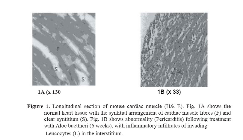
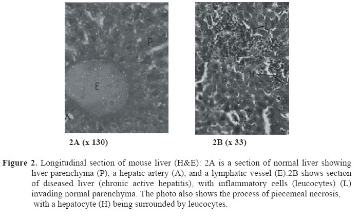
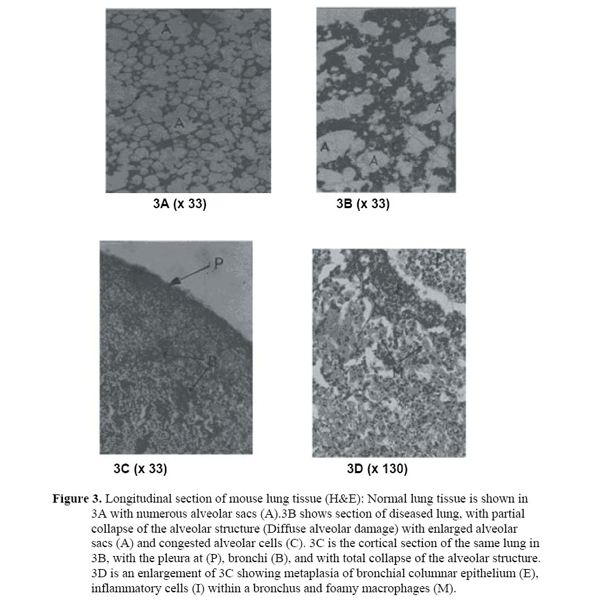
Statistically different relative to control, *p<0.05, **p<0.01; N, number of rats. Histological analysis revealed pathological signs in heart, lung and liver but not in the spleen, kidneys or testes. The cardiac histological lesion, described as pericarditis, was manifested as an inflammatory infiltrate in the cardiac interstitium. There was also proliferation without any degenerative signs in the myocardial cells (Figure 1). Liver pathology was manifested by liver cell death (both piece-meal necrosis and focal necrosis), with degenerative changes diagnosed as chronic active hepatitis (CAH) (Figure 2). In the process of piece meal necrosis, invading inflammatory cells (leucocytes) systematically surround and destroy parenchymal cells one by one over a diffuse area. However, in focal necrosis there is a localised destruction of a group of cells in a unique area of the hepatic parenchyma unlike the diffuse attack observed in piece meal necrosis.The extract caused diffuse alveolar damage (DAD) in the lung. This was manifested by a congestion of alveolar cells, proliferation of interstitial cells and a partial to total collapse of the normal alveolar structure. Metaplasia (change in structure and function) of the bronchial columnar epithelial cells was also observed, with inflammatory cells present within the bronchus, and foamy macrophages within the collapsed interalveolar compartment (Figure 3). Discussion Antiulcer studies The extract of A. buettneri dose-dependently prevented the formation of gastric ulcers induced by HCl/ethanol solution. Products that have gastric protective effects against similar gastric irritant substances are said to possess cytoprotective potential (Miller et al, 1982). The HCl/ethanol solution directly irritates the stomach mucosa, reduces mucosal resistance and erodes the mucosal barrier. Pre-treatment with indomethacin exacerbated the irritant effect of the HCl/ethanol solution. Indomethacin is known to inhibit gastro-duodenal bicarbonate secretion as well as endogenous prostaglandin (PG) secretion (Miller et al, 1983). Inhibition of PG predisposes the stomach and duodenum to mucosal damage by irritants whereas the stimulation of PG can be protective (Kontureck et al., 1981; Kontureck et al, 1982). Results of the nature obtained in the present experiment are interpreted (Yamamoto et al, 1982) to suggest that the anti-ulcer agent is acting through the intermediary of endogenous PG. For the A. buettneri extract, this interpretation is supported by the significant increase in mucus production in animals that received the extract compared with the controls. Mice pre-treated with indomethacin had gastric pyloro-antral surfaces deprived of a measurable mucus lining. The pylorus ligation technique showed that the extract of A. buettneri does not possess antisecretory properties. However, the gastric acidity, which remained within the 79 to 88 mEq/L range after treatment with the extract, did not result in significant gastric mucosal ulceration. Gastric acidity above 60 mEq/L has been shown to produce extensive damage to the gastric mucosa in rats (Martin et al., 1983; Marhuenda et al, 1993; Tan et al, 1996; Tan et al, 2000). In the absence of an anti-secretory effect, the gastric protective activity of A. buettneri extract may occur through reduced pepsin activity in addition to the observed re-enforcement of the mucosal barrier through increased mucus production. Single administration of the extract in dose levels of 4,000 to 60,000 mg/kg did not reveal any serious toxic signs. LD50 and LD100 could not be determined since it was difficult to administer the extract at dose levels beyond 60,000 mg/kg. This highest dose corresponds to an intake of 4.2 kg of the extract (i.e. 140 kg of the dried Aloe leaf) per day for a 70 kg adult human. For this reason, it is concluded that the extract is non-toxic at the acute level. Sub acute toxicity studiesThe pathological condition (pericarditis) observed in cardiac muscle is usually caused by viral, bacterial and fungal infections. It can also be induced by drugs, radiation, expression of transplant rejection and trauma, and can result in various short-term complications including abscess, acute heart failure, myocarditis, myocardial rupture, chronic heart failure and pericardial fibrosis (Rosai, 1996). Pericanditis can be arrested and stabilized and, in a few cases, it can be reversed. CAH in the liver was manifested by focal or localized necrosis and piecemeal necrosis. In this condition, the infiltrating leucocytes gradually destroy the liver parenchyma. Cells that survive then show a variety of changes including focal necrosis (Rosai, 1996; Scheuer, 1979). Viruses, bacteria, and fungi can cause CAH. It can also be the result of chemotherapy, alcohol and drug intake (Zimmerman, 1993). In particular, herbal medicines have been held responsible for most liver diseases (Scheuer and Lefkowitch, 1994), and the presence of toxic low molecular weight compounds in Aloe vera gel has been confirmed (Avila et al., 1997). Active hepatitis can lead to liver cirrhosis and hepatocellular carcinoma. Cirrhosis develops when liver cell death is sufficiently advanced and the remaining parenchyma undergoes nodular regeneration (Scheuer, 1979). CAH can be arrested and stabilized and is potentially reversible at the pre-cirrhotic stage, but it is difficult to bring back the tissues to their original state after advanced cirrhosis (Baggenstoss et al., 1972). In contrast to our findings, Zhou et al. (2003) did not report any pathological findings in rat liver during a 90-day toxicity test on aloe whole leaf powder at the dose of 8 g/kg. Earlier, Arosio et al. (2000) reported the protective effect of aloe-emodin quinone (50 mg/kg) against hepatocyte death caused by carbon tetrachloride. Fan et al. (1989) reported the protective effect of extracts from Aloe vera on experimental carbon tetrachloride-induced hepatic lesions in dogs, mice and rats. The lung abnormality, DAD, resulting from A. buettneri intake is a condition that can also be caused by shock and radiation (Luna et al., 1972; Myers et al., 1987; Rosemow, 1972; Sostman et al, 1977). DAD can lead to various complications in the body including hypoxia (low oxygen tension), hypercapnia (increased carbon dioxide build-up), pulmonary hypertension (increased pulmonary artery pressure due to compression of the lung vasculature by the collapsed parenchyma) and cardio respiratory arrest. DAD can be arrested and stabilized but it is irreversible. In conclusion, the results show that A. buetmeri extract, when prescribed in ethno medicine, can offer protection to the gastric mucosa against the corrosive action of irritant substances and against gastric mucosal auto-digestion by stomach secretions. This cytoprotective activity appears to be associated with a re-enforcement of gastric mucous defenses through increased mucus secretion possibly mediated by endogenous PGs. While acute toxicity studies suggest that the extract is non-toxic, it is possible that regular consumption of large quantities of the extract may lead, in the long term, to liver, heart and lung disease. Acknowledgement This project was supported by the International Foundation for Science (IFS), Stockholm, Sweden, and the United Nations University Japan through Grant F/2882-2 (PVT) to Dr Paul Vernyuy TANin collaboration with the Committee on Scientific and Technological Cooperation (COMSTECH) of the Organization of Islamic Conference (OIC), Islamabad, Pakistan, and Grant F/2626-2 (BN). Grant FUAR (99/3136) of the University of Yaounde I to PV Tan and SF Kimbu is appreciated. References
© Copyright 2006 - African Journal of Traditional, Complementary and Alternative Medicines The following images related to this document are available:Photo images[tc06029f3.jpg] [tc06029f2.jpg] [tc06029f1.jpg] |
| |||||||||
