 |
| About Bioline | All Journals | Testimonials | Membership | News |
|
||||||
|
||||||
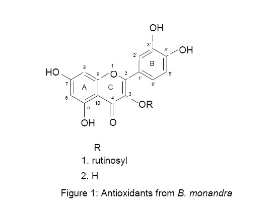
African Journal of Traditional, Complementary and Alternative Medicines, Vol. 3, No. 4, 2006, pp. 59-65 Research Paper ISOLATION OF TWO FLAVONOIDS FROM BAUHINIA MONANDRA (KURZ) LEAVES AND THEIR ANTIOXIDATIVE EFFECTS M. A. Aderogba*1,2, A.O. Ogundaini3 and J. N. Eloff2 1Department
of Chemistry, Obafemi Awolowo University, Ile-Ife, Nigeria., 2Phytomedicine
Programme, Department of Paraclinical Sciences, University of Pretoria, Onderstepoort
0110, South Africa,3Department of Pharmaceutical Chemistry,
Obafemi Awolowo University, Ile-Ife, Nigeria. Code Number: tc06052 Abstract Bauhinia monandra (Kurz), traditionally use in the treatment of diabetes with established significant anti-diabetic activity was investigated for its antioxidant constituents since the activity demonstrated can be linked to the presence of antioxidant compounds. Bioassay directed fractionation of the ethyl acetate soluble leaves extract has led to isolation of two active compounds identified as: Quercetin-3-O-rutinoside (1) and Quercetin (2). The molecular structures elucidations of both compounds were carried out using spectroscopic studies (1H NMR, 13C NMR and MS). These compounds are reported from this species for the first time. A DPPH spectrophotometric assay was used to evaluate the antioxidant potential of the compounds. Compound2 had higher antioxidant activity while Compound 1 had lower activity than L-ascorbic acid which was used as standard. Key words: Bauhinia monandra; antioxidant activity; flavonoid; DPPH. Introduction Flavonoids constitute a major group of phenolic compounds in plants. They provide pigmentation for fruits, flowers and seeds to attract pollinators and seeds dispersers. They assist in plant defense against pathogenic microorganism (Schijlen et al., 2004, Parr and Bowell, 2000). The number of flavonoids is constantly increasing due to the structural variation associated with these compounds. It is well known that antioxidant activity in higher plants has often been associated with phenolic compounds (Thabrew et al., 1998). Bauhinia monandra (Kurz) belongs to the family Fabaceae. It is traditionally used for the treatment of diabetes and as a diuretic (Argolo et al., 2004). Freshly crushed leaves are also used to treat stonefish stings (Hansworth, 1990). Pharmacological reports have shown that the ethanol extract of the leaves exhibited hypoglycaemic activity (Coelho and Silva 2000). Subsequent fractionation of the ethanol extract led to isolation of one of the active compounds which was identified as galactose-specific lectin. (Coelho and Silva, 2000). Oral administration of the stem bark extract in rats (1g/kg), exhibited significant anti-diabetic activity (Abo and Jimoh, 2004). Methanol leaf extracts had no antimicrobial activity against B. substilis, E. coli, S. aureus and P. aeruginosa (Binutu, 1986). Many plant constituents are effective as remedy for some diseases and accounts for large number of pharmaceutical important compounds in Western Pharmacopoeia and a number of important drugs. For example, taxol and artermisinin were reported from plants (Tshibangu et al., 2002). In our quest to finding a novel antioxidant agents from plants, we have carried out bioactivity directed phytochemical study on the leaves extract of B. monandra to isolate and identify the antioxidant constituents because anti-diabetic activity has been linked to antioxidant compounds (McCune and Johns, 2002). Antioxidant potential of the isolated compounds was also evaluated using a DPPH free radical scavenging assay. Material and Methods General Spectroscopic data were obtained from the following instruments: UV- Versa-max® microplate reader, NMR – Varian (1H 300 MHz, 13C 75 MHz), Electron impact mass spectra (EI-MS) – Shimadzu 2010 gcms. L-ascorbic acid (Merck), DPPH 2, 2-diphenyl-1-picryl hydrazyl (Sigma) and absolute methanol (Fluka). All other chemicals used were of analytical grade obtained from BDH Chemicals Ltd, Poole England and Sigma chemical Co. USA. Plant material Leaves of B. monandra were collected at the Obafemi Awolowo University, Ile-Ife, Nigeria, in February 2005. Collected leaves were air dried at room temperature for three weeks. Dr. H.C. Illoh of the Department of Botany, Obafemi Awolowo University, Ile-Ife, authenticated the plant. A voucher specimen was deposited at the Herbarium of the Faculty of Pharmacy, Obafemi Awolowo University with herbarium number FPI 107. Extraction Finely ground leaves (700 g) were extracted with methanol (MeOH). The extract was concentrated to dryness in vacuo at 40°C to remove the methanol. The aqueous extract was made and successively partitioned with hexane (Hex.), dichloromethane (DCM), ethyl acetate (EtOAc) and butanol (BuOH). The combined organic layer of each partition was evaporated to dryness in vacuo at 40°C using rotary evaporatorto afford Hex., DCM, EtOAc and BuOH fractions, Table 1. Isolation of the compounds The EtOAc fraction (5.40 g) was fractionated on silica gel column chromatography using an increasing gradient of EtOAc in chloroform (CHCl3) up to 100%, followed by an increasing gradient of MeOH up to 100%. This gave five pooled fractions A1 – E1. Purification of E1 (40.0 mg) on Sephadex column chromatography starting with DCM - MeOH (7.5:2.5) followed by increasing gradient of MeOH up to 70% afforded compound 1 (19.0 mg). Combination of fractions B1 and C1 (330.0 mg) and subsequent fractionation on Sephadex LH-20 using toluene (Tol.) -MeOH (4:1) gave compound 2 (92.0 mg). Fractionation of BuOH fraction (8.00g) on Sephadex LH-20 column starting with Tol.-EtOH (1:1) followed by increasing gradient of EtOH up to 100% also afforded compound 1 (240.0 mg). Structure Elucidation Quercetin-3-O-rutinoside (1). 13C-NMR (75 MHz, DMSO- d6). 156.4 (C2), 133.3 (C3), 177.4 (C4), 161.2(C5), 98.7 (C6), 164.2 (C7), 93.6 (C8), 156.6 (C9), 103.9 (C10), 121.2 (C1`), 115.2 (C2`), 144.8 (C3`), 148.4 (C4`), 116.3 (C5`), 121.6 (C6`), 101.2 (C1``), 74.1 (C2``), 76.4 (C3``), 70.6 (C4``), 75.9 (C5``), 67.0 (C6``), 100.8 (C1```), 70.4 (C2```), 70.0 (C3```), 71.8 (C4```), 68.3 (C5```), 17.8 (C6```). 1H NMR (300 MHz, DMSO-d6): δ 3.03 - 3.71 {m, rhamnoglucosyl (rutinosyl) - Hs}, 4.37 (1H, s, rhamnosyl, H-1`` ), 5.34 (1H, d, J = 6.9 Hz, glucosyl,H-1```),6.18 (1H, d, J = 1.2 Hz, H-6), 6.37 (1H, bs, H-8),6.84 (1H, d, J = 8.1 Hz, H- 5' ),7.54 (2H, d, H-2' and H-6'), 12.59 (1H, s, 5-OH). EI-MS: m/z 302 {[MH - rutinosyl]+, 100%}, due to loss of sugar(Markham 1982). The spectra data were in agreement with that of quercetin-3-O-rutinoside reported in the literature(Harborne and Mabry, 1982; Markham 1982). Quercetin (2). 13C-NMR (75 MHz, DMSO- d6). 146.8 (C2),135.8 (C3), 175.9(C4), 160.7 (C5), 98.2 (C6), 163.9 (C7), 93.4 (C8), 156.2 (C9), 103.0 (C10), 122.0 (C1`), 115.1 (C2`), 145.1 (C3`), 147.7 (C4`), 115.6 (C5`), 120.0 (C6`). 1H NMR (300 MHz, DMSO-d6): 6.18 (1H, d, J = 1.8 Hz, H-6), 6.40 (1H, d, J = 2.1 Hz, H-8), 6.89 (1H, d, J = 8.4 Hz, H- 5' ), 7.54 (1H, dd, J = 2.1 and 8.7 Hz, H-6'), 7.67 (1H, d, J = 2.1 Hz, H- 1' ), 12.49 (1H, s, 5-OH).EI-MS: m/z 302 {[M] +,100%}.The spectra data were in agreement with that of quercetin reported in the literature (Markham, 1982). Antioxidant activity Qualitative assay screening entailed spraying the TLC chromatograms of the partitioned fractions and the crude extract with 0.2% DPPH in MeOH. This revealed the antioxidant behaviour of the extracts. This was also repeated for the two compounds isolated. Quantitative antioxidant activity was determined spectrophotometrically as described by Mensor et al.,2001, with some modifications. Briefly, the reactions were carried out in 96-well microtitre plates and each compound was tested at 100.0, 50.00, 25.00, 12.50, 6.25 and 3.13 µM. Twenty micro liters of 0.3 mM DPPH in methanol was added to 50 µL of each concentration of sample tested and allowed to react at room temperature in the dark for thirty minute. Blank solutions were prepared with sample solution (50 μL) and 20 μL of methanol only while the negative control was DPPH solution, 20 μL plus 50 μL methanol. The decrease in absorbance was measured at 515 nm on a microplate reader. Values obtained were converted to percentage antioxidant activity (AA%) using the formula: AA% = 100 - {[(Abssample – Absblank) x 100] / Abscontrol} (Abssample is the absorbance of the sample, Absblank is the absorbance of the blank and Abscontrol is the absorbance of the control). L-ascorbic acid (vitamin C) was used as a positive control (antioxidant agent). The EC50 value, defined as the concentration of the sample leading to 50% reduction of the initial DPPH concentration, was calculated from the linear regression of plots of concentration of the test compounds (µM) against the mean percentage of the antioxidant activity obtained from the three replicate assays. Statistical analysis The results were expressed as mean ± SEM and the EC50 values obtained from the regression plots (Sigma PlotsR 2001, SPSS Science) had a good coefficient of correlation, (r2 ≥ 0.955). Result and Discussion Immediate bleaching of the purple DPPH colour by some constituents of EtOAc and BuOH fractions was observed on spraying TLC chromatograms of the solvent fractions of the crude extract with 0.2% DPPH in MeOH. The bleaching effect was also observed with the two compounds isolated. The chemical structures of the two antioxidants: quercetin-3-O-rutinoside (1) and quercetin (2) were established by comparison of their spectral data with those reported in the literature (Harborne and Mabry, 1982; Markham 1982). These compounds were different in their antioxidant activity in quantitative assay, (Table 2). Compound 2 had higher antioxidant activity while Compound 1 had lower activity than L-ascorbic acid which was used as standard. Conditions for effective radicals scavenging activity in flavonoids observed from structure activity relationship studies include the presence of catechols group (3´-OH and 4´-OH) on ring B, the 3-OH group in combination with a C2 C3 double bond and keto group in position 4 (Gilbert et al., 2003, Harborne and Williams, 2000 and Saskia et al., 1996). These structural units fortify the antioxidant activity by increasing the stability of the flavonoids radical after donating phenolic hydrogen.The higher antioxidant activity of compound 2 compared to compound 1 could be due to the presence of free hydroxyl group in position 3 as both compounds have catechol group (3` and 4` di-OH) on ring B. It has been demonstrated that substitution of 3-OH reduces activity, (Op de Beck, 2003). Conclusion Many flavonoids have shown strong antioxidant properties(Harborne and Williams, 2000; Raj and Shalini, 1999). Quercetin-3-O-rutinoside (rutin) and quercetin have been established as strong antioxidant principles and had been used as standards in antioxidant experiments(Braca, et al., 2003; Mensor et al., 2003; Thabrew et al., 1998). Table 1: Weights of the B. monandra leaves extracts
Table 2: Antioxidant potential of B. monandra constituents
The presence of these compounds in abundance in the leaves extracts of B. monandra could provide rationale for the use of this plant in folk medicine. Our results confirmed the earlier qualitative antioxidant analysis of B. monandra leaves extracts in which three major antioxidant compounds (flavonoids and steroid) were identified, (Argolo et al., 2004). We have now isolated, characterised and evaluated the antioxidant potential of the flavonoid constituents. Acknowledgements M.A. Aderogba is grateful to NRF South Africa, for the award of postdoctoral research fellowship and NAPRALERT for literature survey on the plant species. References
© Copyright 2006 - African Journal of Traditional, Complementary and Alternative Medicines The following images related to this document are available:Photo images[tc06052f1.jpg] |
| |||||||||
