 |
| About Bioline | All Journals | Testimonials | Membership | News |
|
||||||
|
||||||
African Journal of Traditional, Complementary and Alternative Medicines, Vol. 3, No. 4, 2006, pp. 78-93 Research Paper SAFETY EVALUATION OF AQUEOUS EXTRACT OF LEAVES OF A PLANT PHYLLANTHUS AMARUS, IN RAT LIVER K. N. S.Sirajudeena*, Siti Amrah Sulaimanb, M. Madhavanc, Zabidah Ismailb, M.Swamya, Md. Lukmi Ismailb, Musa Yaacobd aDepartment of Chemical Pathology, bDepartment of Pharmacology, cDepartment of Pathology, School of Medical Sciences, Universiti Sains Malaysia, 16150-Kubang Kerian, Kelantan, Malaysia, dMalaysian Agricultural Research and Development Institute, Telong, 16310, Kelantan, Malaysia. E-mail: sirajuden@kb.usm.my or knssiraj@yahoo.com Fax:00609-7653370 Ph: 00609-7664745 Code Number: tc06055 Abstract Phyllanthus amarus (Euphorbiaceae) is used as a folk medicine for jaundice and other diseases in Malaysia and other countries. But, so far, no safety studies have been carried out on this plant with clear documentation, especially with those plants growing in Malaysia. So the aim of this study was to determine the toxic side effects of aqueous extract of leaves of P. amarus (grown in Malaysia) following oral administration in rats. Acute admininstration of P. amarus extract at a dose of 5 g/kg body weight did not produce any signs of toxicity or mortality. In the chronic study, no significant difference (P > 0.05) was observed between the control and P. amarus extract administered (male and female) rats (at the doses of 100, 400 and 800 mg/kg body weight for 6 weeks) in the total body weight gain as well as in the liver marker enzymes analyzed in serum. The non-toxic nature of P. amarus extract administration was confirmed by histological studies [light microscopy, proliferative cell nuclear antigen study and apoptotic study] i.e., no observable changes were found between control and P. amarus extract administered rats. Therefore, acute oral administration of P. amarus extract is non-toxic to the rat liver, even at a dose of 5 g /kg body weight and also the chronic toxicity studies of P. amarus extracts administration showed the absence of cumulative toxicity as reflected by the non-significant change in the parameters studied as well as from the results of the histological studies. Key words: Phyllanthus amarus, Aqueous extract, Liver, Toxicity, Marker enzymes, Histological studies IntroductionMost of the population of the underdeveloped and developing countries depends on some form of traditional and herbal medicines since ancient times. Malaysia, an important nation of biodiversity, is enriched with herbal resources. One of the plant genera widely used traditionally for the treatment of many diseases is Phyllanthus Schum. & Thonn. (Family: Euphorbiaceae) and is distributed in most tropical and subtropical countries and comprise approximately of 550-750 species throughout the world. One of the most studied and widely used species in Malaysia and other countries is P. amarus (Calixto et al., 1998; Muhamad bin Zakaria and Mustafa Ali Mohd, 1994). In Malaysia, the plant is called “Dukung anak”. P. amarus has bitter, astringent, cooling, diuretic, stomachic, antiseptic, antiviral, antidiabetic, hypotensive, antinociceptive, febrifuge properties and is traditionally used in the treatment of jaundice, diarrhea, dysentery, diabetes, fevers, uro-genital diseases, ulcers and wounds (Santos et al, 1995; Calixto et al, 1998). Some reports have shown the antiviral effect of this plant by reducing the detectable hepatitis B surface antigen (HbsAg) of HBV positive patients (Thyagarajan et al., 1988; Ott et al., 1997). But other studies from China, Thailand, and India showed the failure of P. amarus in eradicating the HbsAg in patients with chronic hepatitis B virus (Leelarasamee et al, 1990; Wang et al, 1991, Doshi et al, 1994). The variation in the clinical effect of these studies has been attributed to many factors such as different species, differences in growing condition and different processing methods (Thyagarajan et al., 2002; Wang, 2000). Usually herbal medicines are widely perceived by the public as being natural, healthful and free from side effects, but that is over simplification of the matter. Plants contain hundreds of constituents and some of them may elicit toxic side effects. A number of studies exist reporting the toxic effect of herbal medicines (Shaw et al., 1997; Kaplowitz, 1997; Calixto, 2000). Therefore efficacy and safety study should be performed on these herbs. Even though a large number of clinical trials have been done on the benefits of P. amarus, so far no systematic toxicological investigation has been reported on this plant, especially P. amarus growing in Malaysia. Since the efficacy of Phyllanthus species varies with geographical location and varieties and because of their constituent composition variation, the P. amarus grown in Malaysia (widely used here as the folk medicine for jaundice) has to be ascertained for their bio-safety by conducting acute and chronic toxicity study. So the aim of this study was an attempt to determine the toxic effects of aqueous extract of leaves of P. amarus (grown in Malaysia) following oral administration in rat liver, by assessing the morphological, biochemical and histological changes. Materials and methods Plant material P. amarus was collected from MARDI (Malaysian Agricultural Research and Development Institute, Telung, Bachok, Kelantan, with voucher specimen number KL5241 during the months of July-October and identified by FRIM (Forest Research Institute of Malaysia, Kepong, Selangor). The aqueous extraction of P. amarus leaves was carried out at the Department of Pharmacology, School of Medical Sciences, Universiti Sains Malaysia. Preparation of plant extract The plant material was cleaned and the leaves were separated, oven dried at 50ºC. This was macerated into dry powder. Approximately 4.5 kg of this powder was extracted with distilled water (2:25) using soxhlet apparatus and concentrated by rotary evaporator at 65ºC. This was transferred into a suitable container and freeze dried. The yield of the final crude aqueous P. amarus extract was 6-9% (~380 g). The dried extract was stored in a desicator until its use. The extract was dissolved in distilled water to the desired concentration just before the study. Animal and experimental design Male and female Sprague-Dawley rats weighing (180-220 g) were used as the experimental animals in this study and were obtained from Animal House Facility Unit, Health Campus, Universiti Sains Malaysia, Malaysia and acclimatized for one week prior to start of the experiment. Animals were housed in standard cages at a temperature of 23±2ºC, 45-55 % relative humidity with a 12h light/12h dark cycle. The animals were fed with commercial pellet diet and water ad libitum. Our study protocol was approved by the animal ethical committee, Health Campus, Universiti Sains Malaysia, Malaysia. Acute and chronic studies were carried out (WHO, 1993; Arnold, 1990) and in both studies the rats were administered with extract of leaves of P. amarus orally. Acute study Aqueous leaves extract of P. amarus at a single dose of 5g/Kg body weight was administered orally to six male rats. Another six male rats served as control without P. amarus extract administration. The animals were observed carefully for any visible signs of toxicity and mortality immediately after dosing, at 4h, 24h intervals, during the recovery period of 48h and twice daily up to 14 days. After 14 days, the rats were sacrificed under ether anesthesia. A thorough autopsy was carried out and all organs were observed for any macroscopic changes. Chronic study Animal groups For the chronic study, rats of both sex were grouped into four (ten rats in each group) as follows: For the male rats, Group I served as the control (without plant extract administration), Group II, III and IV were administered with P. amarus extract at the doses of 100, 400 and 800 mg/Kg body wt/day for 6 weeks by gavage, respectively. For the female rats, Group A served as the control (without plant extract administration), Group B, C and D as the groups administered with P. amarus extract at the doses of 100, 400 and 800 mg/Kg body wt/day for 6 weeks by gavage, respectively. The animals were observed daily for any signs of morbidity and mortality and their body weights were measured periodically in the experimental period. Collection of serum and liver samples for analysisAt the end of the experimental period (6 weeks), after an overnight fasting, both the male and female rat controls and P. amarus extract administered groups were sacrificed by decapitation. Blood was collected, allowed to clot and then centrifuged at 3,000 rpm for 15 minutes. Serum samples were separated and used for biochemical analysis. The samples were stored at -80ºC, if not used immediately. A portion of the liver tissue of all the (control and P. amarus extract administered) groups was fixed in 10% formal saline for the histological studies. Biochemical analysis In the collected serum, the total protein and the activities of the liver marker enzymes such as Alanine transaminase (ALT), Aspartate transaminase (AST), Alkaline phosphatase (ALP) and Lactate dehydrogenase (LDH) were assayed by using standard kit (ALT Randox kit, Randox Total protein biuret reagent - Randox Laboratories Ltd, UK; AST,ALP and LDH Roche Kits - Roche diagnostics, GmbH, Germany) in a Hitachi-912 autoanalyser available in the department of chemical Pathology, School of Medical Sciences, Universiti Sains Malaysia, Malaysia. Histological studies A portion of all the liver specimens fixed in 10% formal saline was processed routinely overnight using histokinette. Then they were embedded in paraffin wax. Three sections, each four micron in thickness were cut from each paraffin block. Light Microscopic study One section from each sample was stained with Haematoxylin and Eosin (H&E) stain by the standard method for light microscopic histological examination. Proliferative Cell Nuclear Antigen (PCNA) study Another section from each sample was stained with anti PCNA (DAKO, Denmark) using standard immunohistochemistry procedure and the positive sample was visualized with the chromogen diaminobenzidine (DAB) [DAKO, Denmark] and expressed in percentage. Apoptosis study The third section from each sample was assessed for apoptosis. They were stained with ApopTag – Apoptosis detection kit (Chemicon, USA) whereby the reagents are designed to label the free 3’OH DNA termini in situ with chemically labelled and unlabelled nucleotides and the procedures are followed according to the manufacturer instructions. . Statistical Analysis The results of the biochemical analysis are expressed as mean ± S.D. for ten animals in each group. The difference between the control and P. amarus extract administered groups (for each sex of animals) was analysed by Student’s t-test. P value < 0.05, was considered as significant. Results Acute study During the course of the acute study, no treatment related effect was observed on the general condition or behaviour of the experimental animals. All the rats appeared to be normal, survived during the experimental period and none of them showed any visible signs of toxicity. A thorough autopsy of the P. amarus administered rats revealed no treatment related macroscopic changes. Chronic study Body weight Mean body weight gain of the P. amarus extract administered groups (at the doses of 100, 400 and 800 mg/Kg body weight/day) have shown no appreciable difference when compared to their control (both male and female rats) after 6 weeks duration of the study (Data not shown). The macroscopic appearance of all the organs observed was found to be normal between control and P. amarus extract administered groups. Biochemical study There were no significant differences (P > 0.05) observed in the biochemical parameters studied (ALT, AST, ALP, LDH and total protein) in the serum between controls and P. amarus extract administered groups (of both male and female) (Tables 1 and 2). Table 1: Level of total protein and activities of serum marker enzymes of control and phyllanthus amarus administered groups of male rats
Values are mean ± S.D., n = 10 rats in each group. Group I was the control, Group II, III and IV were the plant extract phyllanthus amarus administered groups at the doses of 100 mg/kg body weight/day, 400mg/Kg body weight/day and 800mg kg body weight/day for 6 weeks respectively. Group II, III and IV are compared with Group I (Control) by student’s t-test and p value <0.05, considered significant. NS p>0.05 , not significant. Table 2: Level of total protein and activities of serum marker enzymes of control and phyllanthus amarus administered groups of female rats
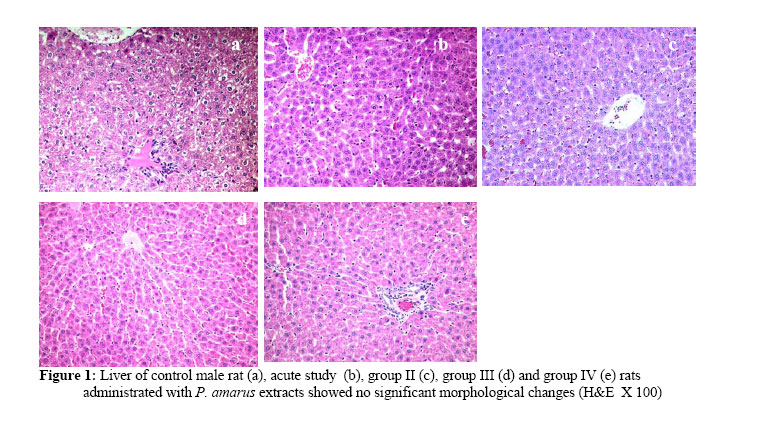
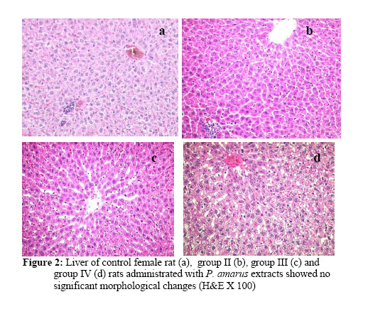
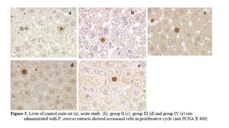
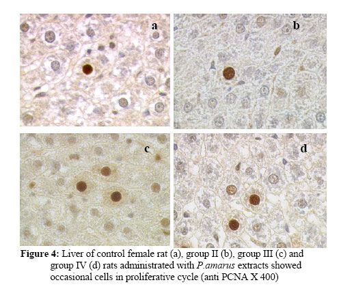
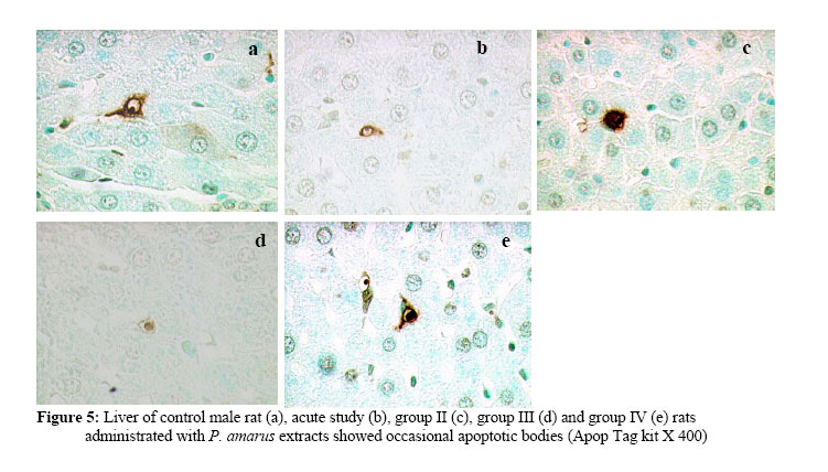
Values are mean ± S.D., n = 10 rats in each group. Group A was the control, Group B, C and D were the plant extract Phyllanthus amarus administered groups at the doses of 100 mg/kg body weight/day, 400mg/Kg body weight/day and 800mg kg body weight/day for 6 weeks respectively. Group B, C and D are compared with Group I (Control) by Student’s t-test and P value < 0.05 was considered significant. NS P > 0.05 , not significant. Histological study Light microscopic study Histological examination of the liver under light microscope showed no significant pathological changes in the P. amarus extract administered groups (at the acute dose of 5g/Kg body weight and at the doses of 100, 400 and 800 mg/Kg body wt/ day, orally, for six weeks to both male and female rats) (Figures 1b - e and 2b -d) when compared to their controls (Figures 1a and 2a). PCNA Study Anti PCNA antibody was used to measure the proliferation of hepatocytes by immunohistochemistry. PCNA of liver from control rat was 1.9% (Figures 3a and Figures 4a) and for the acute dose (5g/Kg body weight) P. amarus extract administered rat was 1.8% (Figures 3b). For the P. amarus extract administered groups at the doses of 100, 400 and 800 mg/Kg body wt/ day, orally, for 6 weeks (both male and female rat livers) showed PCNA of 0.2% - 2.1% (Figures 3c-e and 4b - d) which are similar to that of their controls. Apoptosis study Apoptosis in the liver was measured in the hepatocytes using ApopTag Kit. Apoptosis in the livers from control (both male and female rats) were 0.2% (Figures 5a and 6a). Apoptosis in the livers from P. amarus extract administered groups ranged from 0.2% to 1.5% (Figures 5b - e and 6b - 6d) which are similar to that of their controls. Discussion Herbal medicines are very popular in developing and underdeveloped countries. Reports indicate that the ideal herbal drugs are very safe and free from side effect is false (Shaw et al., 1997; Kaplowitz, 1997; Calixto, 2000). Therefore, clear understanding of potential adverse effect of herbs used by human population is necessary for implementing safety measures to the public. In the case of P. amarus, available reports are not unanimous on the efficacy of the plant grown in different countries but no systematic safety study had been done so far and hence, a study on their toxicity is required. In our present work, we have studied the toxicity of P. amarus (grown in Malaysia) on rat liver. The results of our acute study showed the absence of lethality or toxic side effect on oral administration of P. amarus extract even at a dose of 5g/kg body wt. This clearly indicates the non-toxic nature of the plant extract. Toxicologists agree that any test substance that is not lethal on acute administration at a concentration of 5g/Kg body wt, is essentially non-toxic (OECD, 1981; Brock et al., 1995). In the chronic toxicity study, we did biochemical analysis and histological studies to determine the cumulative toxic effect P. amarus extract on rat liver. Liver is an organ involved in many metabolic functions and is prone to xenobiotic-induced injury because of their central role in xenobiotic metabolism (Sturgill and Lambert, 1997). Liver contains a host of enzymes such as ALT, AST, LDH and ALP. The activities of these enzymes are used to assess the functional status of the liver and as the biochemical markers of liver damage (Moss and Ralph Handerson, 1999). Hepatotoxic drugs cause damage to the liver cell Membrane and these enzymes are leaked out into serum and shows increased activities (Kumar et al, 2004; sturgill and Lambert, 1997). Our study results showed that there were no increased activities of ALT, AST, LDH and ALP on P. amarus extract administration indicating that the plant is non-hepatotoxic in nature. The results of our biochemical estimations were confirmed by the histological studies (light microscopy, PCNA study and apoptosis study). Histological examination of liver specimen is useful in identifying the type of injury present in it (Friedman et al., 1996). Light microscopic study showed no observable changes between control and P. amarus administered groups. Researchers assessed the role of cell proliferation and compensatory tissue repair in response to initial liver injury by PCNA study (Wang et al., 2000). The expression of PCNA by immunohistochemistry is a reliable indicator of both the proliferation rate and location of the compartment (Yamada et al., 1992). We also found no difference between control and P. amarus administered groups in the PCNA study. Experimentally induced T-cell mediated hepatic injury is assessed by apoptosis (Tiegs et al., 1992). In our study, the assessment of apoptosis by using Apop Tag Kit showed similar pattern between control and P. amarus administered groups. ConclusionsAcute oral administration of P. amarus extract is non-toxic to the rat liver, even at a dose of 5 gram /kg body weight. The chronic toxicity studies of P. amarus extracts administration (of 100-800 mg/kg body wt) showed the absence of cumulative toxicity as reflected by the non-significant change in the parameters studied as well as from the results of the histological studies. Further studies are recommended in this plant (grown in Malaysia) to assess their antihepatotoxic, antidiabetic, anticancer and antidiarrheal properties.
Acknowledgement
This project was carried out under the financial support of Universiti Sains Malaysia short-term project grant (304/PPSP/6131166).We would like to acknowledge Professor Khalijah Awang and Ms. Norshita for their help in identification and confirmation of the plant material. References
© Copyright 2006 - African Journal of Traditional, Complementary and Alternative Medicines The following images related to this document are available:Photo images[tc06055f3.jpg] [tc06055f2.jpg] [tc06055f5.jpg] [tc06055f4.jpg] [tc06055f6.jpg] [tc06055f1.jpg] | ||||||||||||||||||||||||||||||||||||||||||||||||||||||||||||||||||||
| |||||||||
