 |
| About Bioline | All Journals | Testimonials | Membership | News |
|
||||||
|
||||||
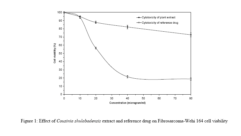
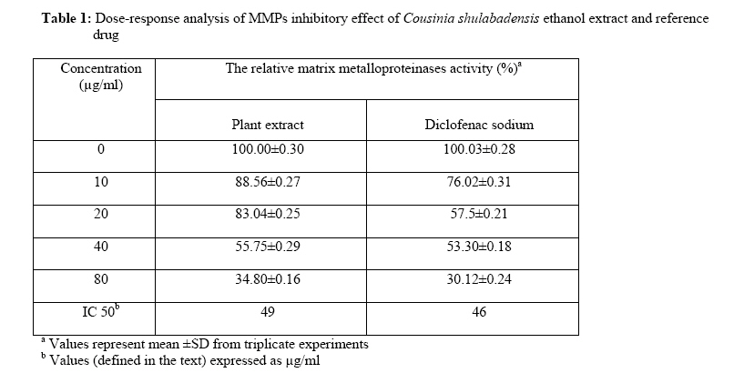
African Journal of Traditional, Complimentary and Alternative Medicines,, Vol.4, No. 1, 2007, pg. 12-16 RESEARCH PAPER CHEMOPREVENTIVE EFFECT OF COUSINIA SHULABADENSIS ATTAR & GHAHRAMAN ETHANOL EXTRACT Ahmad R. Shahverdi 1,*, Mohammad R. Khoramizadeh2, Mohammad H. Ghahramani3, Ardeshir Golyaee3, Farideh Attar4, Ahmad Ghahraman4 1Department of Pharmaceutical Biotechnology, Faculty of Pharmacy, Tehran, University of Medical Sciences, Tehran, Iran Code Number: tc07016 Abstract Matrix metalloprotainases (MMPs) play an important role in several pathologic processes such as malignancy in which they facilitate invasion and metastasis and can be targets for anticancer therapies. Here, in this study, we investigated the cytotoxicity effect of Cousinia shulabadensis Attar & Ghahraman extract as well as its impact on MMPs activity using a model of cell line (Fibrosarcoma-Wehi164). To assess anti-invasiveness potentials, a modified zymoanalysis method was used to measure MMP-2 and MMP-9 activities in the conditioned-media. The concentration necessary to produce 50% cell death was >80µg/ml for ethanol extract of Cousinia shulabadensis, while a 23 µg/ml concentration of the diclofenac sodium produced the same effect. The invasion of WEHI 164 cells was considerably inhibited at concentrations > 20 µg/ml by total plant extract. The total extract of the plant did not show high toxicity at all tested concentrations, but demonstrated significant inhibition of MMP activity in dose-response fashion.Key words: anti-invasive activity; chemoprevention; cytotoxicity; Cousinia shulabadensis; matrix metalloproteinases Introduction Most investigators unanimously admit that matrix metalloproteinases (MMPs) are critical enzymes in tumor growth invasion, metastasis (Stetler-Stevenson et al., 1996; Fidler, 1997; Jones et al., 1999; Tate et al., 2004) and neovascularization (John and Tuszynski, 2001; Nguyen et al., 2001). MMPs are a family of highly homologous, zinc, and calcium dependent endopeptidase that clear most, if not all, components of the extracellular matrix (ECM). The destruction of the extracellular matrix eventually leads to tumor invasion, metastasis, and angiogenesis (Heath and Grochow, 2000). Thus, each component with a potential inhibitory influence on MMP expression, such as a non-steroidal anti-inflammatory agent like diclofenac sodium, is able to reduce the risk of cancer (Saadat et al., 2003). Furthermore several traditional herb medicines, such as phytochemicals from Chinese medicinal herb Eunymus alatus, and the polyphenolics of green tea have been so far reported to exhibit an inhibitory effect on MMPs expression (Tate et al., 2004). In our search for chemopreventive anti-invasive natural products, we have selected an extract prepared from whole plant of Cousinia shulabadensis (Asteraceae). C. shulabadensis is distributed in west-southern part of Iran in the Shulabad region in Lorestan province (Attar and Ghahraman, 2002). Members of the genus of Cousinia are widespeared throughout Iran, especially in Bakhtiari, Khorasan and Lorestan provinces (Attar and Ghahraman, 2002). Here we use enzyme zymography to examine the influence of the ethanol extract of the C. shulabadensis on the expression of MMPs. Furthermore, its cytotoxic effect on a fibrosarcoma cell line was investigated. Materials and methods Plant Collection The whole plant of C. shulabadensis was collected from the Shulabad region in Lorestan province at an altitude of 2600 m, and was identified by Dr. F. Attar. A voucher specimen of the plant (21874-TUH) was deposited in the Central Herbarium of the Tehran University,Tehran, Iran. Extraction procedure The plant were air-dried at room temperature and pulverized. The ethanol (80 % v/v) extract was prepared by maceration of the powder for 72h with three changes of solution at room temperature. The combined solvent extracts were evaporated to yield a brownish viscous residue. All experiments were performed based on the dry mass of concentrated extract.Cell Culture The Fibrosarcoma cell line (WEHI 164) was seeded in 96-well tissue culture plates. Cells were maintained in a RPMI-1640 medium that was supplemented with 5% fetal calf serum, plus antibiotics, at 5% CO2, 37oC, and saturated humidity. The Fibrosarcoma-Wehi 164 cell line was obtained from the National Cell Bank of Iran (NCBI), Pasteur Institute of Iran, Tehran, Iran.Dose-Response Analysis Triplicate, two-fold dilutions of plant extract and diclofenac sodium were transferred to overnight cultured cells. Non-treated cells were used as control. Cells were cultured overnight and were then subjected to colorimetric assay. Cytotoxicity was expressed as the percentage of viable cells at different concentrations of samples. IC 50 was calculated as the dose at which 50% cell death occurred relative to the untreated cells. The corresponding supernatants of the cultured cells were used for zymoanalysis.Colorimetric Assay In the cytotoxicity assay, cells in the exponential phase of growth were incubated for 24h at 37oC with 5% CO2 with a serial dilution of extract. Cell proliferation was evaluated by a modified Crystal Violet colorimetric assay (Saadat et al., 2003). After each experiment, the cells were washed with ice-cold phosphate buffer solution and fixated in a 5% formaldehyde solution. Fixed cells were stained with 1% crystal violet. Stained cells were lysed and solubilized with a 33.3% acetic acid solution. The density of developed purple color was read at 580 nm.Zymoanalysis This technique has been used for the detection of gelatinase (collagenase type-IV or matrix metalloproteinase type-2, MMP-2) and MMP-9 in conditioned media (Heussen and Dowdle, 1980). Briefly, aliquots of conditioned media were subjected to electrophoresis in a gelatin-containing polyacrylamide gel, in the presence of sodium dodecyl sulfate (SDS) under non-reducing conditions. After electrophoresis, SDS was removed by repeated washing with Triton X100. The gel slabs were then incubated at 37oC overnight in a gelatinase-activating buffer and subsequently stained with Coomassie Brilliant Blue R250 (Sigma, MA). After intensive destaining, proteolysis areas appeared as clear bands against a blue background. Using a gel documentation system, quantitativeevaluation of both the surface and intensity of lysis bands, on the basis of grey levels, were compared relative to non-treated control wells and expressed as a percentage of the “Relative Expression” of gelatinolytic activity. The IC50 for the MMP inhibitory effect was calculated as doses at which 50% of MMP inhibition occurred relative to untreated control cells. Statistical Analyses The differences in cell cytotoxicity and gelatinase zymography were compared using the Student’s t test. P values <0.05 were considered significant.Results The cytotoxicity of the total extract of the C. shulabadensis and reference drug were evaluated in vitro against the fibrosarcoma cell line (WEHI 164) at four doses of 10, 20, 40, and 80 µg/ml. Cytotoxicity analysis of the total extract shows a direct dose-response result with the total extract of C. shulabadensis; the higher the concentration, the higher the toxicity (Figure 1). Cell cytotoxicity of diclofenac sodium versus C. shulabadensis is also illustrated in Figure 1. The presence of 80 µg/ml of C. shulabadensis total extract moderately inhibited the growth of the cell line, while lower dose levels (less than 80 µg/ml) showed minimal cytotoxicity with a viability percentage of more than 85%. In contrast, diclofenac sodium showed a high cytotoxic effect, especially at concentrations more than10 µg/ml. The concentration necessary to produce 50% cell death was >80µg/ml for ethanol extract of C. shulabadensis, while a 23 µg/ml concentration of the reference drug produced the same effect (Table 1).Subsequently, the anti-invasive property of the roots of C. shulabadensis was investigated at the different dose levels of 10, 20, 40 and 80 µg/ml. The inhibitions of the total extract of C. shulabadensis on the invasion of the fibrosarcoma-Wehi 164 cells are presented in Table 1. As shown in Table 1, the invasion of WEHI 164 cells was considerably inhibited at concentrations > 20 µg/ml by total plant extract. At 40 µg/ml, the C. shulabadensis extract was able to inhibit the invasion more than 40%, while there was negligible cytotoxicity at this concentration. Based on zymogeraphy analysis of diclofenac sodium, reduction of MMP expression was associated with increasing concentration of this drug in accordance with previous studies (Sadowski and Steinmeyer, 2001; Saadat et al., 2003). But this drug is too toxic to use at high doses, where the reduction of MMP expression is mostly associated with the total cell death. The IC50 values for total extract and reference drug (the concentration that inhibits the invasion of the WEHI 164 cells by 50% relative to untreated control) were calculated as 49 and 46 µg/ml respectively (Table 1). Either the total extract of the plant or the diclofenac sodium showed a significant MMPs inhibitory activity at concentrations of above than 20 µg/ml. However, the reference drug exhibited more cytotoxicity at these concentrations. Discussion The total extract of the plant did not show high toxicity at all tested concentrations, but demonstrated significant inhibition of MMP activity in dose-response fashion. Conversely, these results indicate that ethanol extract of C. sulabadensis is an antitumor agent with low cytotoxicity acting on MMPs. According to the critical role of MMPs in many pathological disorders, this extract represents a promising approach to the treatment of a variety of malignant and inflammatory disorders. Future work, however, should focus on the purification and identification of active compound(s) to gain a better perspective of its properties. Acknowledgments This research was supported by a grant from Vice Chancellor for research, Tehran University of Medical Sciences, Tehran, Iran. We wish to acknowledge Dr. F. Saadat for his excellent technical assistance.References
© Copyright 2007 -African Journal of Traditional, Complementary and Alternative Medicines The following images related to this document are available:Photo images[tc07016t1.jpg] [tc07016f1.jpg] |
| |||||||||
