 |
| About Bioline | All Journals | Testimonials | Membership | News |
|
||||||
|
||||||
African Journal of Traditional, Complimentary and Alternative Medicines, Vol.4, No. 2, 2007, pg. 165-172 Research Paper PREVENTION OF RADIATION INDUCED HEMATOLOGICAL ALTERATIONS BY MEDICINAL PLANT ROSMARINUS OFFICINALIS, IN MICE Garima Sancheti and P. K. *Goyal Radiation
and Cancer Biology Laboratory, Department of Zoology, University of Rajasthan, Jaipur
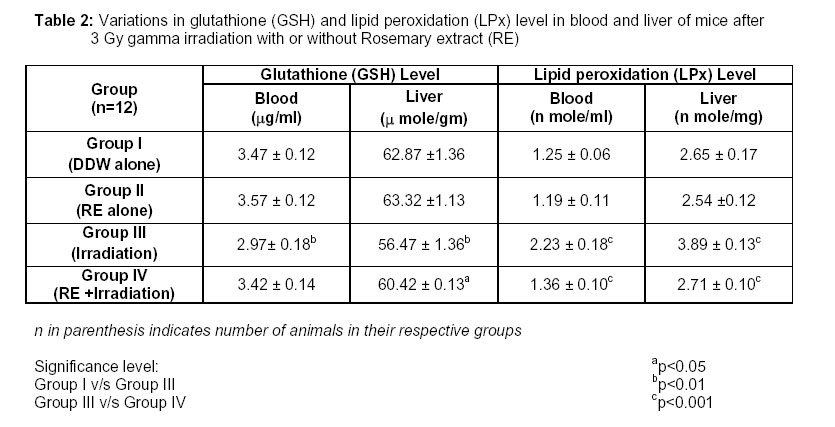
– 302 004 (India) Code Number: tc07029 Abstract The modulatory influence of Rosmarinus officinalis (rosemary) leaves extract was investigated in Swiss albino mice at a dose of 3 Gy gamma radiation. For this purpose, adult Swiss albino mice were irradiated with 3 Gy gamma rays in the presence (experimental) or absence (control) of rosemary (1000 mg/kg body wt.). These animals were necropsied and their blood was collected at days 1, 3, 5, 10, 20 and 30 post-irradiation. A decrease in the number of erythrocyte and leucocyte counts, hemoglobin content and hematocrit percentage was scored in the control group; whereas a recovery pattern was recorded in experimental animals and a normal value of hematological parameters were regained by day 30 post-treatment. In irradiated group, glutathione level was registered low in the blood, whereas a significant elevation was estimated in rosemary pre-treated animals. An increase in lipid peroxidation level above normal was evident in serum of irradiated mice, while a significant decrease in such values was noted in rosemary pretreated group. The present study suggests the possible radioprotective ability of rosemary extract. Key Words: Gamma radiation,Glutathione,Hematology, Lipid peroxidation, Rosmarinus officinalis, Swiss albino mice Introduction Radiation protection concepts and philosophy have been evolving over the past several decades. The inadvertent exposure of human from various sources of radiation causes ionization of molecules, setting off potentially damaging reactions via free radical production. Free radicals are believed to play a role in more than sixty different health conditions, including the ageing process, cancer, radiation damage, atherosclerosis, etc (Ames et al., 1993; LaVerne, 2000). Fortunately, there are many plant derived natural antioxidants that interfere with free radicals before they can damage the body. Antioxidants work in several ways by reducing the energy of the free radicals, stop the free radical from forming in the first place, or interrupt an oxidizing chain reaction to minimize the damage of free radicals. The development of radioprotective agents has been the subject of intense research in view of their potential for use within a radiation environment; however, no ideal, safe synthetic radioprotectors are available to date, so the search for alternative sources, including plants, has been on going for several decades (Scarterrzzimi and Speroni, 2000; Lam and Ng, 2002; Song et al., 2003; Arora et al., 2005). As the utility of medicinal plants suffers from the fact that several of them lack scientific evidences, there is a need to provide scientific back up to justify their potential in treatment of various disorders including radiation damage to living beings (Ammon and Wahl, 1991). Rosemary (Rosmarinus officinalis) belonging to the family Lamiaceae, is best appreciated as a medicinal plant that owes a wide range of biological activities to its antioxidant properties (Al-sereiti et al, 1999). It is indigenous to Southern Europe, particularly on the dry rocky hills of the Mediterranean region. The plant has been used traditionally for curing various disorders among the people around the world for time immemorial. Carribs of Guatemala use this plant to cure various human ailments (Giron et al., 1991). Rosemary has been described as a medicinal plant and wonder-drug in various medieval drug monographs and literature (Selmi, 1967; Grieve, 1971; Zimmermann, 1980). A well-known aromatic culinary herb, rosemary has a long history of medicinal use (Hussain, 1979). This plant has been shown to be safe in toxicity studies in animal models, when added as antioxidant to food (Schuler, 1990). Rosemary has been used as a tonic and stimulant, analgesic, antireumatic, carminative, diuretic, expectorant, anti- epileptic, anti-spasmodic in renal colic, dysmenorrhoea, relieving respiratory disorders effects and for effects on human fertility (Al-Sereiti et al., 1999). Natural products of plant origin may prove to be protective against ionizing irradiation if they counter the harmful effects of radiation-induced free radicals. Evidences support that rosemary is one of the popular antioxidants used for various ailments in different nation’s folk medicine as well as a beverage drink(El-gadi and Bhisna, 1989). Its common usage, wide acceptability and diverse pharmacological and antioxidative properties aroused our interest to obtain insight into the radiomodulatory effect of R. officinalis against gamma exposure in Swiss albino mice. Materials and Methods Animals Animal care and handling were performed according to the guidelines set by the World Health Organization (WHO), Geneva, Switzerland and the INSA (Indian National Science Academy), New Delhi, India. Male Swiss albino mice (Mus musculus), 6-8 weeks old, weighing 20-24 g., from an inbred colony were used for the present study. These animals were maintained under controlled conditions of temperature and light (Light: dark, 10 hrs: 14 hrs.). They were provided standard mice feed (procured from Hindustan Lever Ltd., India) and water ad libitum. Tetracycline water once a fortnight was given as preventive measures against infections. The Departmental Animal Ethical Committee approved the present study. Irradiation Cobalt teletherapy unit (Co-60) at the Cancer Treatment Centre, Radiotherapy Department, SMS Medical College & Hospital, Jaipur was used for irradiation. Unanaesthetised animals were restrained in well-ventilated perspex boxes and exposed to 3 Gy gamma radiation at a distance (SSD) of 80 cm from the source at a dose rate of 0.85 Gy/min. Taxonomic description of the plant It is an evergreen shrub growing to 1.5m by 1.5m at a medium rate. It is in leaf all year, in flower from March to October, and the seeds ripen from August to October. The scented flowers are hermaphrodite. The evergreen leaves of this shrubby herb are about 1 inch long, linear, revolute, dark green above and paler and glandular beneath, with an odour pungently aromatic and somewhat camphoraceous. The flowers are small and pale blue. Much of the active volatile principle(s) resides in their calyces. There are silver and gold-striped varieties, but the green-leaved variety is the kind used medicinally (Grieve, 1971). Preparation of plant extract The identification of the plant Rosmarinus officinalis (family: Lamiaceae, Voucher Specimen No: DDC/2001/DEPTBT/ACHARYA2430) was done by a competent botanist from the Herbarium, Department of Botany, University of Rajasthan, Jaipur (India). The non-infected leaves of the plant were collected, carefully cleaned, shade dried and powdered in a mixer grinder. The plant material was then extracted with double distilled water by refluxing for 36 hrs (12 hrs X 3) at 55 ± 5°C. Pellets of the drug were obtained by evaporation of its liquid contents in an incubator. An approximate 22% yield of the extract was obtained. The required dose for treatment was prepared by dissolving the drug pellets in double distilled water by oral gavage via micropipette in a volume 100 µl/animal at a dose of 1000 mg/ kg body weight. Henceforth, Rosmarinus officinalis leaf extract will be called “RE”. Experimental design Determination of optimum dose of RE against radiation A dose selection of Rosmarinus officinalis (RE) was carried out on the basis of drug tolerance study. For this purpose, various doses of RE extract (100, 200, 400, 800, 1000, 1500 and 2000 mg/kg body weight) were tested for their tolerance (once in a day for 5 consecutive days) in Swiss albino mice. Thus, the most optimum and tolerable dose of RE (1000 mg/ kg b. wt.) was obtained and used for further detailed experimentation. Modification of radiation response A total of 48 animals used for the experiment were assorted into 4 groups of 12 mice each. Animals in group I were administered with double distilled water (DDW), volume equal to RE (1000 mg/ kg body wt. / animal), by oral gavage to serve as normal. Mice in group II were administered orally rosemary extract once daily at a dose of 1000 mg/kg b. wt. / animal for 5 consecutive days. In group III, double distilled water volume equal to RE was administered for 5 consecutive days (as in Group-I). One hour after the last administration of DDW, animals were exposed to 3 Gy gamma rays. Group IV mice were treated with RE, orally for 5 consecutive days (as in Group-II) and were exposed to gamma irradiation one hour after the last administration of RE on day 5. These animals were observed daily for any sign of sickness, morbidity, behavioral toxicity and mortality. A minimum of 6 animals from each group were necropsied on days 1, 3, 5, 10, 20 and 30 post-treatment intervals to evaluate hematological and biochemical parameters. Hematological study For the study, one hour after irradiation, blood was collected from the orbital sinus of animals from each group in a vial containing 0.5 M EDTA. Total number of erythrocytes (RBC), leucocytes (WBC), hematocrit (Hct) and hemoglobin (Hb) percentage were determined by adopting standard procedures. Biochemical determinants Biochemical alterations were studied in animals of all the groups at one hour post-exposure to gamma radiation. The level of glutathione (GSH) was determined in blood and liver by methods of Beutler et al. (1963)and Moron et al. (1979) respectively. The lipid peroxidation (LPx) level in the serum and liver was measured by the assay of thiobarbituric acid reactive substances (TBARS) according to the method of Ohkhawa et al. (1979). Statistical analysis The result for all the groups at various necropsy intervals were expressed as mean ± Standard error (S.E.). To find out whether mean of sample drawn from experimental (group IV) deviates significantly from respective control (group III), Student’s ‘t’ test was used by the method of Bourke et al., (1985). The significance level was set at different levels as P < 0.05, P < 0.01 and P < 0.001. Results General When animals were exposed to a dose of 3 Gy gamma irradiation, no toxic effects in terms of sickness were observed in animals of any of the groups, and also these did not show any mortality and significant change in body weight, urination and defecation pattern. Hematological studies In the present study, alterations in the erythrocytes count, hemoglobin level and hematocrit percentage were found to show a parallel pattern in all the groups. These were markedly suppressed in the 3 Gy irradiated animals and a normal value could not be achieved till the last autopsy interval (i.e., day 30). Prior administration of RE enhanced the recovery in these parameters, and a normal value was registered by day 30th post-interval. Leucocytes were recorded minimum on day 3 post-irradiation with a progressive increase till the last autopsy interval, but a normal value could not be attained. An increasing pattern was also recorded in RE-treated irradiated mice (group IV) and a normal number of leucocytes were regained by day 30th post-treatment (Table 1). Biochemical determinants There was no significant difference in the levels of glutathione (GSH) and lipid peroxidation (LPx) in hepatic as well as blood/ serum content between normal and RE alone treated animals. In concomitant treatment of REand radiation, GSH was found to be further lowered than the radiation treated group. A significant elevation in the values of hepatic and blood GSH as compared to control was estimated in RE pre-treated animals. An increase in LPx level above normal was evident in serum and liver of irradiated mice, while a significant decrease in such values was evident in RE pretreated irradiated group (Table 2). Discussion There is a continual interest worldwide to screen for non-toxic radioprotectors that can be used against harmful effects of radiation in occupation as well as in therapeutic settings for mankind. Despite extensive work done in this field, not a single compound has emerged so far as an effective non-toxic radioprotector for practical purposes (Weiss and Landauer, 2003; Rades et al., 2004; Singh et al., 2005). Therefore, screening of natural products of plant origin represents a major avenue for the discovery of new radioprotective drugs. In this study, we have attempted to evaluate the radiomodulatory effect of R. officinalis in Swiss albino mice. The active constituents of R. officinalis like carnosol, carnosic acid, caffeic acid, rosmarinic acid, ursolic acid, different diterpenes, phenols and flavanoids (Fluck et al., 1976; Al-sereiti et al., 1999; Frost and Tyler, 2000) have been subjected to many pharmacological investigations.Carnosic acid and carnosol, the two major active ingredients of rosemary, were found to exhibit anticarcinogenic activity against the action of oxygen radicals in animals (Minnunni et al., 1992). Furthermore, natural polyphenols found in rosemary have not only potent antioxidant activities but also anticarcinogenic properties (Offord et al., 1997). The present study revealed that after exposure to 3 Gy, the erythrocyte count exhibited a fall that can be attributed to inhibition of new cells entering into blood, loss through haemmorhage and/ or radiation-induced injury (Casarett, 1968). A similar depression was observed in hemoglobinlevel without returning to normal till the last day of experiment. The present investigation suggests that the hemoglobin concentration follows a pattern similar to that of RBC in general. Similar findings were proposed earlier by Daga et al., 1995) who found noticeable depletion in hemoglobin concentration in Swiss albino mice exposed to 3.6 Gy gamma radiation. A depression in the hematocrit value can be attributed to total cell depletion in peripheral blood aided by disturbances in steady state mechanisms in blood forming organs as well as an increase in plasma volume after irradiation. This is in agreement with the recent findings of Nunia and Goyal (2004). The initial rapid fall in leucocytes count was mainly due to a fast decline of lymphocytes in peripheral blood that are the most radiosensitive as revealed by differential leucocyte count in the present study. It is also in favor of earlier findings of Samarth et al. (2001) who reported a depression in the number of leucocytes of gamma irradiated mice. It has been observed that rosmarinic acid is effective in relation to blood circulation and to improve hemodynamics in occlusive arterial diseases (Al-sereiti et al., 1999). Maintenance of the cellular glutathione (GSH), a free radical scavenger, is critical for keeping a check on cellular homeostasis (Munday and Winterbourn, 1989). In group III, glutathione level was found to decrease as compared to RE protected group IV. Oral administration of RE did not influence the endogenous GSH level significantly either in liver or blood. One of the mechanisms of RE protection against radiation can be an elevation in the glutathione level that is mediated through the modulation of cellular antioxidant level. This could have resulted in the reduction in lipid peroxidation level, thereby protecting against damage caused by radiation in the RE pre-treated irradiated group. Rosmarinic acid has been experimentally found to have significant antioxidant role by free radical scavenging activity (Lamaison et al., 1991). The basic effect of radiation on cellular membranes is believed to induce lipid peroxidation by the production of free radicals (Leyko and Bartosz, 1986). The level of radiation-induced LPx increased considerably in 3 Gy irradiated animals whereas a decrease in the values was observed in RE-treated group. This view is supported by the anti-lipoperoxidant activities of the young sprouts of R. officinalis that have shown to reduce the formation of malondialdehyde significantly in rat hepatocytes (Joyeux et al., 1990). Sotelo-Felix et al.(2002)proposed that carnosol could scavenge free radicals induced by carbon tetrachloride, consequently avoiding the propagation of lipid peroxides in the liver of mice. The exact mechanism of action of rosemary is not known; however, it may scavenge free radicals produced by radiation and thus reduce radiation-induced damage to the cellular DNA (Al-sereitiet al., 1999). Furthermore, results of our investigation elucidate the antioxidant effects of rosemary extract that are possibly due to their ability to trigger the endogenous glutathione level and suppress lipid peroxidation in the blood. The role of rosemary can also be attributed to stimulating or protecting hematopoiesis in bone marrow and the subsequent increase of hematological constituents in the peripheral blood. Since significant protection is obtained at a non-toxic low dose, RE may have an advantage over the known radioprotectors. Further investigations are in progress to study the exact mechanism of action and clinical applicability of R. officinalis as a radioprotector. References
© Copyright 2007 - African Journal of Traditional, Complementary and Alternative Medicines The following images related to this document are available:Photo images[tc07029t2.jpg] [tc07029t1.jpg] |
| |||||||||
