 |
| About Bioline | All Journals | Testimonials | Membership | News |
|
||||||
|
||||||
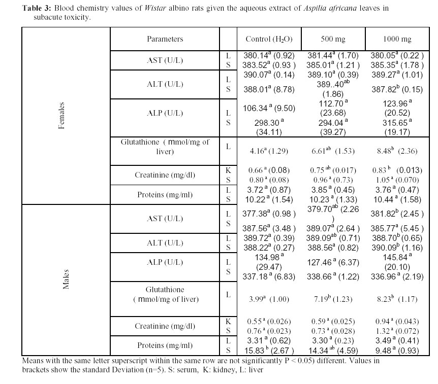
African Journal of Traditional, Complimentary and Alternative Medicines, Vol.4, No. 2, 2007, pg. 127-134 Research Paper ACUTE AND SUBACUTE TOXICITY OF ASPILIA AFRICANA LEAVES Taziebou Lienou C1., Etoa F-X1٭., Nkegoum B.2, Pieme C. A1. Dzeufiet D. P.D3 1Microbiology and Pharmacology Laboratory, Department
of Biochemistry, Faculty of Science, P. O. Box 812, University of Yaounde I ,
Cameroon., 2Pathology Laboratory, Faculty of Medicine and
Biomedical Sciences, P. O. Box 2787, University of Yaounde I, Cameroon., 3Animal Physiology Laboratory, Department of Animal Biology and
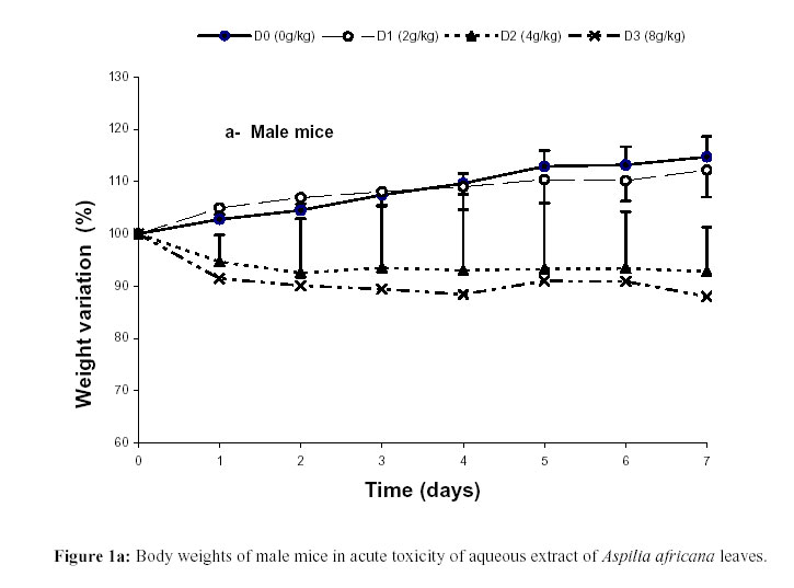
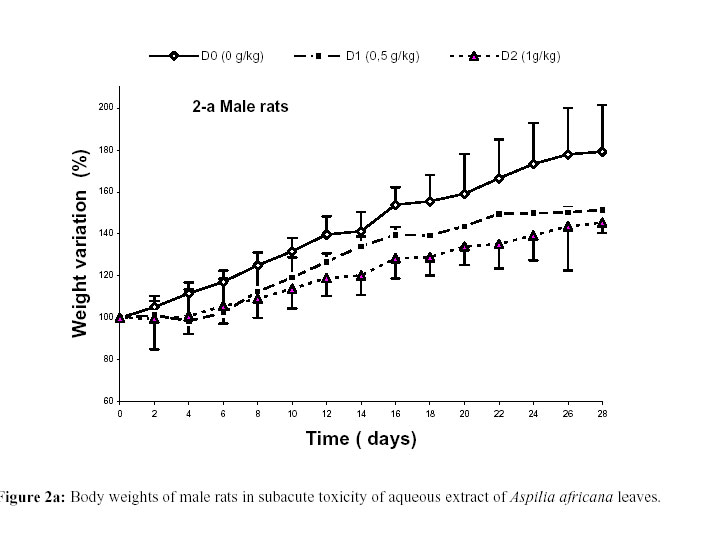
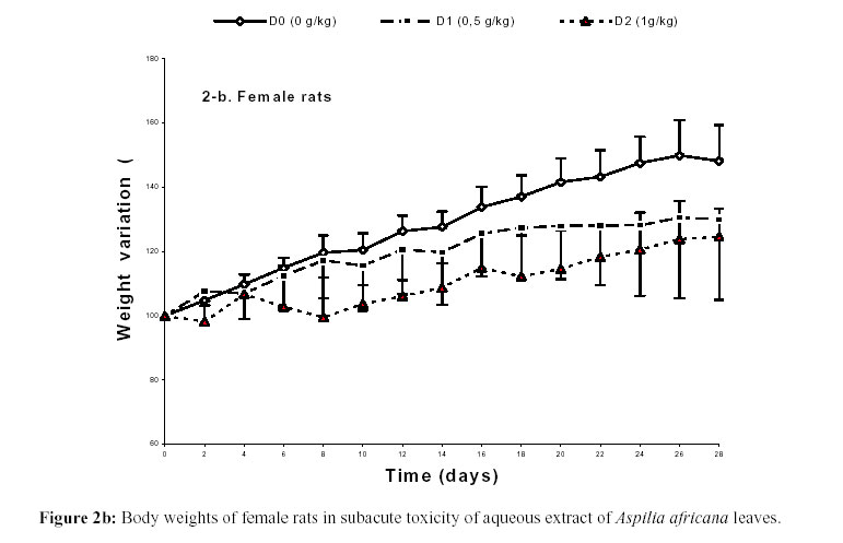
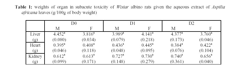
Physiology, Faculty of Science P. O. Box 812, University of Yaounde I, Cameroon. Code Number: tc07034 Abstract This study was designed to evaluate the toxicity of the aqueous extract of Aspilia africana leaves. Oral doses of 500 mg/kg and 1000 mg/kg were administered for 28 days to rats after every 2 days for sub-acute toxicity. For acute toxicity, 5 doses of 2, 4, 8, 12 and 16g/Kg body weight were investigated in mice. The control groups consisted of mice or rats administered with distilled water. The signs of toxicity fluctuated lightly from one mammal to another throughout the experiment. The liver, kidneys and heart weight of rats revealed no significant differences between the test groups and the control. The results indicated that the medium lethal dose (LD50) was found to be greater in females than males with an average of 6.6g/Kg body weight for both sexes. Regardless of the significant differences observed at certain points in some biochemical parameters (ALT, AST, ALP, Creatinine and Glutathione); none showed any linear dose responsiveness. On the other hand, most of the parameters investigated were found to be gender dependent. These results suggested that A Africana can be classified among substances with low toxicity. Key words: Aspilia africana, Asteraceae,toxicities, dose responsiveness. Introduction Natural products have been, and have remained, the cornerstone of health care. Present estimates show that, 80% of the world’s population still rely on traditional medicine for their health care needs (Farnsworth et al., 1985). Unfortunately, most of those who use these plants in our society have not undergone adequate training. Therefore, in order to have standard natural plant products, preliminary studies have to be done in order to evaluate possible risks such as, undesirable effects, overdose or poisoning. Aspilia africana (Pers.) C.D. Adams is not an exception. It is a perennial herb which belongs to the Asteraceae family. Aspilia africana is widely used in African folk medicine to stop bleeding, remove corneal opacities, induce delivery and in the treatment of anaemia and various stomachs complains (Iwu, 1993; Adjanohoun et al., 1996). Phytochemical studies revealed the presence of saponines and tannins as the most abundant compounds in the plant while flavonoids were the least (Obadoni and Ochuko, 1998). Other studies showed that essential oils from the leaves of Aspilia africana were rich in sesquiterpenes and monoterpenes. Also, the presence of precocene1 was found (Kuate et al., 1999). The medicinal plant contained ascorbic acid riboflavin thiamine, and niacinm. These herbs are good sources of minerals such as Ca, P, K,Mg, Na, Fe and Zn (Okwu and Josiah, 2006). Despite the popular use of this plant, no toxicological study has been reported. The present study was undertaken to determine the toxicity of the aqueous extract of Aspilia africana leaves. Materials and Methods Plant material Fresh leaves of Aspilia africana were collected from Simbock, a village in neighbourhood of the capital city of Cameroon. The sample was identified at the National Herbarium where some specimen was already available with the number 6555/SRF/Cam. Preparation of the extract The leaves collected were dried in an air-circulating oven at 38°C ± 2°C. The dry material (300 g) was macerated in 6 litters of distilled water for 48 h at 4°C in a refrigerator. The extract was sieved and the juice was filtered using whatman N°1 filter paper. The filtrate was put in a stainless-steel tray, and concentrated in an air-circulating oven at 42°C until total dryness. The gummy extract was put into small glass dishes and stored at 28°C in an incubator for further studies. Bioassay Young male and female Wistar albino rats (90-110 g) and Swiss albino mice (12-25 g) were bred at the Department of Biochemistry, in the breading house of the Microbiology and Pharmacology Laboratory of the Faculty of Science. They were all clinically healthy and were kept under standard environmental conditions of temperature (27°C ± 2°C). The animals had free access to water and standard diet. The principles of laboratory animal care were approved by the Department’s ethical committee. Acute toxicity The bioassay was conducted according to the World Health Organisations guideline for the evaluation of the safety and efficiency of herbal medicines (O.M.S., 2000). For the study, Swiss albino mice were divided into six groups of 10 animals each (5 males and 5 females). Animals were deprived of food but not water (16-18 h) prior to administration of the extract. Five groups were given single oral doses of 2, 4, 8, 12 and 16 g/kg of the aqueous extract. The last group used as control received distilled water per os in the experiment. Observations were made and recorded systematically as 1, 2, 4 and 24 h after substance administration. The visual observations included motility, respirations, sensitivity to sound and pinch, smelling of food and faeces consistence. The numbers of survivors were recorded after 24 h and the animals were observed daily for the next 7 days. The LD50 was determined based on Behrens and Kaber (1983), and Schorderet (1992) methods. Sub-acute toxicity Three groups of 10 rats each (5 males and 5 females) were given intra-gastric intubation of 500 mg/kg and 1000 mg/kg of the aqueous extract or distilled water (for control) every 48 h for 26 days. Food and water intake as well as body weight were monitored during the period of administration. After 26 days, all surviving animals were allowed to fast overnight and sacrificed by decapitation after anaesthesing with petroleum ether. Blood samples were collected from these mammals into heparinated tubes for haematological analyses and non heparinized centrifuge tubes. The liver, kidneys and heart were collected and weighed. Part of the liver or kidney tissues was also washed and kept in a freezer (-20°C) for further analysis of biochemical parameters. Another part was preserved in 10% formalin for histopathological studies. Biochemical analysis The blood collected into non heparinized tubes was centrifuged at 3000 rpm for 10 min. The serum was separated and liver and kidney homogenates (20%) were analysed for enzymes. Alanine aminotransferase (ALT) and Aspartate aminotransferase (AST) were assayed using the Reitman and Frankel (1957) method; Alkaline phosphatase ( ALP) was analysed by the method of Bessey et al., 1946; Protein by the method of Gornall et al., 1949; Creatinine and Glutathione by Barterls et al., (1972) and Ellman (1959) methods respectively. Haematological studies The blood samples that were collected into heparinized tubes were used for the estimation of white blood cells (WBC), red blood cells (RBC) and platelets by visual methods (Dacie, 1991). Histopathological studies Histopathological analyses of the liver and kidneys were done according to the conventional haematoxylin-eosin technique. Statistical Analysis Statistical analysis was done using ANOVA. The Duncan’s test was used to locate significant differences between means’. Significant differences treatments were accepted at P < 0.05. Data were expressed as means ± standard deviation. Results and Discussions In acute toxicity, a slight but non significant increase in body weight was registered for mice treated with the aqueous extract of Aspilia africana at the dose 2 g/kg (Figure 1a,b). This increase in body weight, which is higher than that of the control group, may be due to stimulation of appetite by the extract, leading to increased food consumption and the body weight observed. Behavioural changes in the mice were linear dose responsiveness. These changes included increased aggressiveness and motility. The changes greatly reduced within 48 h without completely turning to normal and this continued throughout the experimental period with all the animals that survived. This enabled us to suggest that there was an irritating compound in the aqueous extract. The medium lethal dose value (LD50) was 6.1 g/kg and 7.5 g/kg body weight for males and females respectively with an average of 6.6 g/kg body weight. According to Schorderet (1992), substances with LD50 values greater than 5000 mg/kg body weight are classified as substances with low toxicity. Thus, the aqueous extract of Aspilia africana can be considered as a substance with low toxicity. Furthermore, all the male mice died when treated with 12 g/kg body weight dose while the female mice treated with 16 g/kg body weight produced the same result. Thus indicates that the female mice are less sensitive to toxicity than the males. This may be due to the hormonal status which is different in males and females. Similar results were obtained by Solomon et al., (1993) when studying the ethanolic extract of the roots of Plumbogo rosea (Plumbaginaceae). In sub-acute toxicity, the behavioural changes (aggressiveness) observed in acute toxicity were noted with rats during the experiment. As shown in Table 1, there were no significant variations in the weight of the organs of treated animals compared to the control. In the animals tested with the extract there was a slight and time dependent decrease in body weight compared to the control group. This decrease in body weight could be due to reduced appetite or impairment of some nutrients as a result of the extract. The haematological status of the rats, after 28 days of oral administration of the aqueous extract of Aspilia africana is shown in Table 2. Compared to the control, an increase in values of all the parameters studied was registered with the dose of 500 mg/kg body weight. This increase was statistically significant (P < 0.05) for males as far as the platelets are concerned. An increase in platelet numbers led to: chronic myeloproliferative diseases, carcinoma, chronic inflammatory diseases, haemorrhage, sickle cell diseases associated with a non-functioning spleen or after splenectomy, iron deficiency anaemia associated with active bleeding (Cheesbrough, 2001). In females, a significant (P < 0.05) increase was noted with WBC and platelets at the dose of 500 mg/kg body weight. The main causes of increased WBC count are: metabolic disorders, poisoning, acute haemorrhage, leukaemia and myeloproliferative disorders, stress, menstruation and strenuous exercises (Cheesbrough, 2001). At the dose of 1g/kg, a decrease in number of WBC, RBC and platelets was registered for males compared to the control at the dose of 500 mg/kg. The decrease was not significantly different compared to the control. There was a significant difference (P < 0.05) compared to that of the 500 mg dose for platelets only. Causes of raised level of erythropoietin can occur in renal disease, cyanotic heart disease, shock and acute alcohol poisoning (Cheesbrough, 2001). In females, a significant (P < 0.05) increase was noted for platelets and WBC, compared to the control, at 500 mg/kg body weight. For biochemical parameters (Table 3), no significant variations in ALP activity were noted. A significant increase in AST activity was noted in the liver at a dose of 1 g/kg in males only. For serum ALT, a significant (P < 0.05) increase was registered in males. Although AST and ALT are common liver enzymes because of their high concentrations in hepatocytes, only ALT is remarkably specific for the liver function since AST is mostly present in the myocardium, skeletal muscle, brain and kidneys (Sacher and McPherson, 1991, Witthawasku et al., 2003) . Despite the increased level of creatinine observed in the kidneys and serum in both sexes of all the animals with respect to the control groups, a significant increase (P < 0.05) was registered only in females. According to Harper (1971), an increase in creatinine level can be observed in some kidney illnesses, due to loss of its normal excretive function of creatinine, when there is muscular cells damage or following an incompatible medication interfering with normal functioning of the kidneys. The level of glutathione significantly (P < 0.05) increased at the dose of 500 mg/kg in males only, and at 1000mg/kg body weight the doe in males and females. The high concentration of glutathione observed may be due to the effectors effects of some components present in the extract. A histopathological examination of the liver and kidneys showed the presence of necrosis, oedema and inflammatory infiltrations. Conclusion From the results obtained, we can say that, although aqueous extract of Aspilia africana leaves can be classified among substances with low toxicity, dosages of 500mg/kg body-weight or more, can be toxic for a long term treatment when taken orally. Acknowledgement We are grateful to Mrs Achu Mercy Bih for proofreading the manuscripts and making helpful suggestions. References
© Copyright 2007 - African Journal of Traditional, Complementary and Alternative Medicines The following images related to this document are available:Photo images[tc07034f1a.jpg] [tc07034t1.jpg] [tc07034f2b.jpg] [tc07034f2a.jpg] [tc07034t2.jpg] [tc07034f1b.jpg] [tc07034t3.jpg] |
| |||||||||
