 |
| About Bioline | All Journals | Testimonials | Membership | News |
|
||||||
|
||||||
African Journal of Traditional, Complimentary and Alternative Medicines, Vol.4, No. 3, 2007, pg. 362-371 Research Paper ANTIHYPERGLYCEMIC EFFECTS OF SEPARATE AND COMPOSITE EXTRACT OF ROOT OF MUSA PARADISIACA AND LEAF OF COCCINIA INDICA IN STREPTOZOTOCIN-INDUCED DIABETIC MALE ALBINO RAT. Chhanda Mallick a b, Kausik Chatterjee b, Mehuli GuhaBiswas b and Debidas Ghoshb a Reproductive Endocrinology and Family Welfare Research Unit, Department of Human Physiology with Community Health, bBio-Medical
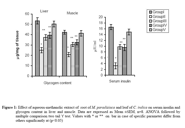
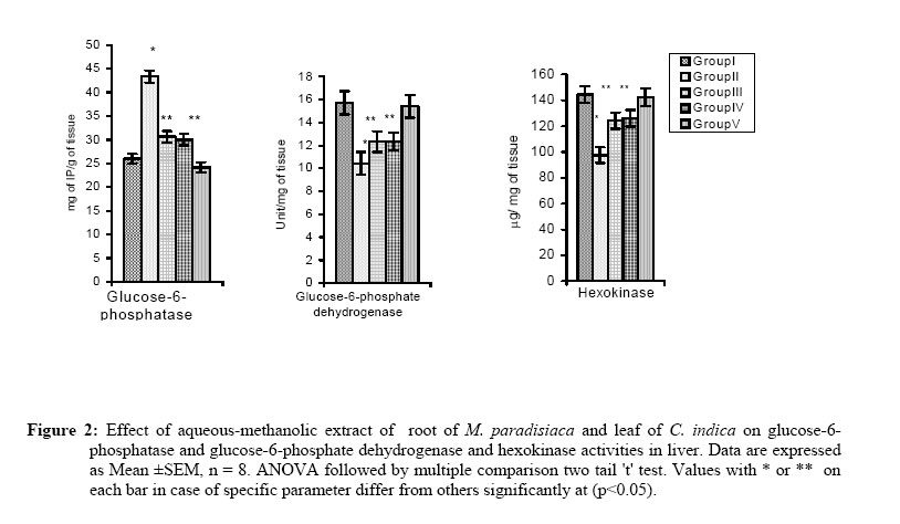
Laboratory Science and Management Code Number: tc07038 Abstract We evaluated the antihyperglycaemic properties of aqueous-methanolic (40:60) extract of root of Musa paradisiaca and leaf of Coccinia indica in separate as well as in composite manner by conducting experiment on streptozotocin-induced diabetic rats. We measured food and water intake ability, the fasting blood glucose level, glucose tolerance, activities of important carbohydrate metabolic enzymes like glucose-6-phosphatase, glucose-6-phosphate dehydrogenase, hexokinase in liver along with quantification of glycogen in liver and in skeletal muscle and serum insulin level. We noted that after treatment of aqueous methanolic extract of above plant parts in separate as well as in composite manner at a concentration of 80mg/100g body weight/day to streptozotocin-induced diabetic rat resulted in a significant remedial effect on blood glucose level as well as carbohydrate metabolic enzymes and the quantity of liver and skeletal muscle glycogen. Serum insulin level that was diminished in streptozotocin-induced diabetic rat recovered significantly after the co-administration of extract of above plant parts. All the above parameters showed a more potent remedial effect after composite extract treatment with respect to separate treatment and none of the extract has any general metabolic toxicity induction. Key words: Diabetes, Glycaemic index, Insulin, Musa paradisiaca, Coccinia indica Introduction Diabetes is recognized as one of the leading causes of mortality and morbidity in the world (Can et al., 2004). It is a heterogeneous group of diseases and at present it is known as syndrome (Zimmet, 1997). The central identifying feature of diabetes mellitus is chronic and substantial elevation in the circulating glucose concentration. The major mode of control of diabetes can be achieved by diet, exercise, insulin replacement therapy and by the use of herbal hypoglycemic agents (Ivorra and Paya, 1989). Diet therapy along with insulin or oral hypoglycemic agent forms an important way of treatment in diabetes (Clark and Lee, 1995) though it has several demerits. The major drawbacks of insulin therapy are the side effects, which include insulin allergy, lipodystrophy and lipoatropy, insulin antibodies, altered metabolic control, placental transfer of insulin antibodies, autoimmunity and other late complications like morphological changes in kidneys and severe vascular complications (Defronzo et al., 1982; Jarvinen and Koivisto, 1984; Jarvinen and Koivisto, 1986). Similarly, the oral hypoglycemic drugs have many side effects such as nausea and vomiting, cholestatic jaundice, agranulocytosis, aplastic and hemolytic anemias, generalized hypersensitivity reactions, dermatological reaction and lactic acidosis (Khan and Shechter, 1991). Plants have been used for the major source of treatment of diabetes mellitus from ancient time in the Indian medicine and in the world. Herbal drugs are mostly out of toxic or side effect than the chemical drug (Rao et al., 2003). From Indian medicinal system it is found that composite extract is most effective than the separate one. On that background this work has been designed to find out the most effective extract of Musa paradisiaca and Coccinia indica in separate and in composite manner to correct the streptozotocin-induced diabetic disorders. From trial and error it has been noted that aqueous-methanolic extract of these two plant parts has a promising antidiabetic effect. . M. paradisiaca is a tree like herb with thick stem composed of convoluted leaf sheaths. Leaves are very large and oblong. This plant belong to the Musaceae family and is distributed throughout the India and Malaysia. Roots of M. paradisiaca are antihelmintic. Flowers are astringent (Joshi, 2000). Fruits are mild laxative. It aids in combating diarrhea and dysentery and promotes healing of intestinal lesions in ulcerative colitis (Joshi, 2000). It is useful in celiac disease, constipation and peptic ulcer (Joshi, 2000). Unripe fruit and cooked flower are useful in diabetes. Different parts of the plant have medicinal value (Kirtikar and Basu, 1991; Joshi, 2000). Coccinia indica is a climbing perennial herb of Cucurbitaceae family and it is distributed widely all over India. Its medicinal importance has been observed earlier by others (Chopra, 1958). Root and leaves of this plant have antilipidemic effects (Eshrat, 2003) as well as antioxidative effects (Venkateswaran and Pari, 2003). Materials and MethodsChemicals Streptozotocin was obtained from spectrochem Pvt. Ltd chemical company (India). Insulin enzyme linked immunosorbant assay (ELISA) kit was purchased from Boehringer Mannheim Diagnostic, Mannheim (Germany). Plant materials and preparation The roots of M. paradisiaca and leaves of C. indica were collected from local area in the month of June and the materials were identified by taxonomist of Botany Department, Vidyasagar University, Midnapore. The voucher specimens with HPCH No: 7, 8 of the plants were deposited in the Department of Botany, Vidyasagar University, Midnapore. Fresh leaves of C. indica and small pieces of fresh roots of M. paradisiaca were dried in an incubator for 2 days at 40 oC, crushed separately in an electric grinder and then powdered. Out of this powder, 100 g was suspended in 500 ml of aqueous-methanol (2:3) mixture and kept in incubator at 37 oC for 36 h. The slurry was stirred intermittently for 2 h and left overnight. The mixture was then filtered and filtrate was dried by low pressure and residue was collected. When required the residue was suspended in olive oil in a fixed dose and used for treatment. Selection of animal and animal care Forty matured normoglycemic Wistar strain male albino rats of 3 month of age weighing about 150.0 ± 10.0 g were used for this experiment. Animals were acclimated for a period of 15 days in our laboratory condition prior to the experiment. Rats were housed at an ambient temperature of 25 ± 2 oC with 12 h light: 12 h dark cycle. Rats have free access to standard food and water ad libitum. The principles of laboratory animal care (NIH 1985) and instruction given by our institutional ethical committee were followed through out the experimental period. Induction of diabetes mellitus Fasting rats for 24 h were subjected to single intramuscular injection of streptozotocin (STZ) at the dose of 4 mg /0.1 ml of citrate buffer/100 g body weight/rat that produced type I diabetes (having fasting blood sugar level more than 250 mg/dl after 24 h of STZ injection. Thirty-two rats were made diabetic in this way. Animal treatmentForty rats were divided into five groups equally as follows and duration of experiment was of 14 days. Group I: (Control group): Rats of this group received single intramuscular injection of citrate buffer (0.1 ml/100 g body weight/rat). Group II: (Diabetic group): Rats of this group were made diabetic by single intramuscular injection of streptozotocin at the dose of 4 mg/0.1 ml citrate buffer/100 g body weight/rat. Group III: (C. indica co-administered group): The diabetic rats of this group were fed by gavage method with aqueous-methanolic extract of leaf of C. indica at the dose of 80 mg/0.5 ml olive oil/100 g body weight/rat/day after 24 h of STZ injection for 14 days at fasting state. Group IV: (M. paradisiaca co-administered group): Streptozotocin-induced diabetic rats were fed by gavage with aqueous-methanolic extract of root of M. paradisiaca at the dose of 80 mg/0.5 ml olive oil/100 g body weight/rat/day for 14 days. Group V: (M. paradisiaca and C.indica co-administered group): The diabetic rats of this group were fed with aqueous-methanolic extract of seeds of M. paradisiaca and leaf of C. indica in composite manner at the dose of 80 mg (1:1)/ 0.5 ml olive oil/100 g body weight / rat for 14 days by gavage. Co-administration of extract in group III, IV and V was performed early in the morning and at fasting state. Animals of control (group I) and diabetic groups (group II) were subjected to forceful feeding of 0.5 ml of olive oil/100 g body weight /day for 14 days at the time of extract co-administration to the animals of group III, IV and V to keep all the animals in same experimental condition. On 16th day of experiment, all the animals were sacrificed by decapitation after recording the final body weight, blood was collected from dorsal aorta and serum was separated by centrifugation at 3000 g for 5 min for the assay of insulin followed by ELISA technique. Liver and skeletal muscle were dissected out and stored at –20 oC for biochemical analysis of enzyme activities of glucose-6-phosphatase, glucose-6-phosphate dehydrogenase, hexokinase in liver as well as glycogen content in liver and skeletal muscle. Testing of fasting blood glucose level Fasting blood glucose (FBG) level was measured from the first day of extract supplementation to diabetic rats. Fasting blood glucose level in all groups was measured by single touch glucometer at the interval of two days. Blood was collected from the tip of the tail vein and FBG level was measured by single touch glucometer. Glucose tolerance test After overnight fasting, on the day the animals were sacrificed, a zero-min blood sample was taken from tip of tail vein of all the rats: control (Group I), diabetic (Group II), diabetic + M. paradisiaca (Group III), diabetic + C. indica(Group IV) and diabetic + M. paradisiaca +C. indica (Group V) . Glucose solution at the dose of 0.5 g /kg body weight/5ml physiological saline was administered intravenously through femoral vein and blood samples were collected at 30th, 60th, 90th and 120th minute for the measurement of glucose levels by single touch glucometer after the administration of glucose (O'Brien et al., 1985). Biochemical assay of glucose-6-phosphatase activity in liver The liver glucose-6-phosphatase activity was measured according to standard protocol (Swanson, 1955). Tissue was homogenized in ice cold of 0.1 M phosphate buffer saline (pH=7.4) at the tissue concentration of 50 mg/ml. In a calibrated centrifuge tube, 0.1ml of 0.1 M glucose-6-phosphate solution and 0.3 ml of 0.5 M maleic acid buffer (pH=6.5) were taken and brought to 37 oC in water bath for 15 min. The reaction was stopped with 1 ml of 10% trichloroacetic acid (TCA) followed by chilling in ice and centrifuged at 3000 × g for 10 min. The optical density was noted at 340 nm. The enzyme activity was expressed as mg of inorganic phosphate liberated per gm of tissue. Biochemical assay of glucose-6- phosphate dehydrogenase in liver The liver glucose-6-phosphate dehydrogenase activity was measured spectrophotometrically (Langdon, 1966). One unit of enzyme activity is defined as that quantity which catalyses the reduction of 1 µM of NADP per minute. Activity of this enzyme was recorded by using glucose-6-phosphate as a substrate and absorbance was measured at 340 nm. Assay of hexokinase in liver The enzyme activity was determined on the basis of reduction of NADPH coupled with hexokinase which was measured spectrophotometrically at 340 nm (Chou and Wilson, 1975). Biochemical assay of glycogen content Glycogen levels in liver and skeletal muscle were measured biochemically (Sadasivam and Manickam, 1996). Tissue homogenized in 80% ethanol, and extract was collected by centrifugation using anthrone reagent, and quantity of glycogen was measured in relation to standards, which was expressed in µg of glucose per mg of tissue. Serum insulin level Serum insulin was measured by enzyme linked imunosorbant assay (ELISA) using the kit (Brugi et al., 1988) (Boehringer Mannheim Diagnostic, Mannheim, Germany). The intra-assay variation was 4.9%. As the samples were run at a time, so there is no inter-assay variation. The insulin level in serum was expressed in µIU/ml. Biochemical assay of glutamate oxalate transaminase (GOT) and glutamate pyruvate transaminase (GPT) Liver and kidney GOT and GPT activities were measured using the kit supplied by Crest Biosystems, Alto Santacruz, Bambolim Complex (Goa, India). The activities of these enzymes were expressed as unit per gram of tissue (Henry et al., 1960). Statistical analysis Analysis of Variance (ANOVA) followed by multiple two-tail ‘t’ test was used for statistical analysis of collected data (Sokal and Rohle, 1997). Differences were considered significant at p< 0.05. Results Body weight, water and food intake Body weight, water and food intake of diabetic animals decreased significantly and after composite extract supplementation there was a significant recovery of these parameters towards the control level although separate or individual extract of the M. paradisiaca and C. indica plants were able to protect these parameters partially (Table 1). Glycemic parameters Streptozotocin injection resulted in a significant elevation in fasting blood glucose level throughout the experiment compared to control and the diabetic-induced animals were unable to tolerate extra glucose load. Treatment of composite extract to streptozotocin diabetic animals resulted in a complete recovery of fasting blood glucose level on 16th day of treatment and the animals were able to tolerate the exogenous glucose load and behaved as control. Individual extract treatment in separate manner to diabetic animal was unable to reverse these parameters to the control level but was able to protect the above parameters partially (Tables 2 and 3). Quantity of glycogen in liver and muscle decreased significantly in diabetic group compared with control group. After composite extract co-administration , the levels of liver and muscle glycogen were reestablished to the control level though the individual extract was unable to reset these values to the control level (Figure 1). Streptozotocin-induced diabetic animal resulted in a significant elevation in glucose-6-phosphatase activity along with diminution in glucose-6-phosphate dehydrogenase and hexokinase activities in liver with respect to control group. Though separate extract co-administration to diabetic rats resulted in a partial protection in these parameters but composite extract resulted in a significant protection and the levels of these parameters reversed to control level (Figure 2). Serum insulin level was decreased significantly in diabetic group in respect to control. After composite extract co-administration, the serum insulin level was restored towards the level of matched control group more effectively than the separate extract co-administration (Figure 1). Table 1: Effect of aqueous-methanolic extract of root of M. paradisiaca and leaf of C. indica on body growth, water and food intake in diabetic rats.
Data are expressed as Mean ± SEM, n=8. Values ANOVA followed by multiple
comparison two tail 't' test. Table 2.: Effect of aqueous-methanolic extract of root of M. paradisiaca and leaf of C. indica on fasting blood glucose level in diabetic rat. Data are expressed as Mean ±SEM, n = 8 Values with * or ** or *** in each column differ from others significantly at (p<0.05)
Table 3: Effect of aqueous-methanolic extract of root of M. paradisiaca and leaf of C. indica on intravenous glucose tolerance in diabetic rats.
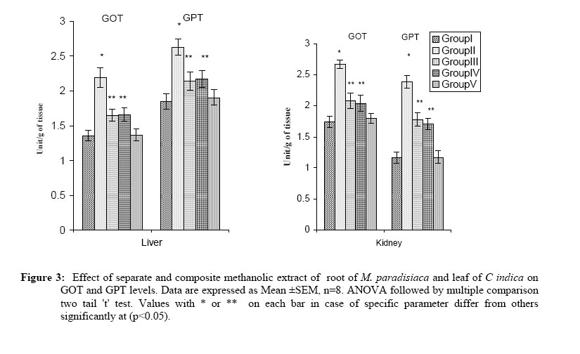
Data are expressed as Mean ±SEM, n=8. ANOVA followed by multiple comparison two tail 't' test Values with * or ** in each column differ from others significantly at (p<0.05) GOT & GPT activities Streptozotocin induced diabetes resulted in an elevation of GOT and GPT activities in liver and kidney at significant level when compared to control. After the treatment of composite extract, both parameters in liver as well as in kidney were in the levels of control group although the individual extract was able to protect these parameters partially (Figure 3). DiscussionStreptozotocin-induced experimental diabetes is a valuable model for induction of type-I diabetes (Junod et al.,1969). The present investigation highlights the antidiabetic efficacy of aqueous-methanol extract of M. paradisiaca and C. indica along with comparative analysis of composite as well as separate extracts of these plant parts. Antihyperglycemic potency of these plant parts has been evaluated here by the measurement of body growth, water and fluid intake, fasting blood glucose level, intravenous glucose tolerance, activities of glucose-6-phosphatase, glucose-6- phosphate dehydrogenase, hexokinase in liver, and quantification of liver and muscle glycogen along with serum insulin level. The present study is important in this respect as this is the first biochemical study on the effects of the extracts of these two plants in the management of type-I diabetes mellitus. Regarding the mode of action of the extract of these plants, the exact mechanism can not be deduced from this experiment but the following hypothesis may be proposed. Glucose-6-phosphate dehydrogenase utilize glucose through pentose phosphate pathway and its activity is under insulin (Tian et al., 1998). Induction of diabetes by STZ resulted in significant diminution in the activity of this enzyme in the liver and this is consistent with our previous report (Mallick et al., 2006). Aqueous-methanolic extract of these plants resulted in a significant recovery of these enzymes which may be one of the ways of antidiabetic efficacy of these plants. Elevation in the activity of glucose-6-phosphatase in STZ-induced diabetic group as reported in the previous studies (Pari and Amarnath, 2004). Extract of these plants resulted in a significant recovery in the activity of this enzyme in liver and this may suggest another possible way of its antidiabetogenic efficacy. The quantity of glycogen in both liver and skeletal muscle was observed to decrease significantly in STZ induced-diabetic rat as STZ selectively destroy β cells of Islets of Langerhance (Kavalali et al., 2002; Palmer et al., 1998) and there by lowering the blood level of insulin has been reported by others (Zhang et al., 2004). This is similar to our observation in this experiment. Separate and composite extract treatment of the above plants resulted in a significant recovery with respect to this effect and this may be due to either stimulation in insulin release from β cells noted in this study or due to insulinomimetic activity of some of the components present in this extract or due to combination of both effects. Protective effect of diabetes of the extract of above plants has been supported with the results obtained with regards to food intake and water intake tending towards the control level in diabetic animal after the extract co-administration. Diabetic condition resulted in liver and renal toxicity induction, which has been observed in this study as shown by GOT and GPT assessment and as reported by others (Ghosh and Suryawanshi, 2001). Both separate and composite extract treatment in diabetic animal resulted in a significant recovery in the levels of GOT and GPT activities in liver and in kidney with respect to diabetic group. As GOT and GPT activities are considered as indicators of general toxicity (Akther et al.,1990; Das et al., 2006) so we suggest that these experimental plant parts have no general toxic effect. From the comparative analysis, it has been revealed that the extract of the studied plants when used in composite manner on streptozotocin-induced diabetic state has a more potent antidiabetogenic effect in comparison to the antidiabetogenic activities of the individual extract of the plants. In conclusion, it may be stated that this composite extract contains the active antihyperglycemic agent (s) that can be used to overcome diabetic complication by pancreatic β cell regeneration or stimulation of insulin secretion or in other ways. AcknowledgementThis research work was funded by University Grants Commission, New Delhi, India. References
© Copyright 2007 -African Journal of Traditional, Complementary and Alternative Medicines ; The following images related to this document are available:Photo images[tc07038t1.jpg] [tc07038t2.jpg] [tc07038t3.jpg] [tc07038f3.jpg] [tc07038f2.jpg] [tc07038f1.jpg] | ||||||||||||||||||||||||||||||||||||||||||||||||||||||||||||||||||||||||||||||||||||||||||||||||||||||||||||||||||||||||||||||||||||||||||||||||||||||||||||||||||||||||||||||||||||||||||||||||||||||||||||||||||||||||||||||||||||||||||||||||||||||||||||||||||||||||||||||||
| |||||||||
