 |
| About Bioline | All Journals | Testimonials | Membership | News |
|
||||||
|
||||||
African Journal of Traditional, Complimentary and Alternative Medicines, Vol.4, No. 3, 2007, pg. 345-351 Research Paper APOPTOSIS INDUCING EFFECT OF ANDROGRAPHOLIDE ON TD-47 HUMAN BREAST CANCER CELL LINE. Sukardiman, Harjotaruno 1, Aty Widyawaruyanti1 , Sismindari 2, Noor Cholies Zaini1 1Department of Natural Product Sciences, Faculty
of Pharmacy Airlangga University Surabaya Indonesia , 2 Department
Pharmaceutical Chemistry Faculty of Pharmacy Gadjahmada University Yogyakarta,
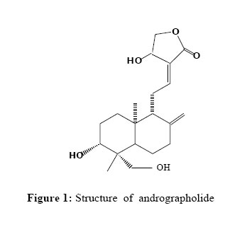
Indonesia Code Number: tc07040 Abstract Andrographolide isolated from Andrographis paniculata Ness (Acanthaceae) at 0.35 mM , 0.70 mM and 1.40 mM induced DNA fragmentation and increased the percentage of apoptotic cells when TD-47 human breast cancer cell line was treated for 24 , 48 and 72 h. The results demonstrated that andrographolide can induce apoptosis in TD-47 human breast cancer cell line in a time and concentration-dependent manner by increase expression of p53, bax , caspase-3 and decrease expression of bcl-2 determined by immunohistochemical analysis. Key words: Andrographis paniculata Ness , andrographolide, TD-47 cell , apoptosis. Introduction Apoptosis is cellular suicide or programmed cell death that which is mediated by activation of an evolutionary conserved intracellular pathway. Recently the relation of apoptosis and cancer has been emphasized and increasing evidence suggests that the processes of neoplastic transformation, progression and metastasis involve alteration of normal apoptotic pathway (Bold et al.,1997). Apoptosis also gives some clues about effective anticancer therapy, and many chemotherapeutic agents were reported to exert their anti-tumor effect inducing apoptosis of cancer cells. Andrographis paniculata Ness (Acanthaceae) is a traditional medical herb, a shrub grown in the moist, shady areas of India, China, Indonesia and throughout Southeast Asia and has been used as immunostimulant (Puri et al., 1993) and used for myocardial ischemic (Guo et al., 1995), pharyngotonsillitis (Thamlikitkul et al.,1991), respiratory tract infections (Coon and Ernst., 2004) and common cold (Melchior et al.,1996). It possesses antimicrobial (Prajjal et al., 2003), anti-inflammatory (Shen et al., 2002), hypotensive ( Zhang and Tan, 1996) , antihyperglycemic ( Yu et al., 2003; Borhanuddin et al ., 1994); oxygen radical scanvenging (Shen et al., 2002), atherosclerotic ( Wang and Zhao.,1994), anti-malarial activity (Dua et al.,2000), anti-HIV (Calabrese et al.,2000), antiplatelet aggregation (Amroyan et al.,1999), hepatic lipid peroxidation protective (Choudhury and Poddar,1984), hepatoprotective (Handa and Sarma ,1990), choleretic effect (Shukla et al., 1992), and anticancer effects (Kumar et al.,2004 ; Rajagopal et al. 2003; Chang, 1987; Matsuda et al., 1994 ). One of the major constituents of A. paniculata Ness is diterpene lactone such as andrographolide, which has anticancer activity in vitro in many tumor cell lines including leukemia, myeloma, HeLa, colon (HT-29), human peripheral blood lymphocytes (HPBLs), and human breast cancer MCF-7 (Satyanaraya et al., 2004). It was found that andrographolide possessed inhibitory effect of DNA Topoisomerase II (Sukardiman et al., 2005). Satyanarayana et al. (2004) reported that andrographolide inhibition of cell cycle from human breast cancer cell MCF-7 by induction of cell-cycle inhibitory protein p27 also decreased expression of cyclin-dependent kinase. This evidence suggests that the inhibitory effect of andrographolide might have contributed to its cytotoxicity by DNA fragmentation and induction of apoptosis. In this study the apoptosis-inducing effect of andrographolide isolated from A. paniculata Ness in T47-D human breast cancer cell line was investigated.. Material and Methods Plant material The aerial parts of Andrographis paniculata Ness were collected in September 2004 from Mojokerto, East Java , Indonesia and identified by Mr I.G.P Santa. A voucher specimen No. A123) was deposited at the Department of Natural Product Sciences, Faculty of Pharmacy Airlangga University Surabaya Indonesia. Isolation of andrographolide The herb of Andrographis paniculata Ness (300 g) was macerated and extracted with methanol. The solvent was concentrated in vacuo to yield methanol extract (30 g), which was diluted with aqua destilate , and then partitioned by ethyl acetate, from which 10g residue was obtained. The ethyl acetic fraction was subjected to silica column chromatography and gradiently eluted with chloroform-methanol to afford andrographolide. The andrographolide crystal was recrystalized from hot methanol. Its IR, 1H-NMR, 13C-NMR and MS data were in accordance with those of andrographolide (Matsuda et al.,1994) and standard (Sigma, USA). Cell Culture T47-D human breast cancer cell line was obtained from NAIST Narita Japan . Cells were grown in RPMI-1640 media (Sigma, US) containing 10% FBS, 100 mg/L Streptomycin and 105 unit Penicillin. A humidified incubator with 5% CO2 was used to row the cell at 37oC. Chemicals The p53, bax and bcl-2 antibody was from Boeringer Mannheim, Mannheim, Germany. All other chemicals were purchased from Sigma, St. Louis, MO. Cell morphology Cell morphology was determined by examining the culture cell under inverted microscope at magnification of 100x . Untreated cells served as control. Determination of Cell Viability Cells were plated in triplicate at a density of 1 x 10⁵ cell/ml in dish. Cells were treated with 0.35 mM , 0.70 mM and 1.40 mM andrographolide for 24, 48 and 72 h incubation. At the end of treatment, cells were harvested and counted using a haematocytometer. Cell viability was assessed by trypan blue exclusion test. Percentage of cell viabity was calculated from the formula :
Determination of Apoptotic Cells
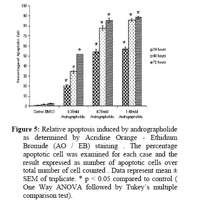
Cells were treated with 0.35 mM, 0.70 mM and 1.40 mM andrographolide for 24, 48 and 72 h incubation. Apoptosis was determined by morphological analysis after Acridine Orange / Ethidium Bromide (AO/EB) staining as described (Spector et al., 1998). A minimum of 300 cells was examined for each case and the result expressed as number of apoptotic cells over total number of cell counted (Spector et al., 1998). Percentage of apoptotic cells was calculated from the formula :
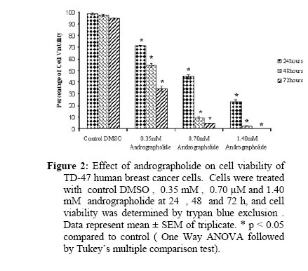
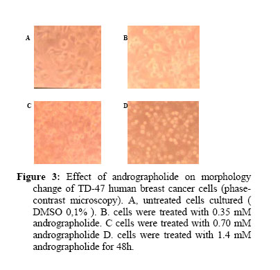
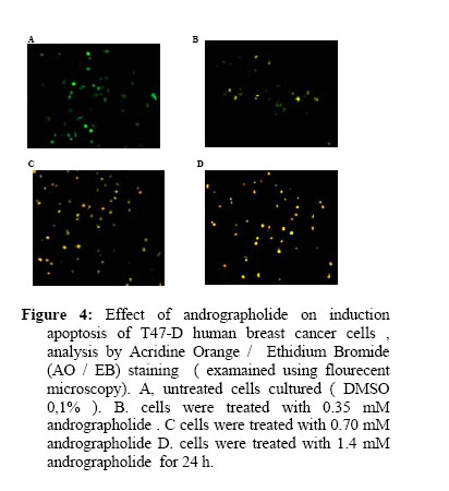
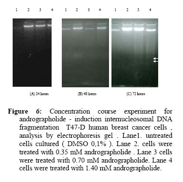
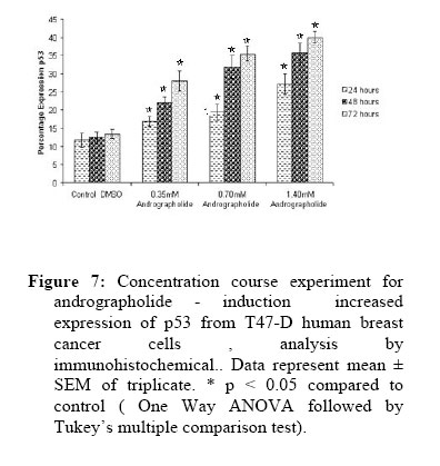
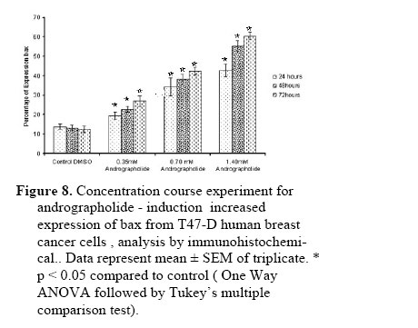
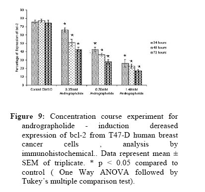
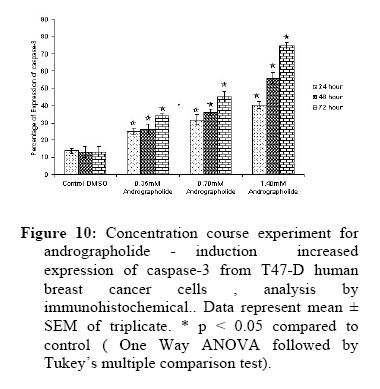
Analysis DNA Fragmentation Cells were treated with 0.35 mM, 0.70 mM and 1.40 mM andrographolide for 24, 48 and 72 h incubation. Cells were harveseted and suspended in 500 µl of lysis buffer containing 20 mM Tris-HCl (pH 7.4), 4 mM EDTA, 0,4%(v/v) Triton X, and incubated on ice for 30 min. After centrifugation for 5 min at 13.000 rpm using Eppendorf tube centrifuge, supernatant was extracted with phenol three times and once with chloroform. Then DNA was precipitated by incubating at -80oC for 30 min after the addition of 1 µg of glycogen, 100 µl of 5 M NaCl and 700 µl of isopropanol to each sample. DNA was collected by centrifuging at 13.000 rpm and washed once with 70% ethanol. DNA pellets were dissolved in 30 µl of TE buffer containing 10 µg/ml RNase A, and incubated at 37oC for 30 min. Ten µl of each DNA sample was loaded on 1.8% agarose gel. Immunohistochemical analysis for p53, bax, bcl-2 and caspase-3 Cells were treated with 0.35 mM, 0.75 mM and 1.40 mM andrographolide for 24, 48 and 72 h incubation. Cancer cells after treatment were studied by IHC using monoclonal antibodies to p53, bax , bcl-2 and caspase-3 (Boeringer Mannheim, Mannheim, Germany). Using methods described ( Santini et al.,1993), the cell were treated using the streptavidin-biotin-peroxidase complex method with the following modifications. Endogenous peroxidase activity was blocked with 3% H2O2 in methanol for 30 min. Sections were blocked with 20% horse serum for 1 h to prevent nonspecific binding. Primary antibody at a 1:50 dilution was applied overnight at 4°C, with appropriate negative and positive controls. Secondary biotinylated antibody at 1:200 was applied for 1 h to visualize bound antibody. SABC reaction was performed for 1 h at room temperature. The peroxidase activity was developed by incubation in 0.05% 3,3'-diaminobenzidine for 5 min, and slides were counterstained with hematoxylin. Immunoreactive score was determined semiquantitatively by means of a visual grading system in which staining intensity was categorized and graded by assessing the following categories: 0 (0-4%), 1 (5-24%), 2 (25 - 49%), 3 (50 - 74%), or 4 (75 - 100%) according to the previously reported criteria (Katja et al.,1999). Statistical analysis The data of viability cell and apoptotic cell were expressed as mean ± SEM of triplicate. Results were analysed statistically by One-way AOVA followed by Turkey,s multiple comparasion using SPSS software Student’s version 12.0. The difference was considered significant if p<0.05. Results and Discussion Andrographolide (Figure 1) showed a significant inhibitory effect on viability of TD-47 human breast cancer cells in vitro. The inhibitory action was noticed to be concentration and time-dependent. The maximum inhibitory effect of andrographolide was obtained at concentration of 1.40mM at 72h, where the percentage cell viability was 0.28%. On the other hand , after 0.35mM andrographolide treatment at 24h , percentage cell viability was 71.45% and significantly decreased after 48 and 72h , when percentage cell viability were 44.83% and 23.52% respectively. After 0.75mM andrographolide treatment at 24h , percentage cell viability was 54.45% and significantly decreased after 48 and 72h, when percentage cell viability were 9.45% and 2.5% respectively. On the other hand , after 1.40mM andrographolide treatment at 24h , percentage cell viability was 34.35% which significantly decreased after 48 and 72hour , when percentage cell viability were 4.36% and 0.28 % respectively, as shown in Figure 2. When TD-47 human breast cancer cells treated with various concentration of andrographolide for 48 h, were examined under a phase contrast microscope they exhibited distinct morphological features of apoptosis, such as cellular shrinkage and formation of apoptotic bodies, as shown in Figure 3. Apoptotic cells can be recognized by stereotypical morphological changes: the cell shrinks, shows deformation and looses contact to its neighbouring cells. Its chromatin condenses and marginates at the nuclear membrane, the plasma membrane is blebbing or budding, and finally the cell is fragmented into compact membrane-enclosed structures, called 'apoptotic bodies' which contain cytosol, the condensed chromatin and organelles. The apoptotic bodies are engulfed by macrophages and thus are removed from the tissue without causing an inflammatory response. These morphological changes are a consequence of characteristic molecular and biochemical events occurring in an apoptotic cell, most notably the activation of proteolytic enzymes which eventually mediate the cleavage of DNA into oligonucleosomal fragments as well as the cleavage of a multitude of specific protein substrates which usually determine the integrity and shape of the cytoplasm or organelles (Saraste and Pulkki , 2000). After 24 h treatment of TD-47 human breast cancer cell with 0.35 mM , 0.70 µM and 1.40 mM andrographolide , nuclear structure using acridine orange/ethidium bromide (AO / EB) staining, and examined using fluorescent microscopy exhibited condensation and fragmentation of some nuclei, as shown in Figure 4. Live cell appeared uniformly green.. Early apoptotic cell stained green an contain bright green dots in the nuclei as a consequence chromatin condensation and nuclear fragmentation. Late apoptotic cell showed condensed and often fragmented nuclei and the incorporated ethidium bromide stained orange ( Spector et al., 1998). After 24, 48 and 72 h treatment of TD-47 human breast cancer cell with 0.35 mM ; 0.70 µM and 1.40 mM andrographolide , nuclear structure using acridine orange/ethidium bromide (AO / EB) staining, examained using flourecent microscopy, exhibited increased apoptotic cells in time- and concentration - related manner (Figure 5). After 72 h treatment of TD-47 human breast cancer cell with concentration 0.70 mM and 1.40 mM of andrographolide, it was found that it induced internucleosomal DNA fragmentation , one of biochemical hall mark of apoptosis, as shown in Figure 6. Comparable findings were reported in the study by Chopin et al. (2002) where they did not find internucleosomal DNA fragmentation from TD-47 human breast cancer cell when treated with 2.5mM sodium butyrate for 48h (that it possesses inhibitory effect of DNA Topoisomerase). When TD-47 human breast cancer cell was treated with various concentration of andrographolide from A. paniculata for 24h, 48h and 72h , the expression p53 increased in time and concentration-related manner as shown in Figure 7. Common features of many anti-cancer drugs such as camptothecin, etoposide were induction of DNA damage followed by the activation of tumour suppressor protein p53 as mediator of their cellular effects. The accumulation and activated of wild-type p53 results in least two pathways, cell cycles and apoptosis. (Bates and Vousden, 1994). After TD-47 human breast cancer cell was treated with various concentration of andrographolide from A. paniculata for 24h, 48h and 72h , the expression bax increased in a concentration-related manner as shown in Figure 8. When TD-47 human breast cancer cell was treated with various concentration of andrographolide from Andrographis paniculata Ness for 24h, 48h and 72h , the expression bcl-2 decreased in a concentration-related manner analysed by immunohistochemical method , as shown in Figure 9. Apoptosis is controlled by ratio of various bcl-2 family members (Read et al., 1996). When level of apoptosis promoters bax increase, apoptosis is accelerated, whereas when the inhibitor of apoptosis bcl-2 increase , the cell are predisposed to be resistant to apoptosis in response stimuli. Previous report have demonstrated that changes in the ratio of protoapoptotic to antiapoptotic protein result in susceptibility to apoptosis , and high bax : bcl-2 ratio has been found to correlate with etoposide-induced apoptosis (Chresta et al., 1996). After TD-47 human breast cancer cell was treated with various concentration of andrographolide from Andrographis paniculata Ness for 24h, 48h and 72h, the expression caspase-3 increased in a concentration-related manner analysed by immunohistochemical metdhod , as shown in Figure 10. From the results it could be concluded that the andrographolide isolated from Andrographis paniculata Ness (Acanthaceae) induced apoptosis in TD-47 human breast cancer cell line in a time and concentration-dependent manner by increase expression of p53 bax , caspase-3 and decrease expression of bcl-2. Further studies should be performed on experimental animals to spotlight the mechanism of action of andrographolide at the molecular level of the tumor cells. Acknowledgments This work was supported by the Research Grant of the Program Hibah Kompetisi B ( Program of Found Competition) 2006 , Directorate of General of Higher Education , Department of National Education , Indonesia. References
© Copyright 2007 -African Journal of Traditional, Complementary and Alternative Medicines ; The following images related to this document are available:Photo images[tc07040f8.jpg] [tc07040f4.jpg] [tc07040f7.jpg] [tc07040f9.jpg] [tc07040f2.jpg] [tc07040f1.jpg] [tc07040f10.jpg] [tc07040f3.jpg] [tc07040f6.jpg] [tc07040f5.jpg] |
| |||||||||


