 |
| About Bioline | All Journals | Testimonials | Membership | News |
|
||||||
|
||||||
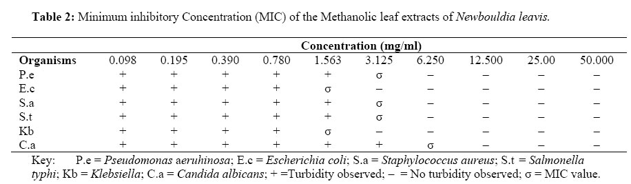
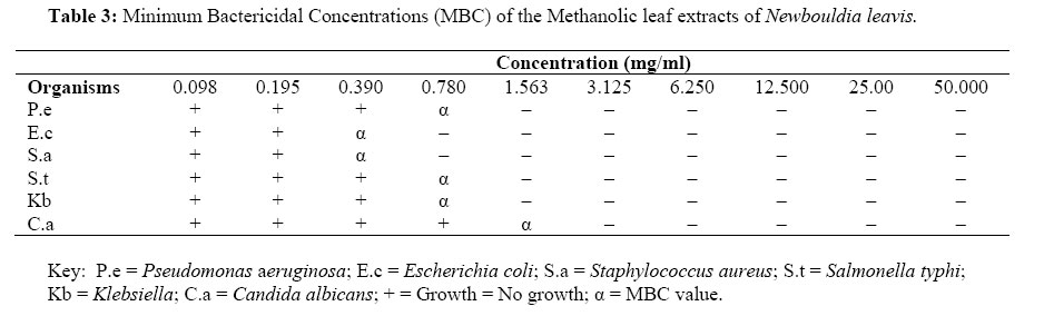
African Journal of Traditional, Complimentary and Alternative Medicines, Vol.4, No. 4, 2007, pg. 476 – 480 Research Paper PHYTOCHEMICAL AND IN VITRO ANTIMICROBIAL ASSAY OF THE LEAF EXTRACT OF NEWBOULDIA LAEVIS *Usman, H. and Osuji, J. C. Department of Chemistry, University of Maiduguri, Maiduguri - Nigeria. E-mail: husman321@yahoo.com Code Number: tc07063 AbstractThe methanolic leaf extract of Newbouldia laevis was subjected to preliminary phytochemical screening and in-vitro antimicrobial tests. The extract revealed the presence of flavonoids, tannins, terpenes, steroidal and cardiac glycosides. The antimicrobial activity of the plant extract was assayed by the agar plate disc diffusion and nutrient broth dilution techniques. Test microorganisms were Pseudomonas aeruginosa, Escherichia coli, Staphylococcus aureus, Salmonella typhi, Klebsiella spp. and Candida albicans; all the organisms were laboratory isolates. The extract inhibited the growth of all the test organisms especially against Klebsiella spp. and S. aureus which had mean inhibition zone of 42.3±1.5 and 32.3±1.5 mm respectively. The results showed minimum inhibitory concentration (MIC) of 1.563 mg/ml against Escherichia coli and Klebsiella spp. and 3.125 mg/ml against Pseudomonas aeruginosa, Staphylococcus aureus and Salmonella typhi. The minimal bactericidal concentration (MBC) against Escherichia coli and Staphylococcus aureus was 0.39 mg/ml. This study has justified the traditional use of this plant for the treatment of stomach discomfort, diarrhea, dysentery and as a remedy for wound healing whose causative agents are some of the organisms used in this study. Key words: Antimicrobial, Leaf Extracts, In vitro, Phytochemical, Newbouldia leavis IntroductionThe use of plants as medicine is an ancient practice common to all societies especially the African society. This practice continues to exist in the developing nations. It is on this basis that researchers keep on working on medicinal plants in order to produce/develop the best medicines for physiological uses. Newbouldia laevis (P. Beauv) seem or boundary tree called variously as; 'Aduruku' in Hausa, 'Ogirisi' in Igbo and 'Akoko' in Yoruba languages (Hutchinson and Dalziel, 1963) is a medium sized angiosperm which belongs to the Bignoniaceae family. It grows to a height of about 7 - 8 (up to 15) metres, more usually a shrub of 2 - 3 metres, many-stemmed forming clumps of gnarled branches (Arbonnier, 2004). Newbouldia laevis is native to tropical Africa and grows from Guinea Savannahs to dense forests, on moist and well-drained soils. It inhabits the secondary forest extending from Senegal to Cameroon, Gabon, Democratic Republic of Congo, Angola (Arbonnier, 2004). In Nigeria, the bark is chewed and swallowed for stomach pains, diarrhea and toothache (Lewis and Manony, 1977). The plant has been found to be effective in the treatment of elephantiasis, dysentery, rheumatic swellings, syphilis, constipation, pile and as a vermifuge to round worms. It has also been found useful for earache, sore feet, chest pain, epilepsy and children's convulsion (Akunyili, 2000). The leaf, stem and fruits have been used for febrifuge; wound dressing and stomach ache (Iwu, 2000). Earlier studies on the leaves and bark of Congolese Newbouldia leavis revealed the absence of flavonoids; saponins, quinones, terpenes or steroids (Oliver-Bever, 1986). Recent phytochemical studies on the root, root bark and stem of this plant revealed the presence of alkaloids, quinoid and phenylpropanoid amongst others (Gafner et al., 1997; Aladesanmi et al., 1998; Germann et al., 2006). There was no extensive report on the presence of compounds from the leaves of this species. In this investigation, the in vitro antimicrobial effects of the crude methanolic leaf extract of this plant against the organisms found commonly responsible for the ailments aforementioned; including Staphylococcus aureus whose related infections are one of the most common cause of noscomical (hospital acquired) infections were investigated. Materials and MethodsThe leaf of Newbouldia laevis was collected from the premises of City Girls Secondary School Enugu, Enugu State, Nigeria in the early morning of 11th February, 2006. The herbarium specimen was identified by Mr. A. O. Ozioko of the International Centre for Ethnomedicine and Drug Discovery (INTERCEDD), Nsukka, Enugu State, Nigeria where a voucher (No. BDCP/INTERCEDD 033) specimen was deposited. The leaves of N. laevis was dried under shade and pulverized into fine powder. About 300 g of the powdered form was extracted with 95% (v/v) methanol in H2O employing the reflux method. The extract was concentrated under reduced pressure to yield a dark green mass weighing 21.1 g (7.03 % w/w). The crude extract was then coded 'CME' and stored aseptically in a desiccator until required. Phytochemical ScreeningThe phytochemical analyses of the crude methanolic extract was carried out in order to ascertain the presence of its constituents such as flavonoids, alkaloids, saponins, steroidal nucleus, tannins, cardio-active glycosides utilizing standard methods of analyses (Sofowora, 1993; Trease and Evans, 2002). OrganismsStaphylococcus aureus, Escherichia coli, Klebsiella spp., Pseudomonas aeruginosa and Salmonella typhi. The only fungus utilized was Candida albicans. Most of the isolates were obtained from the Department of Veterinary Microbiology & Parasitology, University of Maiduguri; Staphylococcus aureus and Candida albicans were obtained from the Department of Microbiology, University of Maiduguri Teaching Hospital, Maiduguri, Nigeria. Antimicrobial TestsThe antimicrobial susceptibility test was conducted using the method earlier described by Sidney et al., (1978); Vollekovà et al., (2001) with little modification by Usman et al., (2005). The tests were carried out using a stock concentration of 100 mg/ml prepared by dissolving 1 g of the crude extract into 10 ml of sterile distilled water. The dilution ratio for Gram-positive bacteria and Gram-negative bacteria was 1:1000 and 1:5000 respectively using peptone water (Usman et al., 2005). About 0.5 ml of the dilute cultures was aseptically inoculated on the surface of sterile Petri-dishes containing sterile solid nutrient agar. Discs impregnated with the crude extract at the concentration of 5 mg/disc were aseptically mounted on agar and thereafter incubated at 37 oC for 24 h, the inhibition zone was observed and then recorded in millimeters using a transparent metre rule. The tests were conducted in triplicate and results presented as mean ± SEM. Standard antimicrobial disc used were Amoxiclive (30 µg), Ceflunat (30 µg), Levoxine (5 µg), Ofloxacine (5 µg) and Peflotab (5 µg). Minimum Inhibitory ConcentrationMIC was defined as the lowest concentration where no visible turbidity was observed in the test tubes. The concentrations were determined as earlier described by Vollekovà et al., (2001) with some modification by Usman et al., (2005). The MIC was determined for the micro-organisms that showed reasonable sensitivity to the test extracts. In this test, the micro-organisms were prepared using the broth dilution technique. The stock extract concentration of 100 mg/ml was made by dissolving 1 g of the extract in 10 ml of sterile distilled water and the working concentrations prepared by two-fold serial dilution technique that ranged from 0.195 mg/ml to 50 mg/ml using nutrient broth and later inoculated with 0.2 ml suspension of the test organisms. After 24 h. incubation at 37 oC, the tubes were observed for turbidity. The lowest concentrations where no turbidity were observed was determined and noted (Usman et al., 2005). Minimum Bactericidal ConcentrationThe minimal bactericidal concentration was determined from broth dilution test resulting from the MIC tubes as described previously (Vollekovà et al., 2001; Usman et al., 2005) by inoculating the content of each test tube on a nutrient agar plate. The plates were then incubated at 37 oC for 24 h.. The lowest concentration of the extract that showed no growth was noted and recorded as the minimum bactericidal concentration Results and DiscussionsThe phytochemical screening of the crude methanolic leaf extracts of N. laevis revealed the presence of flavonoids, tannins, terpenes, steroidal and cardiac glycosides; alkaloids and saponins were found to be absent. These classes of compound are known to show curative activity against several pathogens and therefore could explain its use traditionally for the treatment of wide array of illnesses (Hassan et al., 2004; Usman, et al; 2005). Cardiac glycosides, flavonoids, tannins and terpenoids were detected The in vitro antimicrobial screening presented in Table 1 showed the susceptibility test against Grams- positive and negative organisms and a fungal species. The extract exhibited considerable level of inhibition against all the test organisms with the highest activity on Klebsiella (42.3 ± 1.5 mm) and the lowest against Candida albicans (14.3 ± 2.0 mm). The zone of inhibition produced by most antibiotic discs against some of the organisms were found to be appreciable in relation to those activities produced by most organisms under study though not statistically comparatively to that presented by the extract. However, it is suggested that diameters of zones of inhibition > 10 mm were considered active (Zwadyk et al., 1972; Usman et al., 2005). From the results of the minimum inhibitory concentration (MIC) and minimum bactericidal concentration (MBC) presented in Tables 2 and 3 respectively; it was observed that the broadest activity of the extract was against most Gram-negative organisms studied (E. coli, Klebsiella spp. and P. aeruginosa) while lower activity was noticed against Candida albicans. The extract exhibited considerable activity against S. aureus, a pyogenic Gram-positive bacterium known to play a significant role in invasive skin diseases including superficial and deep follicular lesion (Usman et al., 2005). It also showed appreciable activity against E.coli. The high antibacterial activities of this extracts could not be unrelated to the presence of the plant secondary metabolites detected. In line with these findings, it has been reported that tannins had been widely used as an application to sprains, bruises and superficial wounds (Usman et al., 2005). In conclusion, the fact that the extracts produced inhibitory activities against almost all test organisms particularly Gram - negative organisms and marked higher activities (though not of comparative concentrations) than most of the reference drugs could provides some scientific basis for some of the folkloric claims. We, therefore, suggest the isolation and possible characterization of the bioactive constituent(s) from the extracts of this plant species as a possible antibacterial agent especially now that numbers of reports of methicilin-resistant S. aureus (MRSA) and vancomycine-resistant S. aureus (VRSA) is on the increase (Hiramatsu et al., 1997; Fridkin, 2001). AcknowledgementsThe authors are grateful to the Department of Chemistry, University of Maiduguri for providing some of the reagents and apparatus for the work and Messrs Samson Amali and Samson Gamache of the Department of Veterinary Microbiology University of Maiduguri and Department of Microbiology, University of Maiduguri Teaching Hospital respectively for their technical assistance. References
© Copyright 2007 - African Journal of Traditional, Complementary and Alternative Medicines The following images related to this document are available:Photo images[tc07063t3.jpg] [tc07063t1.jpg] [tc07063t2.jpg] |
| |||||||||
